. 2008 Jun;59(6):1347-54.
doi: 10.1002/mrm.21577.

AxCaliber: a method for measuring axon diameter distribution from diffusion MRI

Affiliations

AxCaliber: a method for measuring axon diameter distribution from diffusion MRI

Yaniv Assaf et al. Magn Reson Med. 2008 Jun.

Abstract

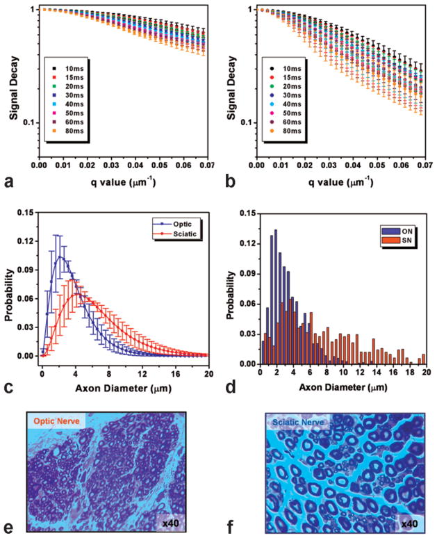
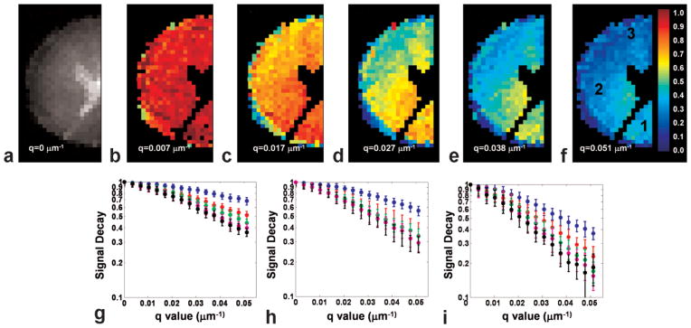
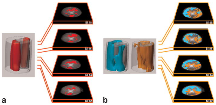
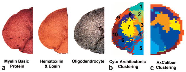
The diameter of a myelinated nerve axon is directly proportional to its conduction velocity, so the axon diameter distribution helps determine the channel capacity of nervous transmission along fascicles in the central (CNS) and peripheral nervous systems (PNS). Previously, this histological information could only be obtained using invasive tissue biopsies. Here we propose a new NMR-based approach that employs a model of water diffusion within "restricted" cylindrical axons to estimate their diameter distribution within a nerve bundle. This approach can be combined with MRI to furnish an estimate of the axon diameter distribution within each voxel. This method is validated by comparing the diameter distributions measured using the NMR and histological techniques on sciatic and optic nerve tissue specimens. The axon diameter distribution measured in each voxel of porcine spinal cord using MRI and using histological methods were similar. Applications are expected in longitudinal studies designed to follow nerve growth in normal and abnormal development, as well as in diagnosing disorders and diseases affecting specific populations of axons in the CNS and PNS.


Figures

FIG. 1
FIG. 1
AxCaliber of porcine optic and sciatic nerves. a: Multi diffusion time diffusion spectroscopy signal decay of an optic nerve sample. b: Multi diffusion time diffusion spectroscopy signal decay of a sciatic nerve sample. c: Extracted AxCaliber axon diameter distribution based on the signal decays given in (a) and (b). d: Axon diameter distribution derived from electron microscopy section of the two nerve samples. e,f: Electron microscope section of one optic nerve (e) and one sciatic nerve samples upon which the data in (a–d) is based. Note the large difference in axonal morphometry between the two nerves.
FIG. 2
FIG. 2
AxCaliber MRI dataset of porcine spinal cord. a: Half spinal cord diffusion-weighted MRI with q = 0 representing the T2 weighting of the sample. b–f: Diffusion-weighted images at different q values and diffusion time of 120 ms; all images are normalized to the q = 0 image (given in a), thus the color-scale represents the normalized decay. Note the differentiation to at least visual three regions in the most diffusion-weighted image. f: The fasciculus gracilis (dorsal region, 1), spinocerebellar and corticospinal fasciculi (lateral region, 2), and at the spinotectalis, reticulospinal, and vestibulospinal fasciculi (rostral region, 3). g–i: The corresponding diffusion signal decay from region of interest in the areas of the aforementioned regions with (g) corresponding to region 1, (h) to region 2, and (i) to region 3.
FIG. 3
FIG. 3
3D clusters of AxCaliber MRI of porcine spinal cord. a: Cluster 1 depicts mostly the dorsal fascicules gracilis. Note the similarity in the location of these regions along the spinal cord slices. b: Clusters 2 and 3 depict mostly the spinotectalis, reticulospinal, vestibulospinal fascicles (cyan) and the spinocerebellar and corticospinal fascicles (orange).
FIG. 4
FIG. 4
Comparison of AxCaliber with histology. a: Histological staining of spinal cord section with MBP, cell body (H&E), and oligodendrocytes markers. b: Digitized cyto-architectonic analysis of the histological sections in (a) with the following fascicles identified: 1. Anterior spino-thalamic; 2. Reticulo-spinal; 3. Anterior cortico-spinal; 4. Gray matter; 5. Fasciculus gracilis and cuneatus; 6. Lateral cortico-spinal; 7. Lateral spino-thalamic; 8. Spino-tectalis; 9. Ventral spino-cerebellar; 10. Dorsal spino-cerebellar. c: AxCaliber clusters of the same spinal cord sections strongly resemble those obtained using histological analysis.

References

    1. Hursh JB. The properties of growing nerve fibers. Am J Physiol. 1939;127:140–153.
    1. Ritchie JM. On the relation between fibre diameter and conduction velocity in myelinated nerve fibres. Proc R Soc Lond B Biol Sci. 1982;217:29–35. - PubMed
    1. Waxman SG. Determinants of conduction velocity in myelinated nerve fibers. Muscle Nerve. 1980;3:141–150. - PubMed
    1. Tasaki I, Ishii K, Ito H. On the relation between the conduction-rate, the fiber-diameter and the internodal distance of the medullated nerve fiber. Jpn J Med Sci III Biophys. 1943;9:189–199.
    1. Nicholls JG, Martin AR, Wallace BG, Fuchs PA. From neuron to brain. Sunderland, MA: Sinauer Associates; 2001.

Publication types

MeSH terms