Comparative Study
. 2008 Jun;47(6):2059-67.
doi: 10.1002/hep.22283.

Transforming growth factor-beta gene expression signature in mouse hepatocytes predicts clinical outcome in human cancer

Affiliations
Comparative Study

Transforming growth factor-beta gene expression signature in mouse hepatocytes predicts clinical outcome in human cancer

Cédric Coulouarn et al. Hepatology. 2008 Jun.

Abstract

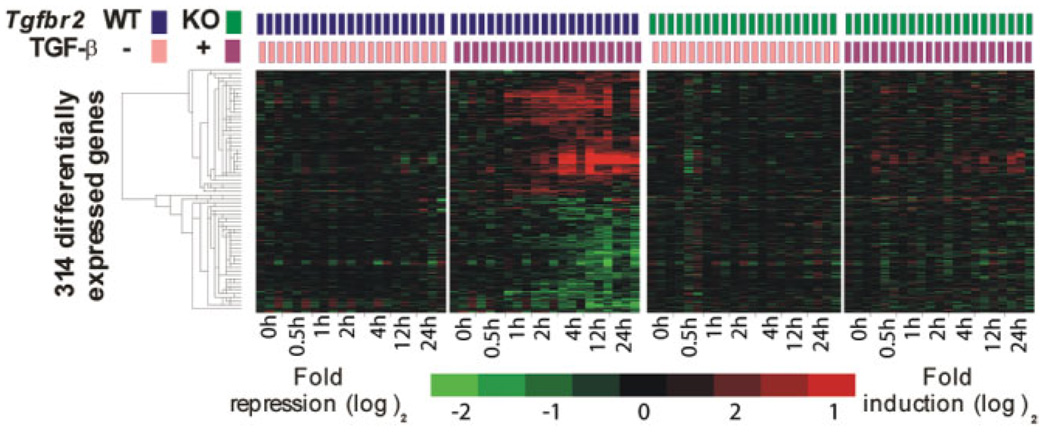
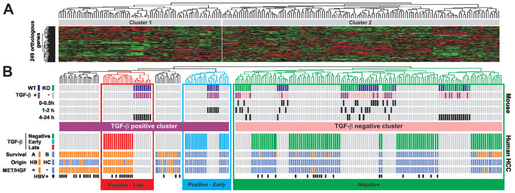
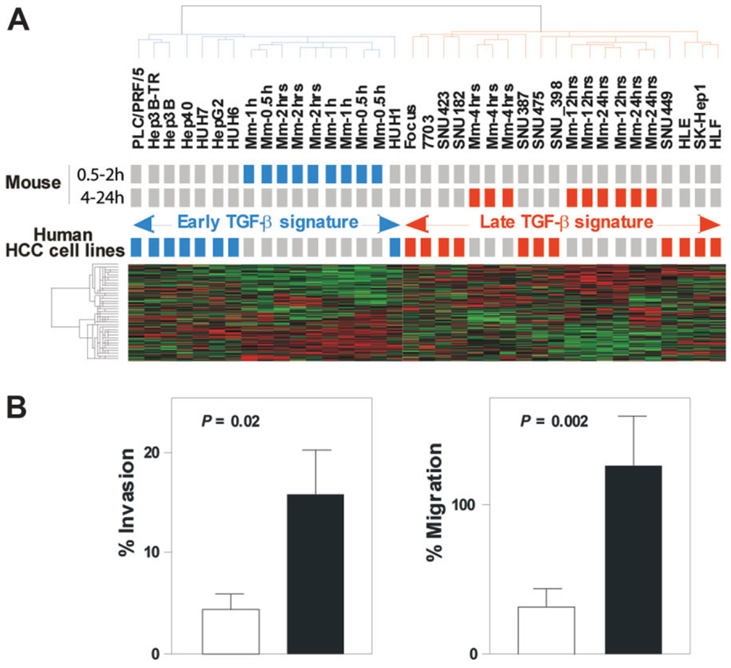
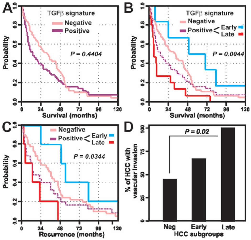
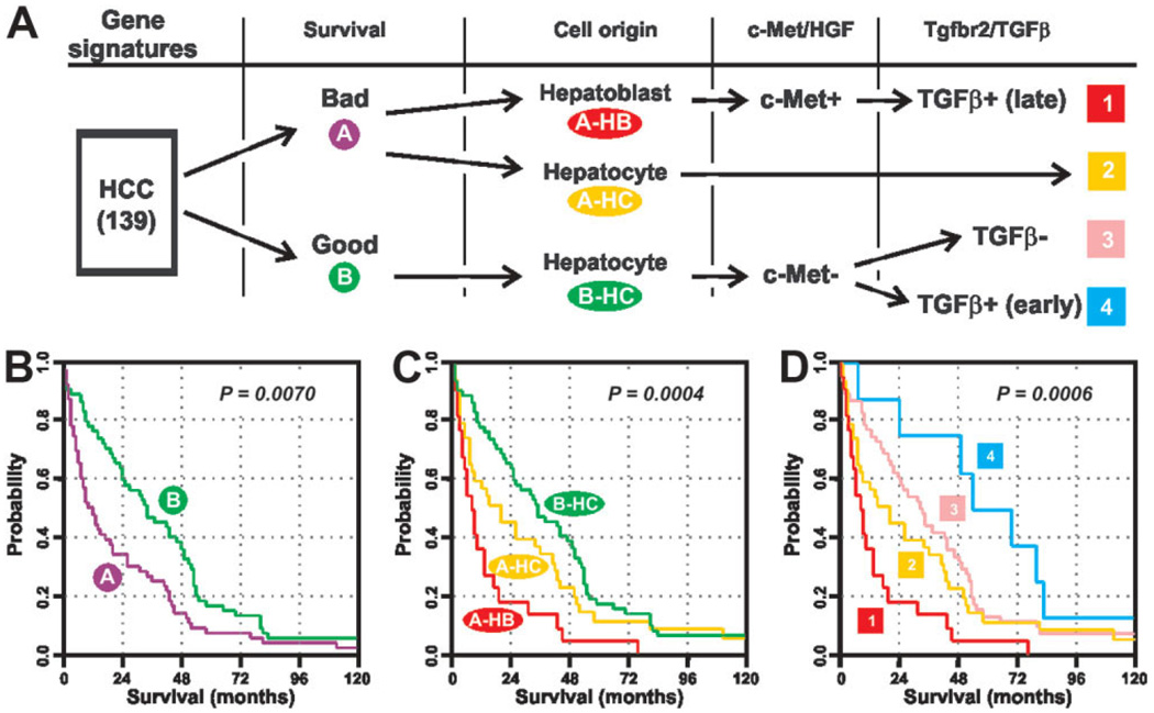
Hepatocellular carcinoma (HCC) is one of the most common cancers in the world. The clinical heterogeneity of HCC, and the lack of good diagnostic markers and treatment strategies, has rendered the disease a major challenge. Patients with HCC have a highly variable clinical course, indicating that HCC comprises several biologically distinctive subgroups reflecting a molecular heterogeneity of the tumors. Transforming growth factor beta (TGF-beta) is known to exhibit tumor stage dependent suppressive (that is, growth inhibition) and oncogenic (that is, invasiveness) properties. Here, we asked if a TGF-beta specific gene expression signature could refine the classification and prognostic predictions for HCC patients. Applying a comparative functional genomics approach we demonstrated that a temporal TGF-beta gene expression signature established in mouse primary hepatocytes successfully discriminated distinct subgroups of HCC. The TGF-beta positive cluster included two novel homogeneous groups of HCC associated with early and late TGF-beta signatures. Kaplan-Meier plots and log-rank statistics indicated that the patients with a late TGF-beta signature showed significantly (P < 0.005) shortened mean survival time (16.2 +/- 5.3 months) compared to the patients with an early (60.7 +/- 16.1 months) TGF-beta signature. Also, tumors expressing late TGF-beta-responsive genes displayed invasive phenotype and increased tumor recurrence. We also showed that the late TGF-beta signature accurately predicted liver metastasis and discriminated HCC cell lines by degree of invasiveness. Finally, we established that the TGF-beta gene expression signature possessed a predictive value for tumors other than HCC.

Conclusion: These data demonstrate the clinical significance of the genes embedded in TGF-beta expression signature for the molecular classification of HCC.


Conflict of interest statement

Potential conflict of interest: Nothing to report.

Figures

Fig. 1
Fig. 1
Hierarchical cluster analysis of 314 genes included in the mouse TGF-β signature. Data are presented in a matrix format in which rows and columns represent genes and samples, respectively. Hepatocytes were isolated from Tgfbr2+/+/AlbCre+/− (WT, two left panels) and Tgfbr2fl/fl/AlbCre+/− (KO, two right panels) mice. Time-course gene expression profiling was performed on primary cultures challenged with TGF-β (+) versus vehicle alone (−) up to 24 hours. Genes included in the TGF-β signature were significantly up-regulated or down-regulated by TGF-β in WT hepatocytes (second panel) but no fluctuation was observed in KO hepatocytes (two right panels).
Fig. 2
Fig. 2
Discrete temporal mouse TGF-β gene expression signatures define distinct subtypes of human HCC. (A) Dendrogram and heat-map overview of mouse data set integrated with 139 cases of human HCC. Clustering was based on the expression of 249 orthologous genes and the data are presented in a matrix format in which rows and columns represent genes and samples, respectively. (B) Detailed view of mouse and human samples cluster analysis. Upper part: Clustering of mouse samples corresponding to WTand KO hepatocytes challenged (+) or not (−) with TGF-β for a given time identified at the beginning of each row. Clustering analysis identified a TGF-β-positive cluster corresponding to WT-treated hepatocytes and a TGF-β-negative cluster corresponding to untreated and treated KO hepatocytes as well as untreated WT hepatocytes. Lower part: Clustering of human HCC samples. Integration of mouse and human samples sorted human HCC into TGF-β-positive and TGF-β-negative clusters. Within the TGF-β-positive cluster, the analysis of dendrogram branches revealed two homogeneous subtypes of HCC associated with WT hepatocytes treated for 1 to 2 hours and 4 to 24 hours and annotated Positive-Early and Positive-Late, respectively. Distribution of HCC between previously described subgroups with respect to survival (A, bad prognosis versus B, good prognosis), cell origin (HB, hepatoblast versus HC, hepatocyte), c-Met/hepatocyte growth factor (HGF) signature (− versus +) or HBV infection status is indicated at the beginning of each row.
Fig. 3
Fig. 3
Early and late mouse TGF-β gene expression signatures discriminate two subtypes of human HCC-derived cell lines with distinct invasiveness behaviors. (A) Dendrogram and heat-map overview of early and late mouse TGF-β signatures integrated with the gene expression profiles of 19 human HCC-derived cell lines. Clustering was based on the expression of 249 orthologous genes and the data are presented in a matrix format in which rows and columns represent genes and samples, respectively. Cell lines that cluster with mouse samples corresponding to WT hepato-cytes challenged with TGF-β for 0.5 to 2 hours (early signature) or for 4 to 24 hours (late signature) are colored in blue and red, respectively. (B) Early and late TGF-β signatures predict invasiveness of HCC-derived cell lines. Invasive and migrating activity of HCC cell lines was measured by using the BD BioCoat Matrigel Invasion Chambers as described in Materials and Methods. Invasion and migration of HCC cell lines defined by the late TGF-β signature (black column) were higher than those defined by the early TGF-β signature (white column). Columns, mean; bars, standard error of the mean (SEM), n = 3.
Fig. 4
Fig. 4
Clinical relevance of the HCC subtypes defined by the TGF-β signatures. (A) Kaplan-Meier plots and log-rank statistics analysis of overall survival do not reveal significant differences between patients defined by positive and negative TGF-β signatures. (B) Within the TGF-β-positive group (dashed purple line) the subtypes of HCC defined by early (blue line) and late (red line) TGF-β signatures show marked differences in the overall survival of patients. (C) Similar results are obtained by the analysis of patient recurrence. (D) Vascular invasion rate in HCC defined by negative, early, and late TGF-β signatures.
Fig. 5
Fig. 5
Comparative and integrative functional genomic strategy improves the molecular prognostication of liver cancer. (A) Stratification of a cohort of 139 cases of human HCC based on the successive integration of gene expression signatures covering relevant genes with respect to survival (A, bad prognosis versus B, good prognosis), cell origin (HB, hepatoblast versus HC, hepatocyte), c-Met/hepatocyte growth factor (HGF), and Tgfbr2/TGF-β signaling pathways. (B-D) Kaplan-Meier plots and log-rank statistics demonstrate the difference in the overall survival between individuals with distinct subtypes of HCC defined by the successive integration of gene expression signatures.

References

    1. Parkin DM, Bray F, Ferlay J, Pisani P. Global cancer statistics, 2002. CA Cancer J Clin. 2005;55:74–108. - PubMed
    1. El Serag HB. Hepatocellular carcinoma: recent trends in the United States. Gastroenterology. 2004;127(5 Suppl 1):S27–S34. - PubMed
    1. Villanueva A, Newell P, Chiang DY, Friedman SL, Llovet JM. Genomics and signaling pathways in hepatocellular carcinoma. Semin Liver Dis. 2007;27:55–76. - PubMed
    1. Bruix J, Boix L, Sala M, Llovet JM. Focus on hepatocellular carcinoma. Cancer Cell. 2004;5:215–219. - PubMed
    1. Llovet JM, Burroughs A, Bruix J. Hepatocellular carcinoma. Lancet. 2003;362:1907–1917. - PubMed

Publication types

MeSH terms

Substances