. 2008 May 27;5(5):e112.
doi: 10.1371/journal.pmed.0050112.

Decreased brain volume in adults with childhood lead exposure

Affiliations

Decreased brain volume in adults with childhood lead exposure

Kim M Cecil et al. PLoS Med. .

Abstract

Background: Although environmental lead exposure is associated with significant deficits in cognition, executive functions, social behaviors, and motor abilities, the neuroanatomical basis for these impairments remains poorly understood. In this study, we examined the relationship between childhood lead exposure and adult brain volume using magnetic resonance imaging (MRI). We also explored how volume changes correlate with historic neuropsychological assessments.

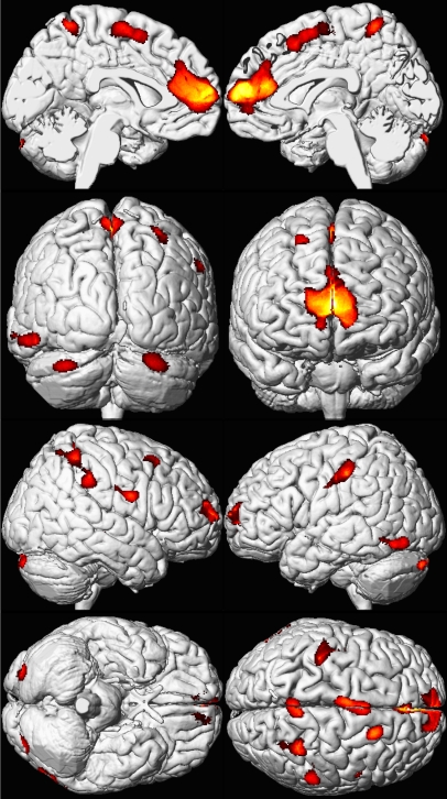
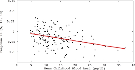
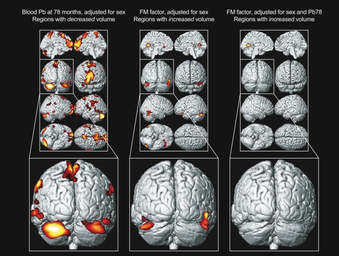
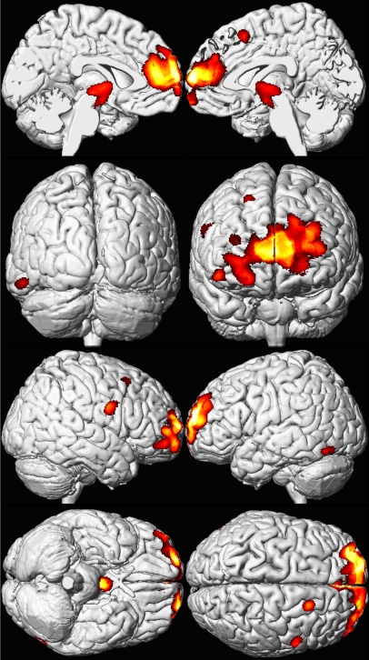
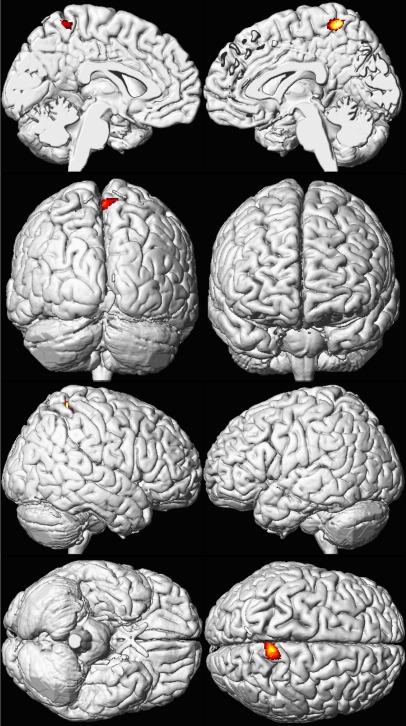
Methods and findings: Volumetric analyses of whole brain MRI data revealed significant decreases in brain volume associated with childhood blood lead concentrations. Using conservative, minimum contiguous cluster size and statistical criteria (700 voxels, unadjusted p < 0.001), approximately 1.2% of the total gray matter was significantly and inversely associated with mean childhood blood lead concentration. The most affected regions included frontal gray matter, specifically the anterior cingulate cortex (ACC). Areas of lead-associated gray matter volume loss were much larger and more significant in men than women. We found that fine motor factor scores positively correlated with gray matter volume in the cerebellar hemispheres; adding blood lead concentrations as a variable to the model attenuated this correlation.

Conclusions: Childhood lead exposure is associated with region-specific reductions in adult gray matter volume. Affected regions include the portions of the prefrontal cortex and ACC responsible for executive functions, mood regulation, and decision-making. These neuroanatomical findings were more pronounced for males, suggesting that lead-related atrophic changes have a disparate impact across sexes. This analysis suggests that adverse cognitive and behavioral outcomes may be related to lead's effect on brain development producing persistent alterations in structure. Using a simple model, we found that blood lead concentration mediates brain volume and fine motor function.


Conflict of interest statement

Competing Interests: Two of the study's authors, BPL and RH, are on the editorial board of PLoS Medicine. BPL and KND sporadically serve as expert witnesses without personal financial gain.

Figures

Figure 1
Figure 1. Regional Brain Volume Loss for the Cincinnati Lead Study Participants
A composite representation of regions with significant volume loss for male and female CLS participants (n = 157) associated with mean childhood blood lead concentrations is shown with red and yellow clusters overlaid upon a standard brain template (seen at multiple angles; the first row presents views from the midline of the left and right hemispheres, respectively; the second row demonstrates views from the back and front of the cerebrum, respectively; the third row shows the lateral right and left hemispheres; and the fourth row shows views from below and above the cerebrum. Brain template source reference [51].
Figure 2
Figure 2. Regional Brain Volume Loss for the Cincinnati Lead Study Participants
The relationship of individual brain volume with mean childhood blood lead concentrations within a medial frontal cluster is illustrated by this plot. The model is adjusted for age at time of scanning and birth weight, using a cluster threshold of 700 voxels and unadjusted p ≤ 0.001.
Figure 3
Figure 3. Structure-Function Relationship
Left, a composite representation of regions with significant volume loss for CLS participants associated with blood lead concentration at 78 mo of age and adjusted for sex is shown in red and yellow overlaid upon a standard brain template with the posterior coronal view highlighted below. Middle, analogous composite representation of regions with significant volume gain associated with fine motor factor scores and adjusted for sex. Right, analogous composite representation of regions with significant volume gain associated with fine motor factor scores, adjusted for sex and blood lead concentration at 78 mo of age. Brain template source reference [51].
Figure 4
Figure 4. Sex Influences Brain Volume Loss Associated with Lead Exposure (Males)
A composite representation of regions with significant volume loss for CLS participants associated with mean childhood blood lead concentrations is shown in red and yellow overlaid upon a standard brain template for males (n = 83). The model is adjusted for age at time of scanning and birth weight, using a cluster threshold of 700 voxels and unadjusted p ≤ 0.001. Views are the same as shown in Figure 1; brain template source reference [51].
Figure 5
Figure 5. Sex Influences Brain Volume Loss Associated with Lead Exposure (Females)
A composite representation of regions with significant volume loss for CLS participants associated with mean childhood blood lead concentrations is shown in red and yellow overlaid upon a standard brain template for females (n = 74). The model is adjusted for age at time of scanning and birth weight, using a cluster threshold of 700 voxels and unadjusted p ≤ 0.001. Views are the same as shown in Figure 1; brain template source reference [51].

Comment in

References

    1. Staudinger KC, Roth VS. Occupational Lead Poisoning. American Family Physician. 1998;57:719–726. - PubMed
    1. al Khayat A, Menon NS, Alidina MR. Acute lead encephalopathy in early infancy–clinical presentation and outcome. Ann Trop Paediatr. 1997;17:39–44. - PubMed
    1. Mani J, Chaudhary N, Kanjalkar M, Shah PU. Cerebellar ataxia due to lead encephalopathy in an adult. J Neurol Neurosurg Psychiatry. 1998;65:797. - PMC - PubMed
    1. Atre AL, Shinde PR, Shinde SN, Wadia RS, Nanivadekar AA, et al. Pre- and posttreatment MR imaging findings in lead encephalopathy. AJNR Am J Neuroradiol. 2006;27:902–903. - PMC - PubMed
    1. Tuzun M, Tuzun D, Salan A, Hekimoglu B. Lead encephalopathy: CT and MR findings. J Comput Assist Tomogr. 2002;26:479–481. - PubMed

Publication types