. 2008 May 27;6(5):e123.
doi: 10.1371/journal.pbio.0060123.

Notch-deficient skin induces a lethal systemic B-lymphoproliferative disorder by secreting TSLP, a sentinel for epidermal integrity

Affiliations

Notch-deficient skin induces a lethal systemic B-lymphoproliferative disorder by secreting TSLP, a sentinel for epidermal integrity

Shadmehr Demehri et al. PLoS Biol. .

Abstract

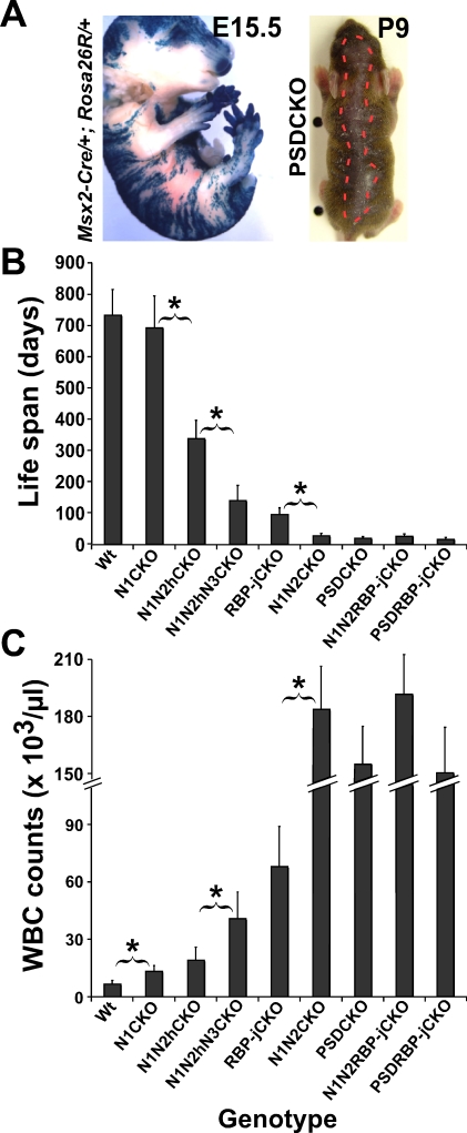
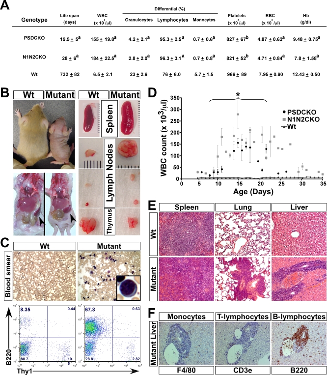
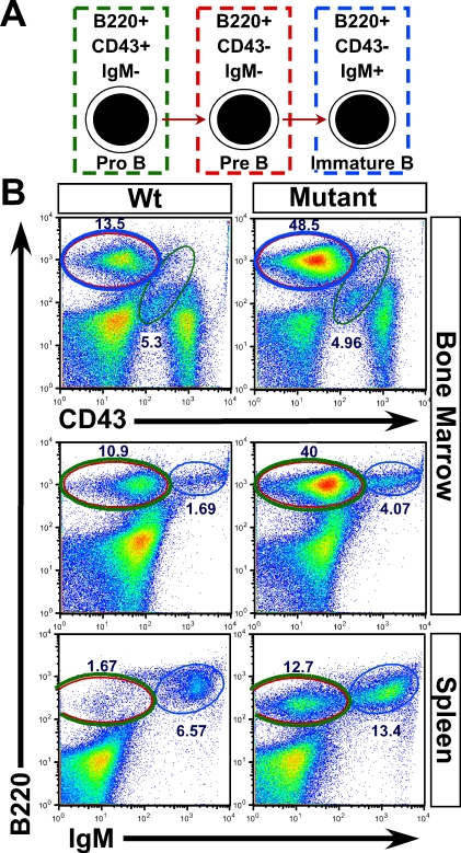
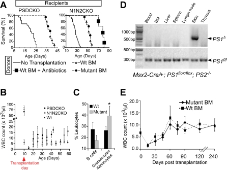
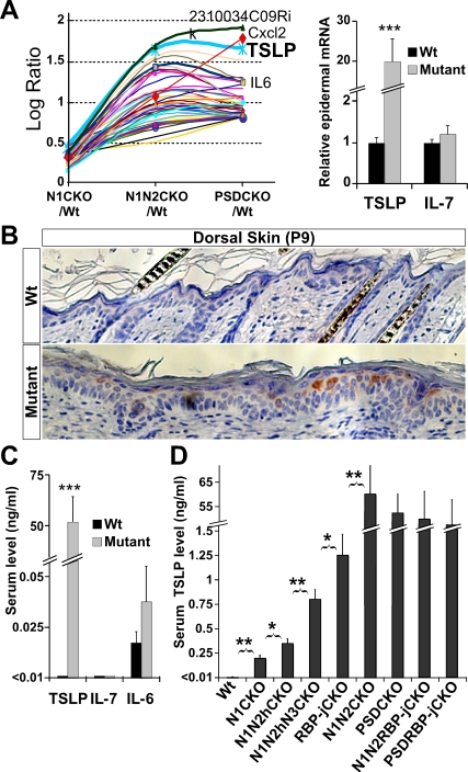
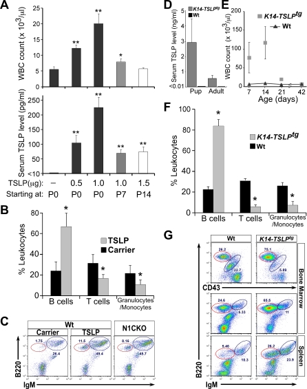
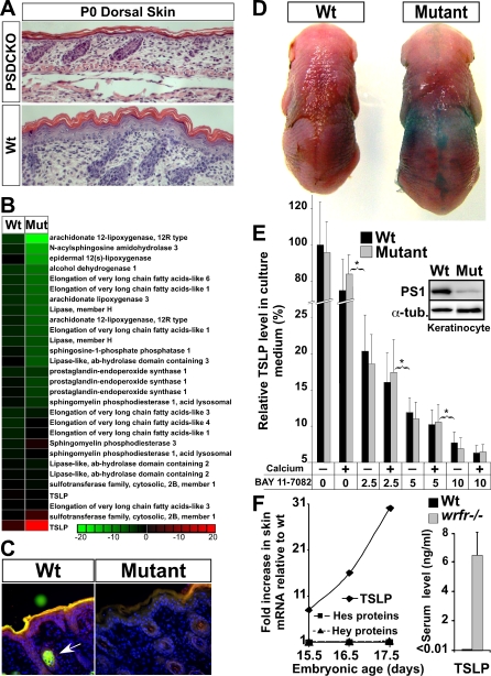
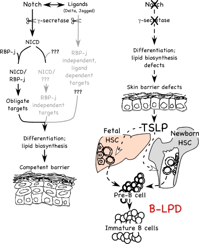
Epidermal keratinocytes form a highly organized stratified epithelium and sustain a competent barrier function together with dermal and hematopoietic cells. The Notch signaling pathway is a critical regulator of epidermal integrity. Here, we show that keratinocyte-specific deletion of total Notch signaling triggered a severe systemic B-lymphoproliferative disorder, causing death. RBP-j is the DNA binding partner of Notch, but both RBP-j-dependent and independent Notch signaling were necessary for proper epidermal differentiation and lipid deposition. Loss of both pathways caused a persistent defect in skin differentiation/barrier formation. In response, high levels of thymic stromal lymphopoietin (TSLP) were released into systemic circulation by Notch-deficient keratinocytes that failed to differentiate, starting in utero. Exposure to high TSLP levels during neonatal hematopoiesis resulted in drastic expansion of peripheral pre- and immature B-lymphocytes, causing B-lymphoproliferative disorder associated with major organ infiltration and subsequent death, a previously unappreciated systemic effect of TSLP. These observations demonstrate that local skin perturbations can drive a lethal systemic disease and have important implications for a wide range of humoral and autoimmune diseases with skin manifestations.


Conflict of interest statement

Competing interests. The authors have declared that no competing interests exist.

Figures

Figure 1
Figure 1. Notch Dosage in the Skin, the Animals' Life Span, and WBC Count Are Tightly Correlated
(A) Chimeric pattern of Msx2-Cre activity is shown on the left by X-gal staining of an E15.5 Msx2-Cre/+; Rosa26R/+ embryo (also see [14,15]). On the right, the dorsal patch of hair loss in P9 Msx2-Cre/+; PS1flox /flox; PS2 –/– (PSDCKO) mice (border is highlighted by red broken line) matches the area of Cre activity. Progressive (B) shortening of life span and (C) increase in peak WBC count measured during the second week of life are caused by stepwise removal of Notch proteins, RBP-j, Presenilins, or certain combinations of those. Abbreviations: wild type (Wt), Msx2-Cre/+; N1flox /flox (N1CKO), Msx2-Cre/+; N1flox /flox; N2flox /+ (N1N2hCKO), Msx2-Cre/+; N1flox /flox; N2flox /+; N3 –/– (N1N2hN3CKO), Msx2-Cre/+; N1flox /flox; N2flox /flox (N1N2CKO), PSDCKO, Msx2-Cre/+; RBP-jflox /flox (RBP-jCKO), Msx2-Cre/+; PS1flox /flox; PS2 –/–; RBP-jflox /flox; (PSDRBP-jCKO), and Msx2-Cre/+; N1flox /flox; N2flox /flox; RBP-jflox /flox (N1N2RBP-jCKO). Note that N1N2CKO, PSDCKO, PSDRBP-jCKO, and N1N2RBP-jCKO animals live only a few weeks and experience severe leukocytosis. Data are collected from lifelong monitoring of 10 to 20 animals from each genotype. Significant difference (p < 0.05) between adjacent genotypes is highlighted by an asterisk.
Figure 2
Figure 2. Mice Lacking Total Notch Signaling in the Skin Develop Severe B-LPD
(A) Table shows the peripheral blood analysis of the mutant mice during the second week of life when WBC counts are at their maximum (n = 6, for each group). The values are presented as mean ± standard deviation (“a” indicates p < 0.001 and “b” indicates p < 0.05 (compared to wild-type control)). (B) Macroscopic examination of a P14 mutant (N1N2CKO) compared to its wild-type littermate reveals the mutant's smaller body size yet significantly larger spleen and lymph nodes. (C) Peripheral blood smear (Giemsa-stained; 250× magnification) and FC analysis show the appearance of lymphoblasts (inset) and the expansion of B220+ B cells in the mutant blood, respectively. (D) Monitoring the mutant animals' WBC counts over their life span (n = 10 for each genotype) demonstrates a surge during the first 2 weeks and plateau during the third week of life, when most mutants die (asterisk). Note the trend of WBC counts toward normalization in a few mice that live a few days longer. Severe B-LPD leads to infiltration of liver, lung, and spleen of the mutant animals with B220+ B cells shown on (E) hematoxylin-and-eosin-stained tissue sections and (F) antibody-stained liver sections (200× magnification).
Figure 3
Figure 3. Expanding Pre- and Immature B Cells Cause B-LPD in the Mutant Animals
(A) Schematic representation of how surface markers applied (B220, CD43, and IgM) identify pro-, pre-, and immature B cells. (B) FC analyses of BM and spleen cells demonstrate a clear expansion of pre- (red circle) and immature (blue circle) B cells in both central and peripheral compartments of the mutant animals. More than three mice from each genotype (PSDCKO, N1N2CKO, and wild-type littermates) are analyzed. Representative plots are shown.
Figure 4
Figure 4. BMT Cures the Lethal B-LPD in the Mutant Animals and Confirms the Local Deletion of Notch Pathway in the Skin as the Sole Driver of B-LPD
(A) Both PSDCKO and N1N2CKO mice, lethally irradiated and transplanted with BM from their wild-type (n = 4) or mutant (n = 3) littermates (at ∼P10), live significantly longer than their untransplanted mutant counterparts (n = 20; p < 0.001, log rank test). In the N1N2CKO group, three mice that are transplanted with wild-type BM also receive daily antibiotic treatment after BMT, leading to their significantly longer life span compared to other transplanted mutants (p < 0.001, log rank test). B-LPD does not recur in the transplanted mutant animals shown by (B) their persistently low WBC count during their post-BMT life (data are drawn from four animals in each genetic group) and (C) on average lower B220+ B cell percentage in their blood as compared to the wild-type recipients, measured 25 d after transplantation (four PSDCKO and four wild-type littermates are compared). Note that at this stage the transplanted PSDCKO animals develop granulocytosis with significantly higher granulocyte/monocyte percentage (*, p < 0.05). (D) Msx2-Cre-mediated PS1 gene deletion (PS1 Δ) is only detectable in the skin of the PSDCKO animals. The data are representative of three independent tests. DNA samples are prepared from PSDCKO animals with WBC count >100,000 cells/μl. BM samples are unfractionated and include BM stroma. (E) Wild-type mice (∼P10) receiving BMT from their mutant (n = 12, both N1N2CKO and PSDCKO are included) or wild-type (n = 12) littermates reconstitute normal hematopoiesis and have a normal life span without developing B-LPD.
Figure 5
Figure 5. TSLP Is Highly Expressed in the Skin Lacking Total Notch Signaling, Leading to Its High Systemic Levels
Such elevated serum levels inversely correlate with Notch dosage in the skin. (A) Modified trend analysis performed on microarray data from P9 mutant and wild-type total-skin RNA samples identifies TSLP as the most up-regulated cytokine gene in the mutants. The epidermal overexpression of TSLP in the mutant mice is confirmed by qRT-PCR. (B) TSLP is mainly produced by suprabasal keratinocytes in the mutant epidermis (200× magnification). (C) Unlike IL-6 and IL-7, TSLP serum protein levels measured on ELISA are highly elevated in the mutant animals. (D) Serum analyses during the second week of life show that TSLP levels increase as more alleles of Notch are removed from the skin. Data are extracted from comparing three mutant (of each genotype) and three wild-type littermates (*, p < 0.05, **, p < 0.01, ***, p < 0.0001).
Figure 6
Figure 6. TSLP Is Sufficient To Drive B-LPD in Newborn Animals
(A) Wild-type mice injected intravascularly with carrier alone (PBS), 0.5, 1, or 1.5 μg of recombinant mouse TSLP daily for 7 days show a dose-dependent increase in serum TSLP levels and WBC counts (measured 12 h after the last dosing) only if the treatment starts at P0. The impact of elevated serum TSLP on WBC count is diminished or absent when the treatment starts on P7 or P14, respectively (n = 3, for each condition; *, p < 0.05 and **, p < 0.01). (B and C) FC analysis on peripheral blood from mice receiving 1 μg of TSLP daily for 7 days starting at P0 confirms that the increase in WBC count is because of the expansion of pre- (red circles) and immature B cells in the periphery, identical to N1CKO newborns that have similar serum TSLP levels and WBC counts (n = 3, for each group; *, p < 0.05). (D) K14-TSLPtg mice phenocopy the neonatal B-LPD in RBP-jCKO animals; they have highly elevated serum TSLP levels at several time points during the first two weeks of life (averaged as pup). Adult animals (>P21) also have elevated TSLP in their serum but to a lesser degree than in the pups. (E and F) In addition, K14-TSLPtg mice have high WBC counts due to B-LPD during the first few weeks of life (*, p < 0.05). (G) FC analysis identifies the expanding population of cells causing B-LPD as pre- (red circle) and immature B cells. TSLP measurements and FC analyses are performed on three K14-TSLPtg mice and three wild-type littermates. WBC counts are obtained from six mice in each group.
Figure 7
Figure 7. Skin-Barrier Defect Explains How Loss of Notch Signaling in the Skin Leads to Elevated TSLP Production
(A) P0 PSDCKO dorsal skin shows a severe epidermal differentiation defect (loss of spinous and granular layers) compared to wild-type littermate (200× magnification). (B) Heat map of microarray data on P9 epidermal RNA shows down-regulation (green) of genes involved in epidermal lipid biogenesis in the mutants versus their wild-type littermates. Note that TSLP probes are included as a control. (C) The Nile Red florescent staining of the polar (red) and non-polar (green) lipids in the epidermis of a P5 PSDCKO mouse demonstrates a deficient lipid coat in γ-secretase-deficient (mutant) versus unaffected areas (wild type) of the epidermis [48]. Note the prominent green florescent staining of the sebaceous gland in the wild-type section (arrow), which is absent in the skin lacking Notch signaling [14]. (D) The dye penetration assay shows a defect in the skin barrier of the mutant animal at E18.5. (E) Keratinocytes cultured from the mutant (PSDCKO) or wild-type littermates in the presence or absence of calcium release similar yet significant amounts of TSLP into their medium. However, keratinocyte TSLP production decreases by inhibiting NFκB signaling (BAY 11–7082: 2.5, 5, 10 μM; *: p < 0.01) in a dose-dependent manner (data are accumulated from three independent experiments; TSLP levels are presented relative to that of wild-type cells in the absence of calcium and inhibitor). The immunoblot panel confirms that the mutant keratinocyte are deficient in PS1 protein. (F) The wrfr –/– embryos show a surge in the skin TSLP expression around the time of skin-barrier formation, as detected by microarray. Note that known Notch targets, Hes and Hey proteins, are not altered in wrfr –/– mice. TSLP up-regulation translates into its highly elevated serum levels in wrfr –/– mice at birth (n = 3; p < 0.0001).
Figure 8
Figure 8. Model of the Role of the Notch Signaling Pathway in Setting up Competent Skin Barrier and Preventing B-LPD in Newborn Animals
RBP-j-dependent and independent Notch signaling is required for proper epidermal differentiation and lipid biogenesis in the skin to form a competent barrier. The RBP-j-independent signals may be non-cell autonomous. Embryonic loss of Notch signaling leads to aberrant epidermal differentiation/lipid biogenesis and, therefore, the defective barrier formation. In response, TSLP is released by keratinocytes that fail to differentiate, triggering a systemic B-LPD in newborns that results in neonatal lethality at the highest level.

Comment in

  • When skin damage causes death.
    Gross L. Gross L. PLoS Biol. 2008 May;6(5):e133. doi: 10.1371/journal.pbio.0060133. Epub 2008 May 27. PLoS Biol. 2008. PMID: 20076711 Free PMC article. No abstract available.

References

    1. Morasso MI, Tomic-Canic M. Epidermal stem cells: the cradle of epidermal determination, differentiation and wound healing. Biol Cell. 2005;97:173–183. - PMC - PubMed
    1. Clayton E, Doupe DP, Klein AM, Winton DJ, Simons BD, et al. A single type of progenitor cell maintains normal epidermis. Nature. 2007;446:185–189. - PubMed
    1. Jamora C, Fuchs E. Intercellular adhesion, signalling and the cytoskeleton. Nat Cell Biol. 2002;4:E101–E108. - PubMed
    1. Fuchs E, Raghavan S. Getting under the skin of epidermal morphogenesis. Nat Rev Genet. 2002;3:199–209. - PubMed
    1. Segre JA. Epidermal barrier formation and recovery in skin disorders. J Clin Invest. 2006;116:1150–1158. - PMC - PubMed

Publication types

MeSH terms

Substances

LinkOut - more resources