. 2008 May 28:5:21.
doi: 10.1186/1742-2094-5-21.

Squamosamide derivative FLZ protects dopaminergic neurons against inflammation-mediated neurodegeneration through the inhibition of NADPH oxidase activity

Affiliations

Squamosamide derivative FLZ protects dopaminergic neurons against inflammation-mediated neurodegeneration through the inhibition of NADPH oxidase activity

Dan Zhang et al. J Neuroinflammation. .

Abstract

Background: Inflammation plays an important role in the pathogenesis of Parkinson's disease (PD) through over-activation of microglia, which consequently causes the excessive production of proinflammatory and neurotoxic factors, and impacts surrounding neurons and eventually induces neurodegeneration. Hence, prevention of microglial over-activation has been shown to be a prime target for the development of therapeutic agents for inflammation-mediated neurodegenerative diseases.

Methods: For in vitro studies, mesencephalic neuron-glia cultures and reconstituted cultures were used to investigate the molecular mechanism by which FLZ, a squamosamide derivative, mediates anti-inflammatory and neuroprotective effects in both lipopolysaccharide-(LPS)- and 1-methyl-4-phenylpyridinium-(MPP+)-mediated models of PD. For in vivo studies, a 1-methyl-4-phenyl-1, 2, 3, 6-tetrahydropyridine-(MPTP-) induced PD mouse model was used.

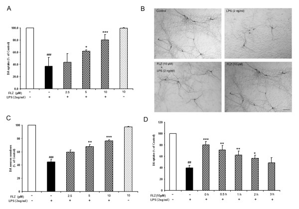
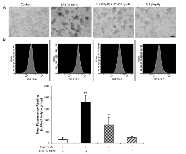
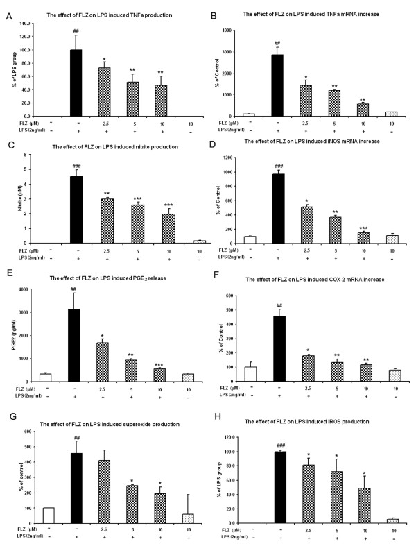
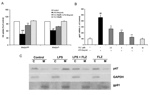
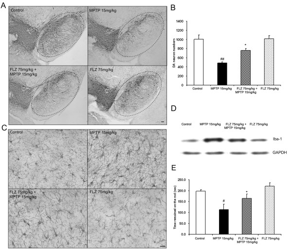
Results: FLZ showed potent efficacy in protecting dopaminergic (DA) neurons against LPS-induced neurotoxicity, as shown in rat and mouse primary mesencephalic neuronal-glial cultures by DA uptake and tyrosine hydroxylase (TH) immunohistochemical results. The neuroprotective effect of FLZ was attributed to a reduction in LPS-induced microglial production of proinflammatory factors such as superoxide, tumor necrosis factor-alpha (TNF-alpha), nitric oxide (NO) and prostaglandin E2 (PGE2). Mechanistic studies revealed that the anti-inflammatory properties of FLZ were mediated through inhibition of NADPH oxidase (PHOX), the key microglial superoxide-producing enzyme. A critical role for PHOX in FLZ-elicited neuroprotection was further supported by the findings that 1) FLZ's protective effect was reduced in cultures from PHOX-/- mice, and 2) FLZ inhibited LPS-induced translocation of the cytosolic subunit of p47PHOX to the membrane and thus inhibited the activation of PHOX. The neuroprotective effect of FLZ demonstrated in primary neuronal-glial cultures was further substantiated by an in vivo study, which showed that FLZ significantly protected against MPTP-induced DA neuronal loss, microglial activation and behavioral changes.

Conclusion: Taken together, our results clearly demonstrate that FLZ is effective in protecting against LPS- and MPTP-induced neurotoxicity, and the mechanism of this protection appears to be due, at least in part, to inhibition of PHOX activity and to prevention of microglial activation.


Figures

Figure 1
Figure 1
The chemical structure of FLZ.
Figure 2
Figure 2
FLZ functionally and morphologically protects DA neurons from LPS-induced neurotoxicity in rat mid-brain neuron-glia culture. Neuronal-glial cultures were pre-treated with different concentrations of FLZ for 1 h followed by 2 ng/ml LPS; 7 days later, DA neurotoxicity was measured by [3H]-DA uptake assay (A) and by immunocytochemical analysis. Representative pictures of immunoreactions are shown in (B) and TH neuron counts are shown in (C). For assessment of FLZ treatment following neurotoxic insult, neuronal-glial cultures were first treated with 2 ng/ml LPS. Then, 0, 0.5, 1, 2, and 3 h later, 10 μM FLZ was added to the cultures. DA neurotoxicity was measured by [3H]-DA uptake assay 7 days later (D). The data are expressed as percentages of control culture values, and represent the mean ± S.E.M. for three independent experiments, each performed with triplicate samples. ## p < 0.01 and ### p < 0.001 compared with vehicle-treated control cultures; * p < 0.05, ** p < 0.01 and *** p < 0.001 compared to LPS treatment group. Scale bar, 100 μm.
Figure 3
Figure 3
Microglia (but not astroglia) mediate the neuroprotection of FLZ. Different kinds of cultures were pretreated with 10 μM FLZ for 1 h, and then 0.25 μM MPP+ was added. DA neurotoxicity was measured by [3H]-DA uptake assay 7 days later. The data are expressed as percentages of control culture values, and represent the mean ± S.E.M. for three independent experiments, each performed with triplicate samples. ### p < 0.001 compared with vehicle-treated control cultures; *** p < 0.001 compared to MPP+ treatment group.
Figure 4
Figure 4
FLZ attenuates LPS-induced microglia activation. (A): Neuronal-glial cultures were pretreated with 10 μM FLZ for 1 h followed by 2 ng/ml LPS; 24 h later, activation of microglia was assessed by OX-42 immunohistochemistry. Representative results are illustrated. (B): HAPI microglia cells were pretreated with 10 μM FLZ for 1 h followed by stimulation with 10 ng/ml LPS for 24 h. Expression of MHC class II antigen was detected by flow cytometry. The cells were analyzed on a FACS Calibur, and the MFI of experimental groups was determined by subtracting the MFI of the isotype control from the MFI of each group. The data are expressed as mean ± S.E.M. for three independent experiments, each performed with triplicate samples. ## p < 0.01 compared with vehicle-treated control group; * p < 0.05 compared to LPS treatment group. Scale bar, 50 μm.
Figure 5
Figure 5
FLZ inhibites LPS-induced production of proinflammatory factors and their gene expression in microglia. The effects of FLZ on LPS-induced production of TNF-α, NO, and PGE2 (A, C, E, respectively) or TNF-α, iNOS, and COX-2 mRNA expression (B, D, F, respectively) and the production of superoxide (G), and intracellular ROS (H) are shown. Enriched microglia cells were treated with different concentrations of FLZ or with vehicle for 1 h before addition of 2 ng/ml LPS. For mRNA analysis, total RNA was harvested 3 h after LPS treatment, followed by real-time RT-PCR analysis of iNOS, TNF-α, COX-2 and GAPDH using specific primers. The data are expressed as mean ± S.E.M. for three independent experiments, each performed with triplicate samples. ## p < 0.01 and ### p < 0.001 compared with vehicle-treated control cultures; * p < 0.05, ** p < 0.01 and *** p < 0.001 compared to LPS treatment group.
Figure 6
Figure 6
Microglial PHOX is the target of FLZ's inhibition in LPS-induced neurotoxicity. (A): The role of PHOX in FLZ's protective effect. PHOX+/+ and PHOX-/- mouse neuronal-glial cultures were pretreated with vehicle or with 10 μM FLZ for 1 h, followed by 10 ng/ml LPS treatment. Neurotoxicity was assessed by [3H]-DA uptake 7 days later. (B): The effect of FLZ on LPS-induced gp91 mRNA expression. Enriched microglia cells were treated with vehicle or with different concentrations of FLZ for 1 h before the addition of 2 ng/ml LPS. For mRNA analysis, total RNA was harvested 3 h after LPS treatment, followed by real-time reverse transcription-PCR analysis using specific primers. (C): Effect of FLZ on cytosolic p47phox protein translocation. HAPI cells were pretreated with vehicle or with 10 μM FLZ for 1 h followed by 10 ng/ml LPS treatment for 10 min. Subcellular fractions were isolated to perform western blot analysis. C = cytosolic extract; M = membrane extract. GAPDH and gp91phox are used as internal cytosolic and membrane controls, respectively. The data are expressed as percentages of control culture values and represent the mean ± S.E.M. for three independent experiments, each performed with triplicate samples. ## p < 0.01 and ### p < 0.001 compared with vehicle-treated control group; * p < 0.05 and ** p < 0.01 compared to LPS treatment group.
Figure 7
Figure 7
FLZ shows significant protective effect against the toxicity of MPTP in vivo. Eight-week-old male C57BL/6J mice received daily MPTP injections (15 mg/kg, s.c.) for 6 consecutive days. From the third day on, FLZ (75 mg/kg, p.o.) was administered 30 min before every MPTP injection for the last 4 days. DA neurotoxicity was measured by immunohistochemistry using an anti-TH antibody (A) and cell counting of DA neurons (B) in the SNpc of different groups of mice. To assess activation of microglia, SNpc brain sections were stained using Iba-1 antibody, a microglial maker (C). To obtain quantitative data, the midbrains of the mice were dissected out, and levels of Iba-1 in these midbrains were determined using western blot assays (D). To assess the protective effect of FLZ on MPTP-induced motor function deficiency, a rotor-rod assay was used and the time for each mouse to remain on the spinning rod was recorded (E). The data are expressed as mean ± S.E.M. for the results of 6–8 mice. # p < 0.05 and ## p < 0.01 compared with vehicle-treated control group; * p < 0.05 compared to MPTP treatment group. Scale bar, 50 μm.

References

    1. Wyss-Coray T. Inflammation in Alzheimer disease: driving force, bystander or beneficial response? Nat Med. 2006;12:1005–1015. - PubMed
    1. McGeer EG, Klegeris A, McGeer PL. Inflammation, the complement system and the diseases of aging. Neurobiol Aging. 2005;26 Suppl 1:94–97. doi: 10.1016/j.neurobiolaging.2005.08.008. - DOI - PubMed
    1. Bruck W, Stadelmann C. Inflammation and degeneration in multiple sclerosis. Neurol Sci. 2003;24 Suppl 5:S265–7. doi: 10.1007/s10072-003-0170-7. - DOI - PubMed
    1. Barouch FC, Miller JW. The role of inflammation and infection in age-related macular degeneration. Int Ophthalmol Clin. 2007;47:185–197. doi: 10.1097/IIO.0b013e3180377936. - DOI - PubMed
    1. Wilms H, Zecca L, Rosenstiel P, Sievers J, Deuschl G, Lucius R. Inflammation in Parkinson's diseases and other neurodegenerative diseases: cause and therapeutic implications. Curr Pharm Des. 2007;13:1925–1928. doi: 10.2174/138161207780858429. - DOI - PubMed

Publication types

MeSH terms

LinkOut - more resources