

Review
doi: 10.1016/j.addr.2008.03.014.
Epub 2008 Apr 10.
Multifunctional magnetic nanoparticles for targeted imaging and therapy
Affiliations
- PMID: 18508157
- PMCID: PMC2583936
- DOI: 10.1016/j.addr.2008.03.014
Item in Clipboard
Review
Multifunctional magnetic nanoparticles for targeted imaging and therapy
Adv Drug Deliv Rev.
.
Abstract
Magnetic nanoparticles have become important tools for the imaging of prevalent diseases, such as cancer, atherosclerosis, diabetes, and others. While first generation nanoparticles were fairly nonspecific, newer generations have been targeted to specific cell types and molecular targets via affinity ligands. Commonly, these ligands emerge from phage or small molecule screens, or are based on antibodies or aptamers. Secondary reporters and combined therapeutic molecules have further opened potential clinical applications of these materials. This review summarizes some of the recent biomedical applications of these newer magnetic nanomaterials.
Figures
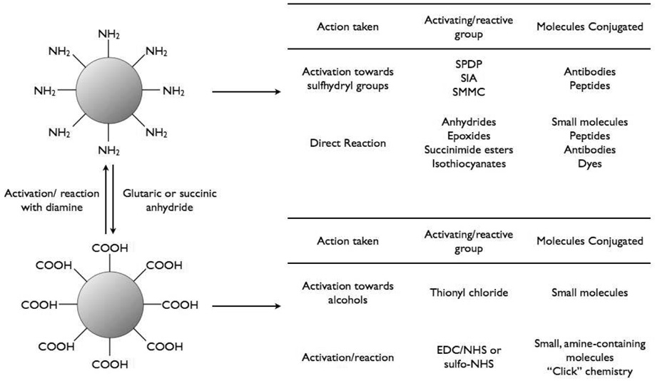
Bioconjugation of iron oxide nanoparticles.
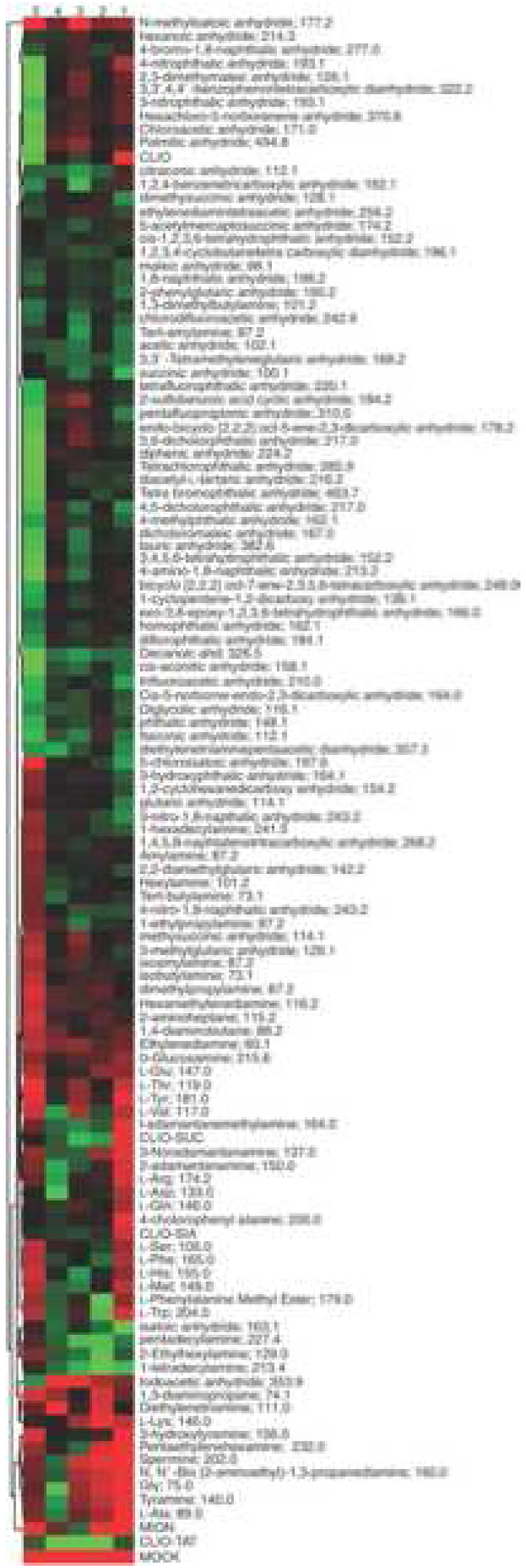
Heat map representing cellular uptake of different nanoparticle preparations. Columns from right to left: 1, pancreatic cancer cells (PaCa-2); 2, macrophage cell line (U937); 3, resting primary human macrophages; 4, activated primary human macrophages; 5, human umbilical vein endothelial cells (HUVEC). Each column represents mean values from six different experiments. Red refers to the lowest accumulation and green refers to the highest accumulation [68].
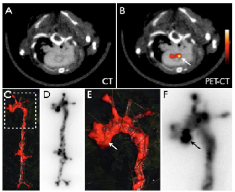
64Cu-CLIO distributes to atherosclerotic lesions. 5A–B: PET-CT shows enhancement of the posterior aortic root (arrow). 5C–F: En face Oil red O staining of the excised aorta depicts plaque loaded vessel segments, which co-localize with areas of high 64Cu-TNP uptake on autoradiography. 5E–F show a zoomed image of the root and arch. The arrows depict a plaque laden segment of the root with high activity, which corresponds to the in vivo signal seen in 5B [84].
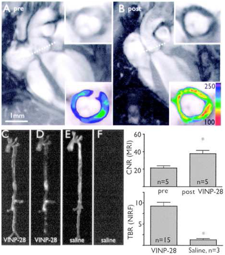
In vivo MR and optical imaging of VCAM-1 expression. A, MRI before injection of targeted nanoagent. Dotted line depicts location of short-axis view (inserts, lower panel with color coded signal intensity). B, Same mouse 48 hours after injection. A marked signal drop in the aortic root wall was noted (insets). The contrast-to-noise ratio (CNR) of the aortic wall was increased significantly after injection of the probe (mean±SD; *P<0.05 before vs after injection). C, E, Light images of excised aortas. D, NIRF image after VINP-28 injection demonstrates distribution of the agent to plaque-bearing segments of the aorta, whereas the aorta of the saline injected apoE−/− shows very little fluorescent signal (F). Both images were acquired with identical exposure times and were identically windowed. The target-to-background ratio (TBR) was significantly higher in the targeted nanoparticle injected mice (*P<0.05) [56].
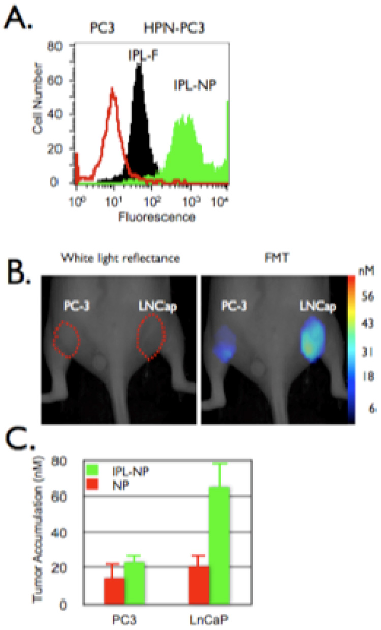
In vivo imaging of prostate cancer. A. HPN-PC3 (black and green histograms) or PC3 (red histogram) cells were incubated with the HPN peptide or HPN peptide labeled nanoparticles (green histogram) then analyzed via flow cytometry. B and C. Mice bearing tumors derived from PC-3 (left flank) or LNCaP (right flank) were co-injected with HPN peptide labeled nanoparticles and a control nanoparticle (red bars) then (B) imaged and (C) accumulation quantified via FMT 24 hours post injection [69].
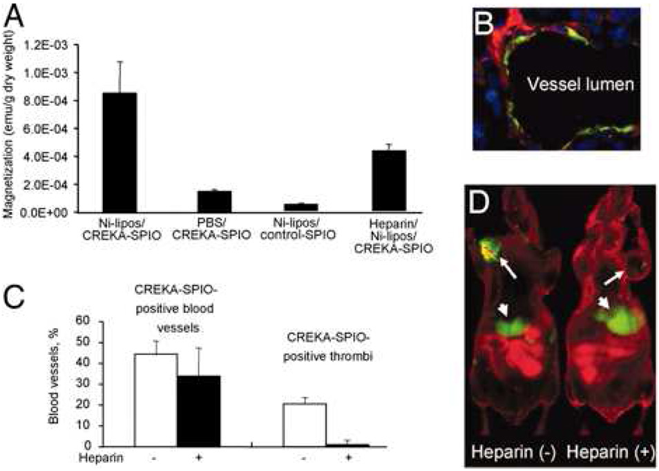
Effect of blood clotting on nanoparticle accumulation in tumors. Mice bearing MDA-MB-435 human breast cancer xenografts were intravenously injected with PBS or heparin, followed by Ni-liposomes (or PBS) and CREKA-modified particles (or control nanoparticles). The mice received additional heparin by i.p. injections (a total of 1,000 units/kg) or PBS throughout the experiment. (A) Tumors were removed 6 h after the nanoparticle injection, and magnetic signal in the tumor after different treatments was determined with SQUID. (B) A representative example of the appearance of CREKA-targeted particles in tumor vessels of mice treated with heparin. (C) Quantification of heparin effect on clotting in blood vessels. Mice were pretreated with PBS (open bars) or heparin (filled bars) as described above, followed by Ni liposomes/CREKA-targeted nanoparticles. Note that heparin did not significantly change the percentage of blood vessels containing particles, but dramatically decreased the incidence of the lumens that are filled with fluorescence. (D) Near-infrared imaging of mice that received Ni-liposomes, followed by Cy7-labeled CREKA-targeted particles, with or without heparin pretreatment. The images were acquired 8 h after the injection of the CREKA-targeted particles. Arrows point to the tumors, and arrowheads point to the liver [70].
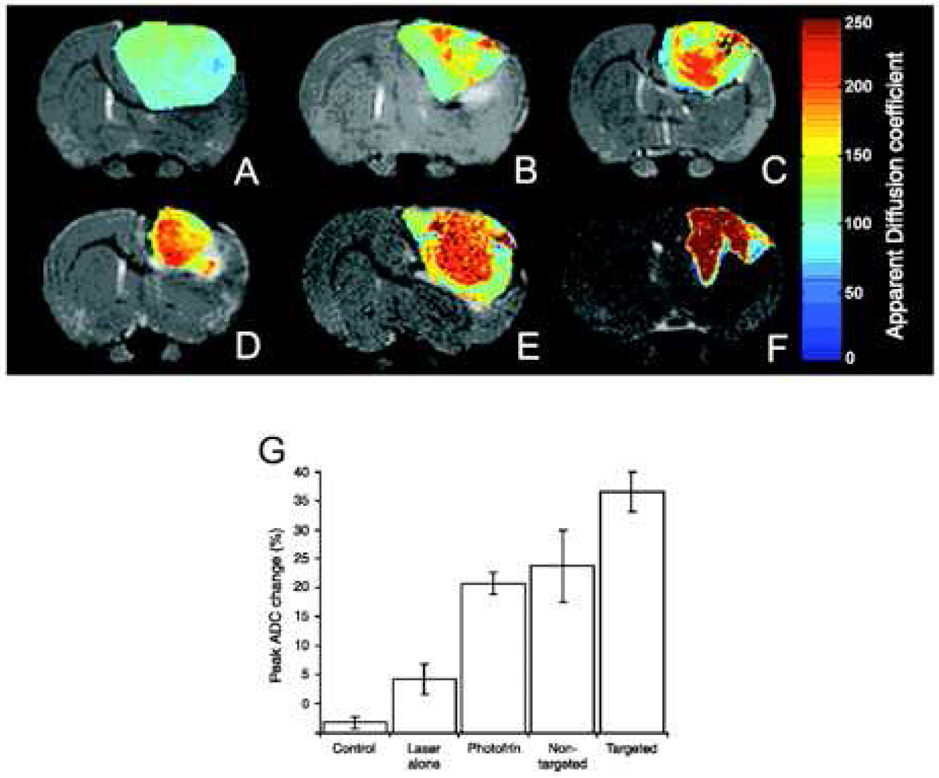
Monitoring of therapeutic efficacy using multifunctional nanoparticles in 9L brain tumors. T2-weighted magnetic resonance images at day 8 after treatment from (C) a representative control i.c. 9L tumor and tumors treated with (D) laser light only, (E) i.v. administration of Photofrin plus laser light, and (F) nontargeted nanoparticles containing Photofrin plus laser light and (G) targeted nanoparticles containing Photofrin plus laser light. The image shown in (H) is from the same tumor shown in (G), which was treated with the F3-targeted nanoparticle preparation but at day 40 after treatment. The color diffusion maps overlaid on top of T2-weighted images represent the apparent diffusion coefficient (ADC) distribution in each tumor slice shown. I, columns, mean peak percentage change in tumor apparent diffusion coefficient values for each of the experimental groups; bars, SE [71].
References
-
- Maeda H, Wu J, Sawa T, Matsumura Y, Hori K. Tumor vascular permeability and the EPR effect in macromolecular therapeutics: A review. J Control Release. 65;2000:271–284. - PubMed
-
- Ohgushi M, Nagayama K, Wada A. Dextran-magnetite: A new relaxation reagent and its application to T2 measurements in gel systems. Journal of Magnetic Resonance (1969–1992) 1978;29:599–601.
-
- Halbreich A, Roger J, Pons JN, Geldwerth D, Da Silva MF, Roudier M, Bacri JC. Biomedical applications of maghemite ferrofluid. Biochimie. 1998;80:379–390. - PubMed
-
- Pankhurst QA, Connolly J, Jones SK, Dobson J. Applications of magnetic nanopartiocles in biomedicine. J Phys D: Appl Phys. 2003;36:R167–R181.
-
- Weissleder R, Stark DD, Compton CC, Wittenberg J, Ferrucci JT. Ferrite-enhanced MR imaging of hepatic lymphoma: An experimental study in rats. AJR Am J Roentgenol. 1987;149:1161–1165. - PubMed
Publication types
MeSH terms
Substances
Grants and funding
LinkOut - more resources
Full Text Sources
Other Literature Sources
