

Ena/VASP: proteins at the tip of the nervous system
- PMID: 18508258
- PMCID: PMC2515615
- DOI: 10.1016/j.conb.2008.05.007
Ena/VASP: proteins at the tip of the nervous system
Abstract
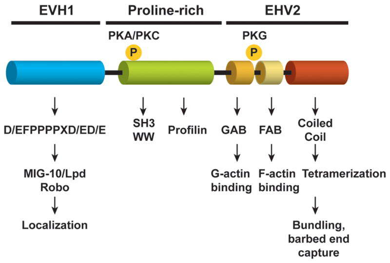
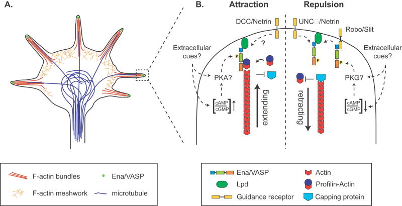
The emergence of neurites from a symmetrical cell body is an essential feature of nervous system development. Neurites are the precursors of axons and dendrites and are tipped by growth cones, motile structures that guide elongating axons in the developing nervous system. Growth cones steer the axon along a defined path to its appropriate target in response to guidance cues. This navigation involves the dynamic extension and withdrawal of actin-filled finger-like protrusions called filopodia that continuously sample their environment. Ena/VASP proteins, a conserved family of actin-regulatory proteins, are crucial for filopodia formation and function downstream of several guidance cues. Here we review recent findings into Ena/VASP function in neurite initiation, axon outgrowth and guidance.
Figures
References
-
- Huber AB, Kolodkin AL, Ginty DD, Cloutier JF. Signaling at the growth cone: ligand-receptor complexes and the control of axon growth and guidance. Annu Rev Neurosci. 2003;26:509–563. - PubMed
-
- Kalil K, Dent EW. Touch and go: guidance cues signal to the growth cone cytoskeleton. Curr Opin Neurobiol. 2005;15:521–526. - PubMed
-
- Gupton SL, Gertler FB. Filopodia: the fingers that do the walking. Sci STKE. 2007;2007:re5. - PubMed
-
- Koleske AJ. Do filopodia enable the growth cone to find its way? Sci STKE. 2003;2003:pe20. - PubMed
-
- Krause M, Dent EW, Bear JE, Loureiro JJ, Gertler FB. Ena/VASP proteins: regulators of the actin cytoskeleton and cell migration. Annu Rev Cell Dev Biol. 2003;19:541–564. - PubMed
Publication types
MeSH terms
Substances
Grants and funding
LinkOut - more resources
Full Text Sources
Other Literature Sources
Medical
