

The determination of projection neuron identity in the developing cerebral cortex
- PMID: 18508260
- PMCID: PMC2483251
- DOI: 10.1016/j.conb.2008.05.006
The determination of projection neuron identity in the developing cerebral cortex
Abstract
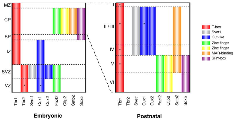
Here we review the mechanisms that determine projection neuron identity during cortical development. Pyramidal neurons in the mammalian cerebral cortex can be classified into two major classes: corticocortical projection neurons, which are concentrated in the upper layers of the cortex, and subcortical projection neurons, which are found in the deep layers. Early progenitor cells in the ventricular zone produce deep layer neurons that express transcription factors including Sox5, Fezf2, and Ctip2, which play important roles in the specification of subcortically projecting axons. Upper layer neurons are produced from progenitors in the subventricular zone, and the expression of Satb2 in these differentiating neurons is required for the formation of axonal projections that connect the two cerebral hemispheres. The Fezf2/Ctip2 and Satb2 pathways appear to be mutually repressive, thus ensuring that individual neurons adopt either a subcortical or callosal projection neuron identity at early times during development. The molecular mechanisms by which Satb2 regulates gene expression involves long-term epigenetic changes in chromatin configuration, which may enable cell fate decisions to be maintained during development.
Figures

References
-
- Butt SJ, Fuccillo M, Nery S, Noctor S, Kriegstein A, Corbin JG, Fishell G. The temporal and spatial origins of cortical interneurons predict their physiological subtype. Neuron. 2005;48:591–604. - PubMed
-
- Wonders CP, Anderson SA. The origin and specification of cortical interneurons. Nat Rev Neurosci. 2006;7:687–696. - PubMed
-
- McConnell SK. The generation of neuronal diversity in the central nervous system. Ann Rev Neurosci. 1991;14:269–300. - PubMed
-
- Bielle F, Griveau A, Narboux-Neme N, Vigneau S, Sigrist M, Arber S, Wassef M, Pierani A. Multiple origins of Cajal-Retzius cells at the borders of the developing pallium. Nat Neurosci. 2005;8:1002–1012. - PubMed
-
- Zhao C, Guan W, Pleasure SJ. A transgenic marker mouse line labels Cajal-Retzius cells from the cortical hem and thalamocortical axons. Brain Res. 2006;1077:48–53. - PubMed
Publication types
MeSH terms
Substances
Grants and funding
LinkOut - more resources
Full Text Sources
Other Literature Sources
Medical
