. 2008 Jul 25;283(30):20874-87.
doi: 10.1074/jbc.M800375200. Epub 2008 May 27.

Multiple intrinsically disordered sequences alter DNA binding by the homeodomain of the Drosophila hox protein ultrabithorax

Affiliations

Multiple intrinsically disordered sequences alter DNA binding by the homeodomain of the Drosophila hox protein ultrabithorax

Ying Liu et al. J Biol Chem. .

Abstract

During animal development, distinct tissues, organs, and appendages are specified through differential gene transcription by Hox transcription factors. However, the conserved Hox homeodomains bind DNA with high affinity yet low specificity. We have therefore explored the structure of the Drosophila melanogaster Hox protein Ultrabithorax and the impact of its nonhomeodomain regions on DNA binding properties. Computational and experimental approaches identified several conserved, intrinsically disordered regions outside the homeodomain of Ultrabithorax that impact DNA binding by the homeodomain. Full-length Ultrabithorax bound to target DNA 2.5-fold weaker than its isolated homeodomain. Using N-terminal and C-terminal deletion mutants, we demonstrate that the YPWM region and the disordered microexons (termed the I1 region) inhibit DNA binding approximately 2-fold, whereas the disordered I2 region inhibits homeodomain-DNA interaction a further approximately 40-fold. Binding is restored almost to homeodomain affinity by the mostly disordered N-terminal 174 amino acids (R region) in a length-dependent manner. Both the I2 and R regions contain portions of the activation domain, functionally linking DNA binding and transcription regulation. Given that (i) the I1 region and a portion of the R region alter homeodomain-DNA binding as a function of pH and (ii) an internal deletion within I1 increases Ultrabithorax-DNA affinity, I1 must directly impact homeodomain-DNA interaction energetics. However, I2 appears to indirectly affect DNA binding in a manner countered by the N terminus. The amino acid sequences of I2 and much of the I1 and R regions vary significantly among Ultrabithorax orthologues, potentially diversifying Hox-DNA interactions.


Figures

FIGURE 1.
FIGURE 1.
UbxIb contains intrinsic disorder. A, schematic depicting the glycine-rich amino acid sequence of UbxIb. Vertical bars indicate individual glycine residues. Key sequence features, including a 13-amino acid-long polyglycine region (G), the YPWM Exd interaction motif (Y; black box), and the DNA-binding homeodomain (HD; black box), are indicated. The activation domain, indicated by a bar above the schematic, can be subdivided into the requisite activation core (AC) and activation enhancement region (AER) (bars below) (54). B–E, extreme sensitivity to native state proteolysis demonstrates that much of Ubx, excluding the homeodomain, is intrinsically disordered. B, a 30-min time course of proteolysis of UbxIb by Proteinase K, trypsin, and α-chymotrypsin at a substrate/enzyme ratio of 1000:1. Uncut UbxIb is marked as 0 min. C, the UbxIb homeodomain is generally not digested by proteases under the same conditions, although trypsin is able to cleave the N-terminal arm of the homeodomain. Both LacI (D) and ApoMb (E) are less susceptible to proteolysis despite the presence of well characterized disordered regions. The relatively rapid degradation of UbxIb is indicative of more prevalent and/or more fluctuating disordered domains. F, CD spectra for UbxIb and UbxHD normalized by the number of amino acids. The troughs at 208 and 222 nm suggest that both full-length UbxIb and UbxHD contain α-helical structure (97, 98). There is substantially less structure per residue in full-length UbxIb than UbxHD, suggesting that the nonhomeodomain regions are largely disordered. The concentrations of UbxIb and UbxHD were measured by magnetic circular dichroism, which determines the concentration of tryptophan in an environmentally independent manner. N-Acetyltryptophanamide was used to generate a standard curve (99).
FIGURE 2.
FIGURE 2.
Location of disorder in UbxIb. A–C, concurrence of computational and experimental approaches on the localization of disordered regions in UbxIb. A, the disorder prediction scores from PONDR (blue), DisEMBL (pink), and IUPred (yellow) are plotted against UbxIb amino acid number. Regions that consistently have scores greater than 0.5 (boundary marked by a dotted line) are predicted to be disordered. B, schematic of the UbxIb sequence with functional domains indicated as follows: transcriptional activation domain, divided into the requisite core region and the activation enhancing region (medium gray); the YPWM Exd interaction motif (Y; light gray), the three alternatively spliced microexons b, mI, and mII (dark gray), the homeodomain (black), and a partial repression domain (medium gray box near the C terminus) (31, 75, 76). Regions of UbxIb predicted to be disordered by all three algorithms are indicated by red lines above the schematic, whereas regions predicted to be ordered using the nnpredict and GOR V algorithms (35, 36) are indicated by light green lines. Note that the predicted structured and disordered regions do not overlap. C, to experimentally corroborate the predicted boundaries of the disordered segments, Ubx proteolytic products, separated by SDS-PAGE, were probed by Western blot using primary monoclonal antibodies to identify different regions of the UbxIb sequence as follows: Ia2D.3 (N-terminal epitope indicated by a dark red line), 10H.7 (orange line), Ia2D.7 (yellow line), J39.2 (turquoise line), 3.11F (blue line), 4A.1 (violet line), and FP3.38 (C-terminal epitope marked by a purple line) (31, 58). Analysis of band size and epitope content allowed classification of most of the potential trypsin target sites in UbxIb. Scaled to the UbxIb schematic in B, filled circles indicate a site cut by trypsin, open circles mark sites protected from trypsin proteolysis by protein structure, and gray circles mark sites that could not be assessed with confidence. The theoretical analysis and experimental protease susceptibility data concur, indicating clearly that a large portion of the UbxIb amino acid sequence is intrinsically disordered.
FIGURE 3.
FIGURE 3.
Impact of sequences outside the UbxHD on DNA affinity. A, DNA binding by full-length UbxIa and isolated UbxHD measured by gel shift experiments. A, equilibrium gel shift images for UbxHD and full-length UbxIa, indicating bound (B) and free (F) DNA bands. Lane 1 for both gels contained no protein, and lanes 2–20 contained UbxHD or UbxIa, increasing from 8 nm to 320 nm and from 50 nm to 2.0 μm, respectively. B, binding curves for UbxIa (formula image) and UbxHD (▪). Each point in a curve was derived from three replicates within a single experiment, and the error bars indicate the S.D. for these replicates. For clarity, 
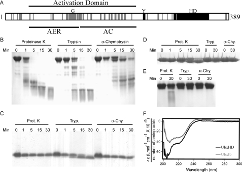
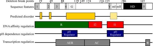
only the fraction of free DNA is shown. C, sequence schematic for 
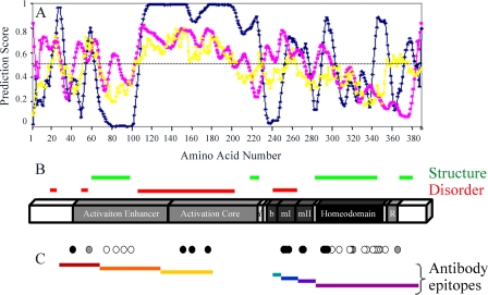
UbxIa and its variants. The homeodomain (HD) and activation domain, which is subdivided into the core region required for function (AC) and the enhancement region that boosts activity (AER), and a partial repression domain (R) (31, 75, 76) are indicated. The highly conserved YPWM (Exd interaction) motif (Y), five moderately conserved motifs (1–5), the polyglycine region (G), and the microexons (mI and mII) are also indicated by bars below. The sequence boundaries for the 11 deletion mutants and UbxHD relative to these domains are also shown. D, note the significant differences in binding affinity. Each value represents at least nine Kd measurements at pH 7.5. The low affinities of N139 and N174 are near the experimental limits of gel retardation assays and therefore have larger errors. Data for UbxIa and the first five N-terminal deletion mutants fit to a line with R2 = 0.92, indicating that affinity is linearly dependent on sequence length for these regions.
FIGURE 4.
FIGURE 4.
Models for different mechanisms by which I1, I2, and R may impact UbxHD-DNA interactions. Because the I1 and I2 DNA binding-inhibitory regions were identified using truncation mutants, the effects of these regions could be either direct or indirect and either independent of or dependent on the N terminus. A, R-independent versus R-dependent models. If the function of either I1 or I2 directly impacts homeodomain function (solid lines), the restoration domain, R, could also act directly on homeodomain or I domain structure or energetics to partially compensate for the loss of binding affinity mediated by the I domain (R-independent model). An internal deletion removing the inhibitory domain should improve binding relative to the full-length protein. In contrast, in the R-dependent model, the I domain interacts with or is restricted or stabilized by the R domain. Removal of this native interaction in the truncation mutant permits the I domain to inhibit binding. Since the inhibition of binding would not occur in monomeric, unmodified full-length protein, an internal mutant would not improve binding affinity. Furthermore, removal of the I-R interaction may also permit nonnative, inhibitory interactions between R and the homeodomain (dashed line). B, distinguishing direct and indirect effects for the R-dependent model. In the R-dependent model, the I domain may inhibit binding by two possible mechanisms, which may be distinguished by their effects on sentinel amino acids in the homeodomain with environmentally sensitive pKa values. If an I domain in a truncated mutant inhibits binding by nonnative, direct interactions with the homeodomain, the pKa values of these amino acids may be altered. If, however, the I domain only impacts DNA binding via increased conformational fluctuations, these pKa values would be unchanged.
FIGURE 5.
FIGURE 5.
pH dependence of full-length UbxIa and its variants. A, pH differentially affects UbxHD and UbxIa interactions with DNA. Binding of UbxHD (▪) (13) and UbxIa (formula image
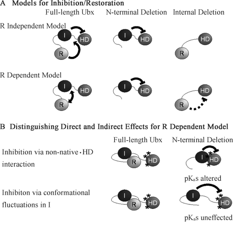
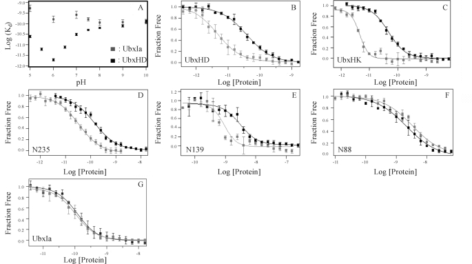
) to 40AB was measured over the pH range 5.0–10.0 in Tris binding buffer at constant ionic strength. Results are similar to measurements in a phosphate-based buffer from 5.0 to 8.0. B and G depict the binding curves of UbxHD, UbxHK, N235, N139, N88, and UbxIa, respectively. The binding affinities of UbxIa and these critical variants were measured at pH 7.5 (▪) and pH 6.0 (formula image) in Tris binding buffer. Binding curves were derived from replicates within a single experiment, which includes three individual affinity measurements, and the error bars indicate the S.D. for these replicates. Data from multiple experiments for each mutant are summarized in Table 1.
FIGURE 6.
FIGURE 6.
Prediction of disordered regions in all Drosophila melanogaster Hox proteins. Disorder tendency was predicted by the IUPred algorithm. Values above 0.5 (black line) indicate predicted disorder in the corresponding region. Homeodomains are enclosed in black boxes. Each graph depicts data for a different Drosophila Hox protein as follows. A, Labial; B, Deformed; C, Sex Combs Reduced; D, Antennapedia; E, Ultrabithorax; F, Abdominal-A; G, Abdominal-B. In general, Hox proteins are highly disordered.
FIGURE 7.
FIGURE 7.
Conservation of the disordered regions in Ubx. An amino acid sequence alignment is shown for Ubx derived from the fruit fly D. melanogaster (DmUbx), the mosquito Anopheles gambiae (AgUbx), the beetle Tribolium castaneum (TcUbx), the butterfly Juonia coenia (JcUbx), the shrimp Artemia franciscana (AfUbx), and the Onychophoran velvet worm Akanthokara kaputensis (AkUbx). The consensus results from the disorder prediction algorithms PONDR, IUPred, and DisEMBL are marked for each Ubx orthologue by red boxes. Identical and conserved residues are marked by black and gray shading, respectively, and previously identified conserved motifs (75, 76) are identified by dashed lines. The YPWM motif and homeodomain are marked by solid lines.
FIGURE 8.
FIGURE 8.
Overlap between intrinsically disordered regions and functional domains within UbxIa. The conserved motifs (1–5), polyglycine region (G), YPWM motif (Y), and homeodomain (HD) are indicated (64, 75, 76). Regions of UbxIa predicted to be disordered by all three algorithms explored in Fig. 2 are indicated by dark yellow boxes. The light yellow box is also disordered in UbxIb and predicted by two of the three algorithms to also be disordered in UbxIa. All three DNA binding regulatory regions contain disordered sequences. High affinity DNA binding by the homeodomain can be directly inhibited by amino acids 235–286 (I1; red), which includes the conserved YPWM motif and Ubx microexons. The I2 inhibitory region (amino acids 174–216; red) inhibits DNA binding via conformational fluctuations in the absence of the R region (amino acids 1–174; green), which restores binding in a length-dependent manner. The I1 region and amino acids 88–139 (blue) regulate the pH dependence of Ubx protein-DNA binding, demonstrating that they can directly impact the pKa of homeodomain residues important for binding and influence access to this region in a pH-dependent manner. Most of the activation domain (activation enhancement region (AER) and activation core (AC)) is encompassed within the R and I2 regions, suggesting that DNA binding and transcription regulation may be functionally linked via overlapping domains.

Similar articles

Cited by

References

    1. Lewis, E. B. (1978) Nature 276 565-570 - PubMed
    1. Hughes, C. L., and Kaufman, T. C. (2002) Evol. Dev. 4 459-499 - PubMed
    1. Weatherbee, S. D., Halder, G., Kim, J., Hudson, A., and Carroll, S. (1998) Genes Dev. 12 1474-1482 - PMC - PubMed
    1. Merabet, S., Saadaoui, M., Sambrani, N., Hudry, B., Pradel, J., Affolter, M., and Graba, Y. (2007) Proc. Natl. Acad. Sci. U. S. A. 104 16946-16951 - PMC - PubMed
    1. Gellon, G., and McGinnis, W. (1998) BioEssays 20 116-125 - PubMed

Publication types

LinkOut - more resources