. 2008 Aug;135(15):2603-14.
doi: 10.1242/dev.019299. Epub 2008 May 28.

Embryonic requirements for ErbB signaling in neural crest development and adult pigment pattern formation

Affiliations

Embryonic requirements for ErbB signaling in neural crest development and adult pigment pattern formation

Erine H Budi et al. Development. 2008 Aug.

Abstract

Vertebrate pigment cells are derived from neural crest cells and are a useful system for studying neural crest-derived traits during post-embryonic development. In zebrafish, neural crest-derived melanophores differentiate during embryogenesis to produce stripes in the early larva. Dramatic changes to the pigment pattern occur subsequently during the larva-to-adult transformation, or metamorphosis. At this time, embryonic melanophores are replaced by newly differentiating metamorphic melanophores that form the adult stripes. Mutants with normal embryonic/early larval pigment patterns but defective adult patterns identify factors required uniquely to establish, maintain or recruit the latent precursors to metamorphic melanophores. We show that one such mutant, picasso, lacks most metamorphic melanophores and results from mutations in the ErbB gene erbb3b, which encodes an EGFR-like receptor tyrosine kinase. To identify critical periods for ErbB activities, we treated fish with pharmacological ErbB inhibitors and also knocked down erbb3b by morpholino injection. These analyses reveal an embryonic critical period for ErbB signaling in promoting later pigment pattern metamorphosis, despite the normal patterning of embryonic/early larval melanophores. We further demonstrate a peak requirement during neural crest migration that correlates with early defects in neural crest pathfinding and peripheral ganglion formation. Finally, we show that erbb3b activities are both autonomous and non-autonomous to the metamorphic melanophore lineage. These data identify a very early, embryonic, requirement for erbb3b in the development of much later metamorphic melanophores, and suggest complex modes by which ErbB signals promote adult pigment pattern development.


Figures

Fig. 1
Fig. 1. Defective adult pigment pattern but normal embryonic/early larval pigment pattern of picasso mutants
(A) Wild-type adult pigment pattern of picasso heterozygote. (B) Defective pigment pattern of picasso homozygote. (C,D) Pigment patterns of wild-type and mutant siblings were indistinguishable at 5 dpf. (E–H) Repeated images of wild-type (picasso/+) larvae revealing normal development of initially dispersed metamorphic melanophores that organize into stripes (arrow, E), as well as metamorphic melanophores that develop already at sites of stripe formation. (I–L) picasso mutant larvae develop very few metamorphic melanophores (arrow, L), and instead many embryonic/early larval melanophores (arrowhead, K) persist into the adult. (E,I) 17 dpf. (F,J) 23 dpf. (G,K) 31 dpf. (H,L) 40 dpf.
Fig. 2
Fig. 2. Regulative posterior adult stripe formation in the picasso mutant
Adult pigment pattern development on the posterior trunk of wild-type (A–D) and picasso mutant (E–H) larvae. Shown are individual, representative larvae of each genotype. (A–D) In wild-type, most dorsal early larval melanophores remained dorsally, though a very few were incorporated into the adult dorsal primary stripe in its posterior region (arrowheads in C,D). (E–H) In picasso mutants, more embryonic/early larval melanophores (e.g., arrowheads in G) were incorporated into an incomplete dorsal primary stripe. Additional metamorphic melanophores differentiated (e.g., arrows in G) where persisting embryonic/early larval melanophores contributed to the adult stripe. Thus, residual adult stripes in the posterior of picasso mutants resulted from an increased contribution of embryonic/early larval melanophores, as well as increased numbers of metamorphic melanophores compared to the mid-trunk. (A,E) 14 dpf. (B,F) 16 dpf. (C,G) 20 dpf. (D,H) 24 dpf.
Fig. 3
Fig. 3. picasso is allelic to erbb3
(A) Schematic of erbb3b cDNA showing picasso lesions. RL, receptor L, ligand-binding domains. C, furin-like, cysteine-rich domains. PK, protein kinase domain. TM, transmembrane domain. Arrow, N835 interruption to kinase domain characteristic of erbb3. (B) Premature stop codon in pcswp.r2e2 (C433T: Q145stop). (C) Premature stop codon in pcsut.r4e1 (T2043A: Y681stop).
Fig. 4
Fig. 4. ErbB gene expression in metamorphosing larvae
(A,B) erbb3 loci are expressed in glia during zebrafish metamorphosis (∼18 dpf). Arrows, glial cells surrounding midbody lateral line. (A) erbb3b. (B) erbb3a. (C) RT-PCR reveals metamorphic melanophore expression of erbb3b and erbb2, but not erbb3a or egfr. All four ErbB genes are expressed in adult fin, comprising chromatophores and their precursors, skin, bone, nerve, and other cell types. mel, melanophore. dct, dopachrome tautomerase, encoding a melanin synthesis enzyme expressed by melanophores and their precursors. dct is more strongly expressed in melanoblasts (present in fin) compared to fully differentiated melanophores (mel). Scale bar: in A, 0.5 mm for A,B.
Fig. 5
Fig. 5. Metamorphic deficiencies for early and late markers of neural crest-derived lineages in picasso mutant larvae
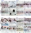
Shown are corresponding regions of the mid-trunk for wild-type (above) and picasso mutant larvae at middle stages of metamorphosis (∼18 dpf). Note that individual melanophores are much more spread in picasso mutants, which is typical of mutants with reduced densities of melanophores and their precursors (Parichy and Turner, 2003b; Parichy et al., 2003). (A,A') crestin marks most or all neural crest-derived cells in embryos (Luo et al., 2001) and identifies a dispersed population of cells in the hypodermis of metamorphosing larvae (arrow) that may be pigment cell precursors. crestin+ cells were dramatically fewer in picasso mutants. (B,B') Higher magnification image of crestin+ cells in wild-type showing typical melanoblast morphology, and their absence in picasso. (C,C') sox10 marks non-ectomesenchymal neural crest-derived cells in embryos, including pigment cell and glial precursors (Dutton et al., 2001; Gilmour et al., 2002), and identifies comparable populations in metamorphosing larvae (Parichy et al., 2003). sox10+ cells were fewer in picasso compared to wild-type. (D,D') Higher magnification showing sox10+ glia along midbody lateral line (arrowhead) and individual sox10+ cells (arrow) in the hypodermis of wild-type, but not picasso. (E,E') mitfa marks melanophore and xanthophore precursors in embryos and is essential for melanoblast specification (Lister et al., 1999; Parichy et al., 2000b). mitfa+ cells are numerous in the hypodermis of wild-type but not picasso larvae. (F,F') dct identifies melanophore precursors (arrow) and melanophores (arrowhead)(Kelsh et al., 2000); dct+ cells are reduced or absent in picasso mutants. (G,G') xanthine dehydrogenase (xdh) encodes an enzyme in the pteridine synthesis pathway of xanthophores (Parichy et al., 2000b). xdh+ cells (arrow) are numerous in wild-type but transiently reduced in number in picasso. (H,H') myelin basic protein (mbp) marks mature glia in the peripheral nervous sytem (Brosamle and Halpern, 2002; Lyons et al., 2005) and mbp+ cells line the midbody lateral line (arrow) as well as ascending and descending nerve fibers (arrowhead) of wild-type larvae. Although mbp+ cells are present in picasso, they are fewer in number and their associated nerves are misrouted.
Fig. 6
Fig. 6. Autonomous and non-autonomous roles for erbb3b in pigment pattern metamorphosis
(A) Wild-type → picasso chimeras frequently developed wild-type melanophores in stripes at high density anteriorly (arrows, left) but at lower density in the mid-trunk (small arrows, right; 75% of chimeras developed donor melanophores; chimeras with donor cells and total reared: n=24, 64, respectively). A wild-type midbody lateral line exhibits misrouting as well, perhaps owing to defects in glial-dependent fasciculation (Gilmour et al. 2002; Lyons et al., 2005). (B). Melanophores at high density anteriorly that are either donor-derived (EGFP+) or host-derived (EGFP). (C) Melanophores in the mid-trunk are more spread, which is typical at low density. In reciprocal picasso → wild-type chimeras, we did not observe donor metamorphic melanophores (n=7, 50). (D) Wild-type → nacre chimeras developed patches of donor-derived metamorphic melanophores that populated stripes (arrow) and scales (84% of chimeras developed metamorphic melanophores; n=75, 155). Persisting embryonic/early larval melanophores (arrowheads) are identififiable by location, large size, and browner color (Quigley et al., 2004). (E) picassonacre chimeras developed melanophores (arrowheads), but did not develop metamorphic melanophores [79% of chimeras developed embryonic/early larval melanophores or fin melanophores (not shown); n=58, 195]. Donor cells in all chimera combinations contributed at similar frequencies to other derivatives, including muscle, epidermis, eye, and neurons of the lateral line. Scale bars: in A, 500 μm; in B, 200 μm for B,C; in D, 1 mm for D,E.
Fig. 7
Fig. 7. Embryonic requirement for ErbB signaling in pigment pattern metamorphosis
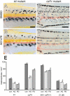
(A) Control wild-type zebrafish treated from embryonic through juvenile stages with DMSO alone. (B) Wild-type treated with AG1478 during embryonic stages (70% epiboly – 4 dpf) shows adult pigment pattern resembling severely affected picasso mutants. (C) Individual treated with AG1478 during the pre-metamorphic, early larval period (5−14 dpf) exhibits a pigment pattern indistinguishable from controls. (D) An individual treated with AG1478 throughout metamorphosis (15−28 dpf) also is indistinguishable from controls. (E) Metamorphic melanophore densities in the mid-trunk (means ± 95% confidence intervals) showing similarities between wild-type (untreated), control (con, DMSO-treated), and fish treated with AG1478 during pre-metamorphosis (pre) and metamorphosis (met), and similar defects between picasso mutants (pcs) and fish treated with AG1478 as embryos (emb). Letters above bars indicate means that are not significantly different from one another by post hoc Tukey-Kramer comparisons. Numbers within bars are samples sizes. (F, G) D. albolineatus normally develop a more uniform melanophore pattern of metamorphic melanophores (arrowheads in F) compared to D. rerio, and exhibit a severe melanophore deficiency when embryos are treated with AG1478 (G). (H, I) In D. nigrofasciatus, adult stripes largely comprise persisting embryonic/early larval melanophores with occasional gaps (arrow in H), and embryonic treatment with AG1478 has relatively subtle effects (arrows in I). (J, K) Embryonic morpholino knockdown of erbb3b results in adult melanophore deficiencies (K) compared to controls (J), providing independent evidence for an early erbb3b dependence of adult pigment pattern formation. Scale bar: in I, 0.5 mm for A–D, F–I, J–K.
Fig 8
Fig 8. Treatment of embryos with ErbB inhibitor PD158780 results in adult pigment pattern defect similar to erbb3b null alleles
(A) Control treated with DMSO alone. (B) Representative individual treated with PD158780 for 2 dpf. Arrow, region deficient for metamorphic melanophores.
Fig 9
Fig 9. ErbB inhibitor treatment for 48 hpf does not enhance the picasso null phenotype
(A) Wild-type control treated with DMSO. (B) Wild-type treated with AG1478 exhibits a severe pigment pattern defect. (C) picasso mutant control treated with DMSO. (D) picasso mutant treated with AG1478 has a pigment pattern defect indistinguishable from that of the control. Scale bar: in D, 0.5 mm for A–D.
Fig. 10
Fig. 10. kit mutant reveals critical period for ErbB activity during neural crest migration
Embryos were reared to juvenile stages after treatment with AG1478 as embryos. (A) Normal kit mutant pigment pattern after AG1478 treatment between 26−30 hpf (stripe break score = 0; for details see Materials and Methods). kit mutant adults exhibit about half as many stripe melanophores as wild-type fish and completely lack melanophores over the dorsum and on scales (Johnson et al., 1995). (B,C) Treatment with AG1478 between 14−18 hpf results in pattern defects that are moderate (B, stripe break score = 3) to severe (C, stripe break score = 4). (D) Distinct melanophore-free patches in kit mutant treated between 8−11 hpf then again between 26−30 hpf. (E,F) Quantitation of stripe break defects in kit mutants treated with AG1478 at various stages of embryonic development in two separate experiments. Shown are median stripe break scores (red bars) and interquartile ranges (50% of scores, black vertical bars). Numbers above each treatment stage are sample sizes of adult fish analyzed. con, DMSO-treated controls. (E) Initial experiment reveals peak sensitivity between 16−22 hpf, with lesser defects apparent between 8−36 hpf (test of differences in median locations across all treatments, Wilcoxon test χ2 approximation = 38.6, d.f.=11, P<0.0001). (F) Second experiment confirms peak sensitivity occurring between ∼14−22 hpf, with lesser defects before and after this period (χ2 = 115.6, d.f.=14, P<0.0001). More severe defects are generated with repeated treatments (right).
Fig. 11
Fig. 11. picasso mutant embryos have defects in neural crest morphogenesis
(A,A') crestin+ cells form segmentally arranged clusters prior to ganglion formation in wild-type embryos (e.g., arrow in A) but appear to migrate past their normal target sites in picasso mutant embryos (e.g., arrowhead in A'). (B,B') mitfa+ cells in wild-type exhibit some segmental patterning and a defect in picasso mutants similar to that of crestin+ cells. (C,C') dct+ melanoblast distributions do not differ consistently between wild-type and picasso mutant embryos. (D,D') Anti-Hu immunoreactivity shows dorsal root ganglia (e.g., arrow in D) and sympathetic ganglia (e.g., arrowhead in D) in wild-type larvae but their absence in picasso mutants. Scale bar: in A', 800 μM for A–C'; in D', 600 μM for D–D'.
Fig. 12
Fig. 12. Cryptic ErbB requirements during metamorphosis revealed by sensitized genetic backgrounds
(A) kit mutants (reared with DMSO) develop fewer metamorphic melanophores than wild-type. (B) kit mutants reared during metamorphosis with AG1478 have an additional metamorphic melanophore deficiency. (C) csf1r mutants have similar numbers of melanophores to wild-type through the middle metamorphic stage shown (Parichy et al., 2000b). (D) csfr1r mutants reared in AG1478 during metamorphosis exhibit a sharp reduction in melanophore numbers as well as increased larval mortality (not shown). (E). Melanophore densities (± 1 s.e.) are moderately or significantly reduced in sensitized backgrounds when treated with AG1478 (AG) or PD158780 (PD) compared to DMSO-only controls (con). Shared letters above bars indicate treatments that are not significantly different from one another (P>0.05) within each genetic background, as determined by post hoc Tukey-Kramer comparisons of means. Treatment sample sizes are shown at the base of each bar. Scale bar: in A, 300 μM for A, B; in C, 300 μM for C, D.

References

    1. Amoh Y, Li L, Katsuoka K, Penman S, Hoffman RM. Multipotent nestin-positive, keratin-negative hair-follicle bulge stem cells can form neurons. Proc Natl Acad Sci U S A. 2005;102:5530–4. - PMC - PubMed
    1. Breuleux M. Role of heregulin in human cancer. Cell Mol Life Sci. 2007;64:2358–77. - PMC - PubMed
    1. Britsch S. The neuregulin-I/ErbB signaling system in development and disease. Adv Anat Embryol Cell Biol. 2007;190:1–65. - PubMed
    1. Britsch S, Li L, Kirchhoff S, Theuring F, Brinkmann V, Birchmeier C, Riethmacher D. The ErbB2 and ErbB3 receptors and their ligand, neuregulin-1, are essential for development of the sympathetic nervous system. Genes Dev. 1998;12:1825–36. - PMC - PubMed
    1. Brosamle C, Halpern ME. Characterization of myelination in the developing zebrafish. Glia. 2002;39:47–57. - PubMed

Publication types

MeSH terms