. 2008 Aug;19(8):3347-56.
doi: 10.1091/mbc.e07-11-1204. Epub 2008 May 28.

Xenopus TACC3/maskin is not required for microtubule stability but is required for anchoring microtubules at the centrosome

Affiliations

Xenopus TACC3/maskin is not required for microtubule stability but is required for anchoring microtubules at the centrosome

Alison J Albee et al. Mol Biol Cell. 2008 Aug.

Abstract

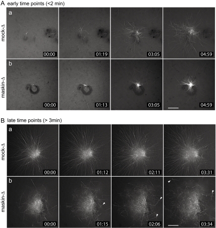
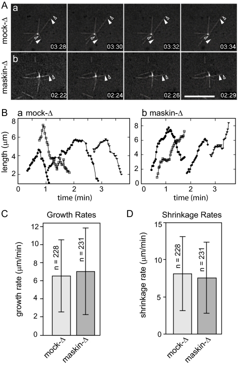
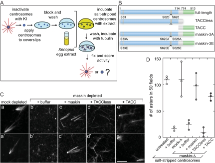
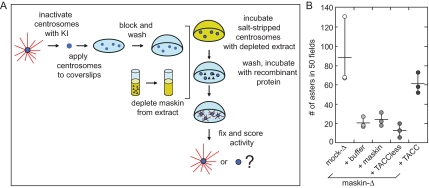
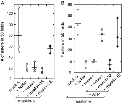
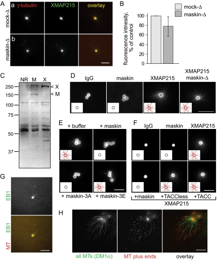
Members of the transforming acidic coiled coil (TACC) protein family are emerging as important mitotic spindle assembly proteins in a variety of organisms. The molecular details of how TACC proteins function are unknown, but TACC proteins have been proposed to recruit microtubule-stabilizing proteins of the tumor overexpressed gene (TOG) family to the centrosome and to facilitate their loading onto newly emerging microtubules. Using Xenopus egg extracts and in vitro assays, we show that the Xenopus TACC protein maskin is required for centrosome function beyond recruiting the Xenopus TOG protein XMAP215. The conserved C-terminal TACC domain of maskin is both necessary and sufficient to restore centrosome function in maskin-depleted extracts, and we provide evidence that the N terminus of maskin inhibits the function of the TACC domain. Time-lapse video microscopy reveals that microtubule dynamics in Xenopus egg extracts are unaffected by maskin depletion. Our results provide direct experimental evidence of a role for maskin in centrosome function and suggest that maskin is required for microtubule anchoring at the centrosome.


Figures

Figure 1.
Figure 1.
Maskin depletion from Xenopus egg extracts results in smaller, disorganized asters. (A and B) Time-lapse fluorescence microscopy of microtubules assembled in mock-depleted (a) or maskin-depleted (b) egg extracts induced to assemble centrosomes and microtubule structures by the addition of demembranated sperm chromatin. The extracts were spiked with a small amount of rhodamine-labeled tubulin to allow visualization of microtubules by fluorescence microscopy. Recording was initiated within the first 2 min (A) or after a minimum of 3 min (B) after warming the reaction mixture to initiate microtubule assembly. Elapsed time is given in the lower right-hand corner of each frame in minutes:seconds. These stills correspond to Supplemental Videos 1 (Aa), 2 (Ab), 3 (Ba), and 4 (Bb). Bars, 10 μm.
Figure 2.
Figure 2.
Microtubule dynamics are not affected by maskin depletion. (A) Representative micrographs from time-lapse series showing microtubule dynamics in mock (a) or maskin-depleted (b) extracts supplemented with centrosomes. Closed arrowhead denotes the position of the microtubule end at the start; open arrowhead denotes the position of the microtubule end at the end of the time sequence. Elapsed time in minutes:seconds after the start of the recording is shown in the lower right of each panel. Corresponding videos are provided as Supplemental Videos 5 and 6. Bar, 10 μm. (B) Microtubule lifetime plots for four representative microtubules in mock-depleted (a) or maskin-depleted (b) egg extracts. (C and D) Quantification of microtubule growth (C) and shrinkage rates (D). Dynamics of 228 (mock-depleted) or 231 (maskin-depleted) microtubules in nine independent experiments were measured. These data are summarized in Table 1.
Figure 3.
Figure 3.
Maskin is required for centrosome function. (A) Schematic diagram of the centrosome complementation assay. Salt-stripped centrosomes are applied to a coverslip and incubated with Xenopus egg extract. The extract is washed away and the centrosomes are challenged with purified bovine tubulin. The samples are then fixed and scored for activity. (B) Schematic diagram of the maskin constructs used in this study. The conserved TACC domain (amino acids 714–931) is shown in green, the TACCless domain (amino acids 1–774) is blue. The overlap between the TACC and TACCless domains is gray. The highlighted Aurora A phosphorylation sites (S33, S620, and S626) were mutated to glutamic acids (maskin-3E) or alanines (maskin-3A). (C) Centrosome activity can be reconstituted if salt-stripped centrosomes are incubated with maskin-depleted extracts containing recombinant full-length maskin or TACC domain. Centrosomes were salt-stripped and incubated with extracts supplemented with recombinant proteins (or buffer) as indicated above the micrographs. Two representative micrographs per complemented centrosomes are shown. Bar, 10 mm. (D) Quantification of centrosome activity in the complementation assay, expressed as the number of asters found in 50 randomly selected microscope fields. The results for three independent experiments are shown (circles). The average is represented as a horizontal line. Vertical lines indicate the spread of the data. Conditions are indicated below the graph.
Figure 4.
Figure 4.
The sequential centrosome complementation assay reveals that full-length maskin needs to be exposed to extract to be functional, whereas the TACC domain of maskin can complement centrosomes independent of extract. (A) Schematic diagram of the sequential complementation assay. Salt-stripped centrosomes are incubated with extract as before. However, the extract is washed away before the centrosomes are incubated with recombinant protein. The recombinant protein is then washed away and the centrosomes are challenged with purified bovine tubulin as before. (B) Quantification of centrosome activity in the sequential assay, expressed as the number of asters found in 50 randomly chosen microscope fields. Centrosomes were incubated with recombinant maskin or truncation mutants as indicated below the graph. The graph represents three independent experiments (indicated by circles).
Figure 5.
Figure 5.
Phosphorylation of maskin restores centrosome function. (A and B) Quantification of centrosome activity in the sequential assay, expressed as the number of asters found in 50 randomly chosen microscope fields. Centrosomes were incubated with recombinant maskin phosphorylation mutants (A), or recombinant maskin in the presence or absence of ATP (B) as indicated below the graphs. Graphs represent three independent experiments (indicated by circles).
Figure 6.
Figure 6.
Maskin is required for centrosomal microtubule assembly independently of XMAP215 localization to the centrosome. (A) Salt-stripped centrosomes incubated in mock (a) or maskin-depleted (b) extract recruit similar amounts of XMAP215. Centrosomes were double labeled for γ-tubulin (red) and XMAP215 (green). Individual channels and overlays are indicated on the micrographs. Bar, 2 μm. (B) Quantification XMAP215 fluorescence intensity (normalized against γ-tubulin fluorescence and expressed as percent of mock-depleted control) for centrosomes incubated in mock-depleted (light bars) or maskin-depleted (dark bars) extracts. n = 3; values are mean ± SD. (C) Coomassie-stained gel of proteins coimmunoprecipitated with antibodies to maskin (M) or XMAP215 (X). Arrowheads mark the positions of XMAP215 (X) and maskin (M). (D) Microtubule asters are able to form around beads coated with XMAP215, but not maskin. Antibodies against random IgG (left), maskin (middle), or XMAP215 (right) were bound to beads and incubated in normal or maskin-depleted extract, as indicated. The beads were isolated, washed, and then challenged with purified tubulin containing a small amount of rhodamine-labeled tubulin to allow visualization in the microscope. The micrographs show representative beads for each sample. The insets show schematics of the beads (circles) with or without microtubules (red lines), to aid visualization. Bar, 10 μm. (E and F) Full-length maskin and maskin-3A, but not truncation mutants or maskin-3E, suppress XMAP215-mediated aster formation. XMAP215-coated beads were challenged with purified tubulin in the presence of 200 nM maskin, maskin-3A, maskin-3E, TACC domain, or TACC-less domain as indicated for each panel. The beads were processed as in D. Insets as in D. Bar, 10 μm. (G and H). XMAP215 attaches to the minus ends of microtubules. (G) XMAP215-coated beads (generated by incubating XMAP215 antibodies with protein A beads and then incubating the antibody-coated beads in egg extract) were incubated in egg extract supplemented with small amounts of rhodamine tubulin to visualize microtubules, and GFP-EB1 to visualize microtubule plus ends. A single timeframe is shown for microtubules (red in the overlay) and GFP-EB1 (green in the overlay). Bar, 10 μm. (H) XMAP215-coated beads (generated as in G) were washed and incubated in unlabeled tubulin for 9 min, followed by incubation in rhodamine-labeled tubulin for 1 min. The microtubules were then fixed, spun onto coverslips, and processed for immunofluorescence to visualize the microtubules. The rhodamine-labeled microtubule plus ends are distal to the beads. Bar, 10 μm.

References

    1. Albee A. J., Tao W., Wiese C. Phosphorylation of maskin by Aurora-A is regulated by RanGTP and importin β. J. Biol. Chem. 2006;281:38293–38301. - PubMed
    1. Andersen J. S., Wilkinson C. J., Mayor T., Mortensen P., Nigg E. A., Mann M. Proteomic characterization of the human centrosome by protein correlation profiling. Nature. 2003;426:570–574. - PubMed
    1. Azimzadeh J., Bornens M. Structure and duplication of the centrosome. J. Cell Sci. 2007;120:2139–2142. - PubMed
    1. Badano J. L., Teslovich T. M., Katsanis N. The centrosome in human genetic disease. Nat. Rev. Genet. 2005;6:194–205. - PubMed
    1. Barros T. P., Kinoshita K., Hyman A. A., Raff J. W. Aurora A activates D-TACC-Msps complexes exclusively at centrosomes to stabilize centrosomal microtubules. J. Cell Biol. 2005;170:1039–1046. - PMC - PubMed

Publication types

LinkOut - more resources