. 2008 Aug;19(8):3299-307.
doi: 10.1091/mbc.e07-11-1125. Epub 2008 May 28.

Fas splicing regulation during early apoptosis is linked to caspase-mediated cleavage of U2AF65

Affiliations

Fas splicing regulation during early apoptosis is linked to caspase-mediated cleavage of U2AF65

José M Izquierdo. Mol Biol Cell. 2008 Aug.

Abstract

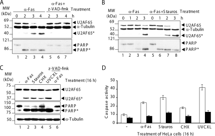
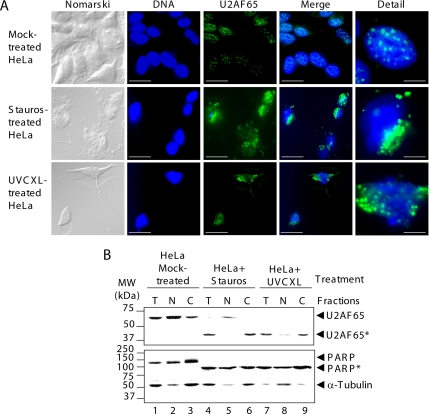
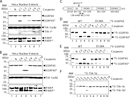
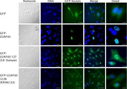
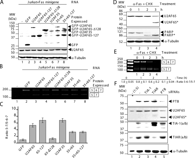
U2 small nuclear ribonucleoprotein (snRNP) auxiliary factor 65 kDa (U2AF65) is an essential splicing factor in the recognition of the pre-mRNA 3' splice sites during the assembly of the splicing commitment complex. We report here that U2AF65 is proteolyzed during apoptosis. This cleavage is group I or III caspase dependent in a noncanonical single site localized around the aspartic acid(128) residue and leads to the separation of the N- and C-terminal parts of U2AF65. The U2AF65 N-terminal fragment mainly accumulates in the nucleus within nuclear bodies (nucleoli-like pattern) and to a much lesser extent in the cytoplasm, whereas the C-terminal fragment is found in the cytoplasm, even in localization studies on apoptosis induction. From a functional viewpoint, the N-terminal fragment promotes Fas exon 6 skipping from a reporter minigene, by acting as a dominant-negative version of U2AF65, whereas the C-terminal fragment has no significant effect. The dominant-negative behavior of the U2AF65 N-terminal fragment can be reverted by U2AF35 overexpression. Interestingly, U2AF65 proteolysis in Jurkat cells on induction of early apoptosis correlates with the down-regulation of endogenous Fas exon 6 inclusion. Thus, these results support a functional link among apoptosis induction, U2AF65 cleavage, and the regulation of Fas alternative splicing.


Figures

Figure 1.
Figure 1.
U2AF65 cleavage occurs during apoptosis. (A) Jurkat cells in the absence (lanes 1–4) or in the presence (lanes 5–7) of z-VAD-fmk were incubated with the anti-Fas antibody. (B) Jurkat cells were incubated with anti-Fas antibody alone (lanes 1–4) or with this antibody plus staurosporine (lanes 5–8). (C) HeLa cells were treated with the indicated apoptotic agents for 16 h. Lane 6, HeLa cells pretreated with z-VAD-fmk as described in A. In A–C, protein extracts were immunoblotted with the indicated antibodies. The identities of molecular mass markers and protein bands are shown. (D) Protein extracts from HeLa cells treated as in described in C were harvested to test caspase-3 or caspase-3-like activity. Where indicated (black bars), 5 μM DEVD-fmk was incubated before the addition of the substrate DEVD-p-NA. Caspase activity was displayed as the number of nanomoles of p-NA released per hour per total milligram of protein as calculated from a standard curve by using free p-NA. The values of caspase activity are means ± SEM for at least two independent samples.
Figure 2.
Figure 2.
Subcellular localization of U2AF65 in apoptotic cells. (A) HeLa cells were mock treated or treated with staurosporine or irradiated with UV light and then stained with anti-U2AF65 mAb MC3. From left to right, images show cells examined using Nomarski method (Nomarski), stained with Hoechst through a 4,6-diamidino-2-phenylindole (DAPI) filter (DNA), or stained with anti-U2AF65 antibody and examined with a tetramethylrhodamine B isothiocyanate filter (U2AF65). The two right-hand images (Merge and Detail) are the sum of DNA and U2AF65 images as well as detailed images. In DNA, U2AF65 and Merge images, bars represent 30 μm. In detailed images, bars represents represent 5 μm. (B) Fractions containing either total protein (T), nuclei (N), or postnuclear supernatant (C) from HeLa cells mock treated (lanes 1–3), treated with staurosporine (lanes 4–6), or irradiated with UV light (lanes 7–9) were analyzed with the indicated antibodies. The migrations of molecular mass markers and protein bands are indicated.
Figure 3.
Figure 3.
U2AF65 is mainly cleaved by group I or III caspases at the aspartate128. (A and B) HeLa nuclear extracts were incubated with either no additional agents (lane 1), buffer alone (lane 2), or buffer plus each of the caspases (lanes 3–7) and analyzed by Western blotting using the antibodies against the indicated proteins. (C) Schematic structure of human U2AF65 protein. The amino acid sequence around the caspase cleavage site is shown in single-letter symbol of amino acids. The RS-rich sequence, U2AF35 binding site and RRM functional domains are indicated by open boxes, and the amino acid number is shown. The epitope recognized by anti-U2AF65 mAb MC3 is indicated. (D and E) 35S-labeled U2AF65 wt (lanes 1–5) or mutant D128A (lanes 6–10) was incubated with either none (lanes 1 and 10), buffer alone (lanes 2), or buffer plus each of the caspases indicated (lanes 3–5 and 7–9). (F) 35S-labeled TIA-1b was incubated with each of the caspases (lanes 1–10) as indicated. In panels, the positions of U2AF65, TIA-1 or PARP and molecular mass markers are shown.
Figure 4.
Figure 4.
Subcellular distribution of the U2AF65 N- and C-terminal fragments as GFP-tagged fusions. HeLa cells were transfected with plasmids expressing GFP (top images), GFP-fused with either U2AF65 (top middle images), the U2AF65 N-terminal fragment (SR domain) (lower middle images), or the U2AF65 C-terminal fragment (RRM123) (bottom images). The left-hand images show cells examined using Nomarski method. Fluorescence from GFP was observed using a fluorescein filter. To visualize nuclei, DNA was stained with Hoechst and examined using a DAPI filter. The two right-hand columns are merged and detailed images, respectively. In DNA, GFP-fusions and Merge images, bars represent 30 μm. In detailed images, bars represent 5 μm.
Figure 5.
Figure 5.
U2AF65 cleavage modulates endogenous Fas alternative splicing in Jurkat cells. (A) Protein extracts from untransfected and transfected Jurkat cells with the plasmids indicated were analyzed by Western blotting using anti-GFP, anti-U2AF65, or α-tubulin antibodies. Ectopic GFP expression from GFP-tagged U2AF65 plasmids and its derivatives is shown in the top panel. Endogenous U2AF65 and α-tubulin expression and equal loading are shown in the bottom panels. (B) Cytoplasmic RNAs from Jurkat cells processed in A were analyzed by RT-PCR using primers PT1 and PT2. (C) The values of ratios between 5–7 and 5-6-7 amplification products are means ± SEM for at least two to three independent experiments. These values were expressed relative to GFP expression (B, lane 2), whose value was fixed arbitrarily to 1. (D and E) Jurkat cells were incubated with the apoptosis-inducers anti-Fas antibody (500 ng/ml) and cycloheximide (10 μg/ml) for the indicated times (0–3 h). (D) Total cell extracts were analyzed by immunoblotting using antibodies against U2AF65, PARP, and α-tubulin. (E) Cytoplasmic RNAs were isolated from apoptotic Jurkat cells in D and analyzed by RT-PCR. The products of amplification were separated by agarose-gel electrophoresis. The values of ratios between 5-6-7 and 5–7 amplification products are means ± SEM for at least two independent experiments. These values were normalized relative to lane 1, whose value was fixed arbitrarily to 1. In D and E, positions of molecular mass markers, protein bands and predicted alternatively spliced products are indicated. (F) The siRNA-mediated down-expression of PTB promotes the U2AF65 cleavage. Western blot analysis of HeLa cell lysates (5 μg [1:5] or 25 μg, lanes 1 and 2–5, respectively) prepared 72 h after transfection with siRNAs against GFP (lanes 1 and 2), TIA-1 (lane 3), TIAR (lane 4), and PTB (lane 5). The blot was probed with antibodies against PTB, U2AF65, TIA-1, TIAR, and α-tubulin proteins, as indicated. Molecular weight markers and the identities of protein bands are indicated.

References

    1. Adams J. M. Ways of dying: multiple pathways to apoptosis. Genes Dev. 2003;17:2481–2495. - PubMed
    1. Banerjee H., Rahn A., Davis W., Singh R. Sex-lethal and U2 small nuclear ribonucleoprotein auxiliary factor (U2AF65) recognize polypyrimidine tracts using multiple modes of binding. RNA. 2003;9:88–99. - PMC - PubMed
    1. Bidere N., Su H. C., Lenardo M. J. Genetic disorders of programmed cell death in the immune system. Annu. Rev. Immunol. 2006;24:321–352. - PubMed
    1. Buendia B., Santa-Maria A., Courvalin J. C. Caspase-dependent proteolysis of integral and peripheral proteins of nuclear membranes and nuclear pore complex proteins during apoptosis. J. Cell Sci. 1999;112:1743–1753. - PubMed
    1. Cheng J., Zhou T., Liu C., Shapiro J. P., Brauer M. J., Kiefer M. C., Barr P. J., Mountz J. D. Protection from Fas-mediated apoptosis by a soluble form of the Fas molecule. Science. 1994;263:1759–1762. - PubMed

Publication types