. 2008 Aug;19(8):3323-33.
doi: 10.1091/mbc.e08-03-0328. Epub 2008 May 28.

The yeast GID complex, a novel ubiquitin ligase (E3) involved in the regulation of carbohydrate metabolism

Affiliations

The yeast GID complex, a novel ubiquitin ligase (E3) involved in the regulation of carbohydrate metabolism

Olivier Santt et al. Mol Biol Cell. 2008 Aug.

Abstract

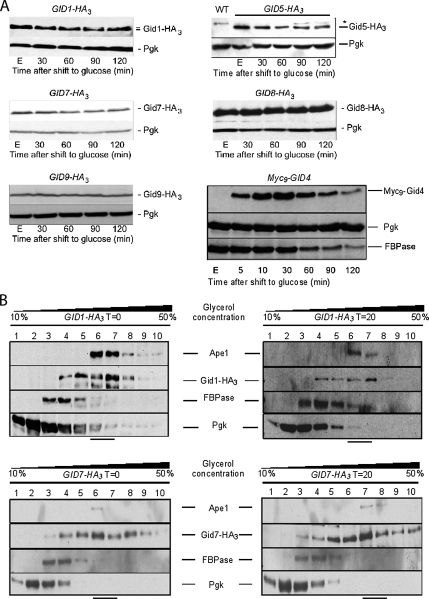
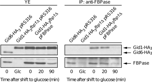
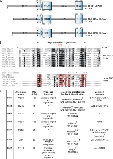
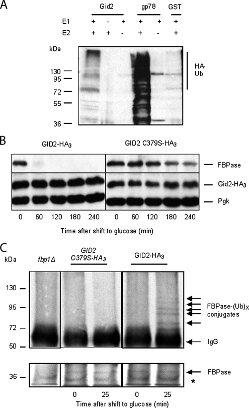
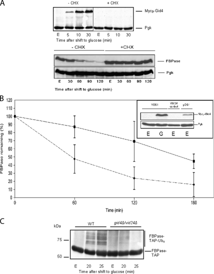
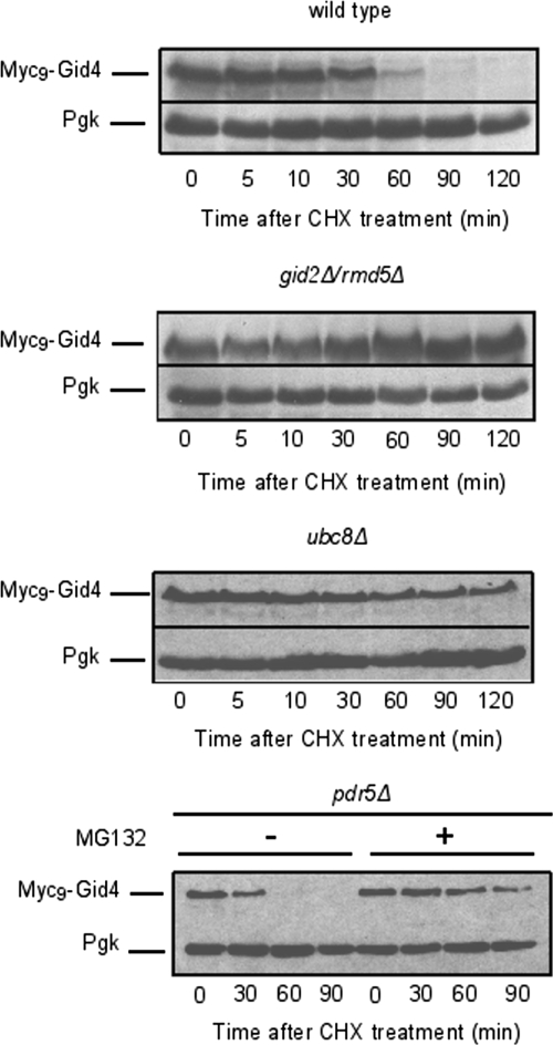
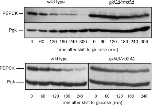
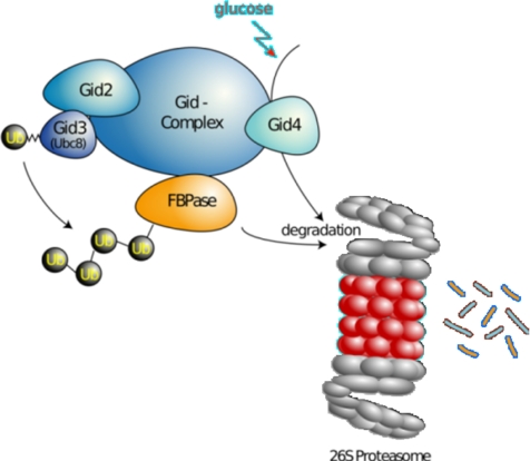
Glucose-dependent regulation of carbon metabolism is a subject of intensive studies. We have previously shown that the switch from gluconeogenesis to glycolysis is associated with ubiquitin-proteasome linked elimination of the key enzyme fructose-1,6-bisphosphatase. Seven glucose induced degradation deficient (Gid)-proteins found previously in a genomic screen were shown to form a complex that binds FBPase. One of the subunits, Gid2/Rmd5, contains a degenerated RING finger domain. In an in vitro assay, heterologous expression of GST-Gid2 leads to polyubiquitination of proteins. In addition, we show that a mutation in the degenerated RING domain of Gid2/Rmd5 abolishes fructose-1,6-bisphosphatase polyubiquitination and elimination in vivo. Six Gid proteins are present in gluconeogenic cells. A seventh protein, Gid4/Vid24, occurs upon glucose addition to gluconeogenic cells and is afterwards eliminated. Forcing abnormal expression of Gid4/Vid24 in gluconeogenic cells leads to fructose-1,6-bisphosphatase degradation. This suggests that Gid4/Vid24 initiates fructose-1,6-bisphosphatase polyubiquitination by the Gid complex and its subsequent elimination by the proteasome. We also show that an additional gluconeogenic enzyme, phosphoenolpyruvate carboxykinase, is subject to Gid complex-dependent degradation. Our study uncovers a new type of ubiquitin ligase complex composed of novel subunits involved in carbohydrate metabolism and identifies Gid4/Vid24 as a major regulator of this E3.


Figures

Figure 1.
Figure 1.
Gid complex subunits are present in gluconeogenic cells. (A) Strains expressing chromosomally tagged Gid proteins with the HA or Myc epitope were grown 16 h in YPethanol (E) and thereafter treated with 2% glucose. Samples were taken at the indicated time points and processed as described in Material and Methods. Proteins were visualized by immunoblotting. *, cross reaction; WT, wild type, control for cross-reaction. (B) Strains bearing a chromosomally expressed HA-tagged Gid protein (Gid1/Vid30 and Gid7) were grown 16 h in YPethanol. Samples were harvested before (T = 0) or 20 min after shift to glucose (T = 20), and proteins were separated on a glycerol step gradient. Ten fractions were collected, and proteins were visualized by immunoblotting (Pgk, 3-phosphoglycerate kinase; Ape1, aminopeptidase I).
Figure 2.
Figure 2.
FBPase binds to Gid1/Vid30. Strains expressing HA-tagged Gid proteins (Gid1/Vid30 and Gid6/Ubp14) and transformed with a plasmid expressing a wild-type FBPase (pFBPase) or its corresponding vector control (pRS316) were grown 16 h on YPethanol. Samples were harvested 0, 20, and 90 min after glucose treatment, and proteins were extracted. Coimmunoprecipitation was performed with FBPase antibody. Protein immunoblot was carried out with FBPase and HA antibodies. The noninteracting Gid6/Ubp14 protein serves as a control (YE, extracts; IP, immunoprecipitations). The FBP1 deletion mutant transformed with the vector control was grown on glucose and provides a control for the specificity of the anti-FBPase antibody.
Figure 3.
Figure 3.
Domain structures of Gid proteins. (A) Structure similarities between the Gid2/Rmd5 (S. cerevisiae) and RMND5A/B (Homo sapiens) proteins. (B) Alignment of the RING domains found in the Gid2/Rmd5 protein family and classic RING-finger proteins. Positions with invariant or conservatively replaced residues in at least 50% of the sequences are printed on black or gray background, respectively (not all sequences analyzed are shown). The leftmost column describes the sequence name and contains the species abbreviation: YEAST, S. cerevisiae; POMBE, S. pombe; CANAL, Candida albicans; HUMAN, H. sapiens; XENLA, Xenopus laevis; DROME, Drospophila melanogaster; CAEEL, Caenorhabditis elegans; TETNG, Tetraodon nigroviridis. Larger insertions are not shown; the number of omitted residues is indicated in parentheses. Asterisks above the Gid2/Rmd5 family alignment correspond to positions homologous to zinc coordinating residues in classic RING finger domains (red). The mutation introduced in the Gid2/Rmd5 degenerated RING domain is highlighted (C379S, red). Above the classic RING-finger proteins alignment, residues of Cbl in contact with UbcH7 are marked by # (Zheng et al., 2000). (C) Overview of the Gid proteins and their counterparts in H. sapiens. The table summarizes the common features in Gid proteins and their human counterparts. aOrthologues identified in a CTLH complex by Kobayashi et al. (2007); bOrthologues found to interact together by Umeda et al. (2003).cMuskelin bears CTLH and Kelch domains, which makes its overall structure similar to Gid7. Other functions and names, SGD, www.yeastgenome.org.
Figure 4.
Figure 4.
Gid2/Rmd5 shows E3 activity. (A) Gid2/Rmd5 ubiquitinates protein in vitro. Lysates of E. coli expressing either Gid2/Rmd5 or the mammalian RING-finger protein gp78 as a positive control and GST as a negative control were incubated with E1, E2 (UbcH5b), ATP and HA-ubiquitin for 2 h at 30°C. To assess the specificity of the reaction, same lysates were incubated without E1 or E2. Polyubiquitination was detected using monoclonal HA antibody. (B) GID2-HA3 and its mutated C379S counterpart-expressing cells were grown 16 h in YPethanol at 25°C and shifted to YPD. Samples were taken at indicated time points and FBPase degradation was monitored by immunoblotting. Pgk, 3-phosphoglycerate kinase, loading control. (C) Point mutation in the Gid2/Rmd5 degenerated RING domain abolishes FBPase polyubiquitination. Chromosomally tagged GID2-HA3, which shows a wild-type phenotype, and a strain bearing the C379S point mutation in a GID2-HA3 strain were grown 16 h in YPethanol at 25°C and shifted to YPD. Samples were taken at indicated time points, and FBPase was immunoprecipitated. Polyubiquitination was detected using monoclonal ubiquitin antibody. fbp1Δ: FBPase deletion. Presence of FBPase in the immunoprecipitates was controlled by immunoblotting with FBPase antibody. *, cross-reaction.
Figure 5.
Figure 5.
Gid4/Vid24 promotes FBPase degradation. (A) De novo protein synthesis is necessary for FBPase degradation and Myc9-Gid4 expression. Wild-type cells were grown 16 h in YPethanol (E) and shifted to YPD with (+CHX) or without (−CHX) cycloheximide (100 μg/ml). FBPase and Myc9-Gid4 levels were monitored at indicated times via immunoblotting with anti-FBPase or anti-Myc antibodies, respectively. (B) Gid4/Vid24 expression triggers FBPase degradation. Myc9-GID4 was cloned under a TetR promoter and expressed in cells growing on YPethanol. Pulse-chase analysis of FBPase in cells bearing either the Myc9-GID4 expressing plasmid (♦) or the respective vector control (■) was carried out (means of three independent experiments, ±confidence interval, α = 0.05). Inset, immunoblot showing the steady-state levels of Myc9-Gid4 in glucose-inactivated cells (30 min, YOS1, G) and in ethanol grown cells (pOS1, E). (C) Gid4/Vid24 is necessary for FBPase-TAP polyubiquitination. A plasmid expressing a FBPase-TAP fusion protein was transformed into W303 (WT) and gid4Δ/vid24Δ strains. Samples were taken at indicated time points, and FBPase was pull-downed using IgG-Sepharose. Polyubiquitination was detected using monoclonal ubiquitin antibody.
Figure 6.
Figure 6.
Gid4/Vid24 degradation depends on Gid2/Rmd5, Ubc8 and the proteasome. W303-1B expressing Myc9-Gid4 was deleted for UBC8, GID2/RMD5, or PDR5 (ubc8Δ, gid2Δ, pdr5Δ, respectively). Strains were grown 16 h on YPethanol, shifted to YPD, and expression of Gid4/Vid24 was allowed to proceed during 30 min. Thereafter, cells were treated with cycloheximide and samples were harvested at the indicated times. Proteins were visualized via immunoblotting. Proteasome involvement in Gid4/Vid24 degradation was analyzed in a pdr5Δ strain using the proteasome inhibitor MG-132.
Figure 7.
Figure 7.
GID deletions impair PEPCK degradation. Wild type, gid2Δ/rmd5Δ, or gid4Δ/vid24Δ cells were grown 16 h on YPethanol, and thereafter shifted onto YPD. Cells were harvested every hour, and extracts were prepared. Proteins were visualized via immunoblotting. Pgk, 3-phosphoglycerate kinase, loading control.
Figure 8.
Figure 8.
The Gid complex, a novel ubiquitin ligase (E3) required for the degradation of the key gluconeogenic enzyme fructose-1,6-bisphosphatase. The Gid complex binds to FBPase, when S. cerevisiae cells are growing on an ethanol-containing medium. On shift of cells to glucose, Gid4/Vid24 occurs and activates the complex, which then polyubiquitinates FBPase before its degradation by the proteasome. Gid4/Vid24 is itself degraded by the proteasome.

References

    1. Amerik A., Swaminathan S., Krantz B. A., Wilkinson K. D., Hochstrasser M. In vivo disassembly of free polyubiquitin chains by yeast Ubp14 modulates rates of protein degradation by the proteasome. EMBO J. 1997;16:4826–4838. - PMC - PubMed
    1. Ausubel F. M., Kingston R. E., Seidman F. G., Struhl K., Moore D. D., Brent R., Smith F. A. Current Protocols in Molecular Biology. New York: Greene; 1992.
    1. Bairoch A., Apweiler R. The SWISS-PROT protein sequence data bank and its supplement TrEMBL. Nucleic Acids Res. 1997;25:31–36. - PMC - PubMed
    1. Balzi E., Wang M., Leterme S., Van Dyck L., Goffeau A. PDR5, a novel yeast multidrug resistance conferring transporter controlled by the transcription regulator PDR1. J. Biol. Chem. 1994;269:2206–2214. - PubMed
    1. Benton D. Recent changes in the GenBank On-line Service. Nucleic Acids Res. 1990;18:1517–1520. - PMC - PubMed

Publication types

MeSH terms

Substances