. 2008 May 28;28(22):5686-90.
doi: 10.1523/JNEUROSCI.0584-08.2008.

Transient spine expansion and learning-induced plasticity in layer 1 primary motor cortex

Affiliations

Transient spine expansion and learning-induced plasticity in layer 1 primary motor cortex

Kimberly J Harms et al. J Neurosci. .

Abstract

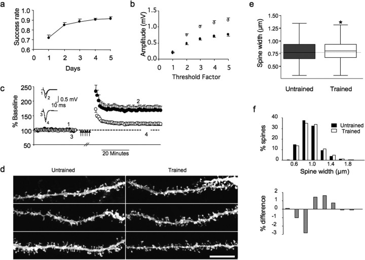
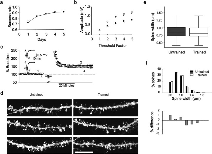
Experience-dependent regulation of synaptic strength in the horizontal connections in layer 1 of the primary motor cortex is likely to play an important role in motor learning. Dendritic spines, the primary sites of excitatory synapses in the brain, are known to change shape in response to various experimental stimuli. We used a rat motor learning model to examine connection strength via field recordings in slices and confocal imaging of labeled spines to explore changes induced solely by learning a simple motor task. We report that motor learning increases response size, while transiently occluding long-term potentiation (LTP) and increasing spine width in layer 1. This demonstrates learning-induced changes in behavior, synaptic responses, and structure in the same animal, suggesting that an LTP-like process in the motor cortex mediates the initial learning of a skilled task.


Figures

Figure 1.
Figure 1.
Motor skill training and slice recording. a, Rats are trained to reach into a food box with one forelimb and grasp small food pellets. The trained hemisphere is contralateral to the trained paw (dashed). b, Extracellular FPs of horizontal connections in M1 are recorded simultaneously in both hemispheres of coronal slices.
Figure 2.
Figure 2.
Five days of training increases synaptic responses and spine size and partially occludes LTP. a, For 5 d, adult rats were trained to reach with a single forepaw through a small aperture in a box and grasp food pellets. Performance improved over the first 3–4 d of training, reaching a plateau by day 5. b, Extracellular FPs were recorded simultaneously in the forelimb area of both hemispheres of coronal brain slices. FP amplitudes were larger in the trained hemisphere across stimulation intensities as revealed by input–output relationships. c, LTP was induced repeatedly (multiple arrows) until responses were saturated in both hemispheres. LTP was reduced in layer 1 of the trained (○) compared with the untrained hemisphere (●; t test, *p = 0.016; n = 5). Inset, Individual traces before and after LTP saturation. d, After recording, slices were fixed, labeled with DiI, and spines in layer 1 imaged with confocal microscopy. n = 6 rats. Scale bar, 10 μm. e, Analysis of morphology showed spines had a greater spine head width in the trained hemisphere (Kruskal–Wallis, *p = 0.02). Note that the whiskers do not represent SD or SEM. f, The trained distribution (□) is shifted to the right of the untrained distribution (■); untrained spines predominate in smaller bins, whereas trained spines prevail in larger bins. Gray bars below represent the percentage of trained and percentage of untrained spines for each bin.
Figure 3.
Figure 3.
Response size increase remains 30 d after training, whereas LTP and spine width recover. a, Rats learned the reach-and-grasp task over 5 d then returned to normal housing for 25–30 d. n = 15. b, FPs recorded simultaneously in both hemispheres indicate that the increase in FP amplitude in the trained hemisphere is maintained 30 d later. c, LTP was induced repeatedly (multiple arrows) until responses were saturated in both hemispheres. LTP after saturation in the trained hemisphere (○) was similar to LTP in the untrained hemisphere (●) 30 d after motor skill training (t test, p = 0.401; n = 5). Inset, Individual traces before and after LTP saturation. d, After recording, slices were fixed, labeled with DiI, and spines in layer 1 were imaged with confocal microscopy. n = 10 rats. Scale bar, 10 μm. e, Thirty days after training, analysis of dendritic spine morphology showed that the dendritic spine width was not different in trained and untrained hemispheres (Kruskal–Wallis, p = 0.19). f, The trained distribution of dendritic spine width is no longer shifted. Gray bars below represent the percentage of trained and percentage of untrained spines for each bin.

References

    1. Bourne J, Harris KM. Do thin spines learn to be mushroom spines that remember? Curr Opin Neurobiol. 2007;17:381–386. - PubMed
    1. Cauller L. Layer I of primary sensory neocortex: where top-down converges upon bottom-up. Behav Brain Res. 1995;71:163–170. - PubMed
    1. Douglas RJ, Martin KA. Neuronal circuits of the neocortex. Annu Rev Neurosci. 2004;27:419–451. - PubMed
    1. Evans HM, Long JA. Characteristic effects upon growth, oestrus and ovulation induced by the intraperitoneal administration of fresh anterior hypophyseal substance. Proc Natl Acad Sci USA. 1922;8:38–39. - PMC - PubMed
    1. Gan WB, Grutzendler J, Wong WT, Wong RO, Lichtman JW. Multicolor “DiOlistic” labeling of the nervous system using lipophilic dye combinations. Neuron. 2000;27:219–225. - PubMed

Publication types

MeSH terms

LinkOut - more resources