. 2008 May 28;28(22):5817-26.
doi: 10.1523/JNEUROSCI.0853-08.2008.

GPR56 regulates pial basement membrane integrity and cortical lamination

Affiliations

GPR56 regulates pial basement membrane integrity and cortical lamination

Shihong Li et al. J Neurosci. .

Abstract

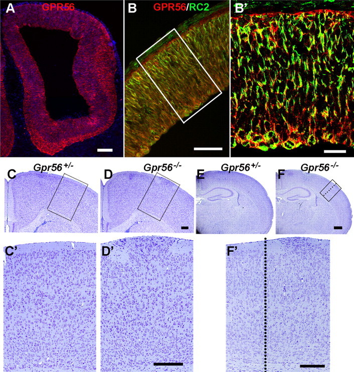
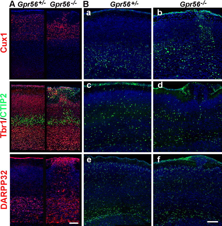
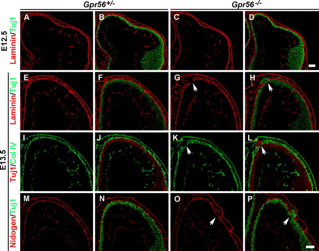
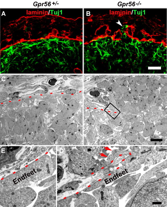
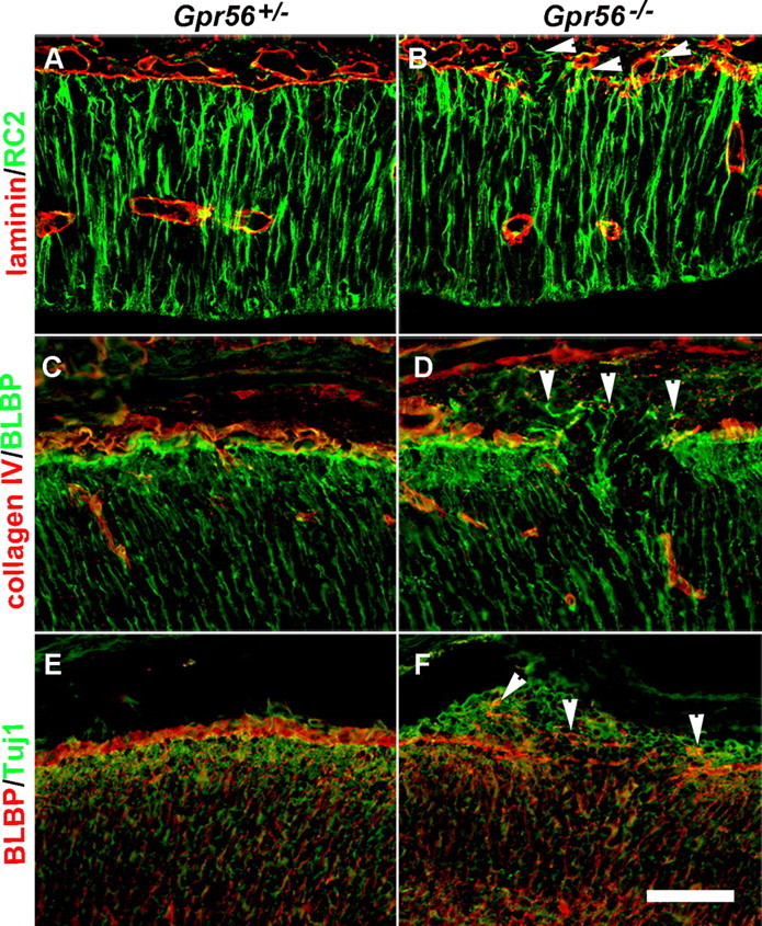
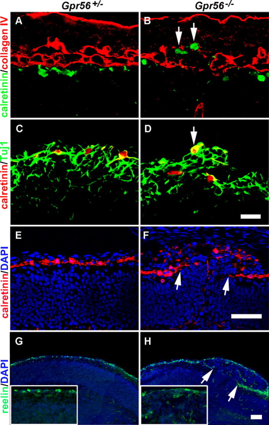
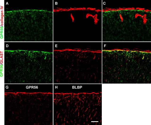
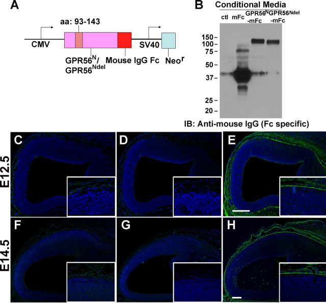
GPR56 is a member of the family of adhesion G-protein-coupled receptors that have a large extracellular region containing a GPS (G-protein proteolytic site) domain. Loss-of-function mutations in the GPR56 gene cause a specific human brain malformation called bilateral frontoparietal polymicrogyria (BFPP). BFPP is a radiological diagnosis and its histopathology remains unclear. This study demonstrates that loss of the mouse Gpr56 gene leads to neuronal ectopia in the cerebral cortex, a cobblestone-like cortical malformation. There are four crucial events in the development of cobblestone cortex, namely defective pial basement membrane (BM), abnormal anchorage of radial glial endfeet, mislocalized Cajal-Retzius cells, and neuronal overmigration. By detailed time course analysis, we reveal that the leading causal events are likely the breaches in the pial BM. We show further that GPR56 is present in abundance in radial glial endfeet. Furthermore, a putative ligand of GPR56 is localized in the marginal zone or overlying extracellular matrix. These observations provide compelling evidence that GPR56 functions in regulating pial BM integrity during cortical development.


Figures

Figure 1.
Figure 1.
GPR56 is detected in neural progenitor cells and deletion of Gpr56 results in neuronal ectopias. A, B, E12.5 coronal sections through mouse forebrains showing GPR56 as visualized by indirect immunofluorescence. Double-label IHC of GPR56 and RC2 antibody (B) performed on adjacent sections, revealing the expression of GPR56 in radial glial cells. B′, Higher-magnification view of the boxed area in B. C–F, Nissl staining of coronal sections from P14 brains of heterozygous and homozygous mutant mice. No cortical defects were observed in heterozygous brains (C, E), whereas neuronal ectopia was seen in homozygous brains (D, F). E, F, The hippocampus was unaffected. C′, D′, F′, Higher-magnification views of the boxed areas in C, D, and F. Scale bars: A, 100 μm; B, 50 μm; B′, 20 μm; (in D′, F′) C, C′, D, D′, F′, 200 μm; (in F) E, F, 500 μm.
Figure 2.
Figure 2.
Abnormal cortical lamination in Gpr56−/− mice. A, Immunostaining for cortical layer-specific markers in P6 mouse brains revealed the disorganized lamination in Gpr56−/− mice. Ectopic cluster comprises neurons from both deep and superficial layers. B, In vivo migration assay using BrdU pulse labeling at E12.5 (a, b), E15.5 (c, d), and E17.5 (e, f) revealed that neuronal proliferation and migration were mostly unaffected in Gpr56−/− cortex, except in the ectopic regions. Both deep and upper layer neurons migrated into the marginal zone. Scale bars, 100 μm.
Figure 3.
Figure 3.
Defective BM leads to neuronal ectopias. A–D, Double-label IHC of Tuj1 and laminin on E12.5 coronal sections revealing intact BM in both control and mutant animals. E–P, Double-label IHC of Tuj1 and various BM constituents on E13.5 coronal sections revealed a continuous thin lining of the BM on the cortical surface in the heterozygous brains (E, I, M) and postmitotic neurons positioned underneath the BM (F, J, N). In contrast, regionally ruptured BM was detected in the homozygous brains (G, K, O, arrows). Tuj1-positive neurons migrated through the defective BM (H, L, P, arrows). Scale bars, 100 μm.
Figure 4.
Figure 4.
Regional breaks of the pial BM in the absence of GPR56. A, B, Double IHC of laminin (red) and Tuj1 (green) at embryonic stage E12.8 revealing the continuous pial BM and the migrating neurons underneath in a heterozygous brain (A), whereas a linear break of the pial BM and ectopic neurons were detected in a Gpr56 mutant (B, arrow). C–E, Electron-microscopic views at E13.5 showing the continuous pial BM in heterozygous brain (C, red dashed line) and ectopic neurons migrating out through the broken pial BM in Gpr56 knock-out brain (D). D′, Higher-magnification view of the boxed area in D showing radial glial endfeet extending out (arrows) through the broken pial BM (red dashed line). E, Higher-magnification view of the normal attachment of radial glial endfeet to the pial BM in heterozygous brain. Scale bars: (in B) A, B, 20 μm; (in D) C, D, 10 μm; (in D′) D′, E, 1 μm.
Figure 5.
Figure 5.
Abnormally positioned radial glial endfeet in Gpr56 mutant mice. A, B, E12.8 coronal section through mouse forebrain showing that radial glial processes (green) extend through the cortical wall and terminate at the continuous BM (red) in Gpr56+/− brain. In contrast, the radial glial processes and endfeet protruded through the defective BM in mutants (B, arrowheads). C, D, Double-label IHC of BLBP (green) and Col IV (red) on E15.5 coronal sections. The endfeet were extended through the defective BM in mutants (D, arrowheads). E, F, Double-label IHC of BLBP (red) and Tuj1 (green) on E15.5 brain sections. The endfeet were extended into the neuronal ectopia in mutants (F, arrowheads). In contrast, a continuous lining of the endfeet on the outer surface of the migrating neurons was found in control animals (E). Scale bar, 50 μm.
Figure 6.
Figure 6.
Displaced CR cells in Gpr56 mutant mice. A, B, Calretinin/Col IV double-label IHC on E12.8 brain sections revealed a single layer of CR cells beneath the BM in the heterozygous brain, whereas ectopically placed CR cells were observed in the subarachnoid space at the region of defective BM in the homozygous brain (B, arrows). C, D, Double-label IHC of calretinin (red) and Tuj1 (green) on coronal sections of E12.8 brains. A single layer of CR cells outlined the surface of Tuj1-positive neurons in heterozygous cortex (C), whereas a bump of overmigrated neurons was seen in homozygous neocortex with one CR cell placed on the peak of the ectopia (D, arrow). E–H, Calretinin and reelin IHC on E16.5 coronal sections of heterozygous and mutant brains showing a single layer of CR cells continuously outlining the cortical surface in control animals (E, G), and in contrast, mutant brain showing scattered CR cells within the ectopia (F, H). No CR cells were detected at the stem, where a stream of neurons were migrating through (between the arrows in F and H). Scale bars: (in D) A–D, 20 μm; (in F, H) E–H, 50 μm.
Figure 7.
Figure 7.
GPR56 protein is present in radial glial endfeet. A–C, Double-label IHC of GPR56 (green) and Col IV (red) on E16.5 wild-type cortex showing GPR56 immunoreactivities terminate at the pial BM. D–F, Double-label IHC of GPR56 (green) and GLAST (red) on E16.5 wild-type cortex. GPR56 immunoreactivities were colocalized with GLAST in the radial glial endfeet (F). G, H, GPR56 and BLBP IHC on adjacent sections of E16.5 wild-type cortex, revealing that GPR56 immunoreactivity (G) was observed at the pial surface, corresponding to the position of radial glial endfeet outlined by BLBP on the adjacent section (H). Scale bar, 20 μm.
Figure 8.
Figure 8.
GPR56 binds a putative ligand in pial basement membrane. A, Schematic drawing of the fusion constructs. An in-frame deletion mutation was created by deleting amino acids 93–143, GPR56Ndel. Mouse IgG Fc tag was fused to the C terminus of GPR56N or GPR56Ndel to form a fusion construct. B, The fusion constructs were transfected into HEK-293T cells. Secreted proteins in the conditioned media were collected, concentrated, and verified by Western blot. C–H, GPR56N-mFc fusion protein in situ. GPR56N-mFc binds a putative ligand in the ECM at both E12.5 (E) and E14.5 (H) brains, whereas mFc (C, F) and GPR56Ndel-mFc (D, G) controls revealed no binding. Nuclear counterstain was performed by Hoechst 33342 (blue). Scale bars, 100 μm.

References

    1. Anton ES, Kreidberg JA, Rakic P. Distinct functions of alpha3 and alpha (v) integrin receptors in neuronal migration and laminar organization of the cerebral cortex. Neuron. 1999;22:277–289. - PubMed
    1. Beggs HE, Schahin-Reed D, Zang K, Goebbels S, Nave KA, Gorski J, Jones KR, Sretavan D, Reichardt LF. FAK deficiency in cells contributing to the basal lamina results in cortical abnormalities resembling congenital muscular dystrophies. Neuron. 2003;40:501–514. - PMC - PubMed
    1. Chang BS, Piao X, Bodell A, Basel-Vanagaite L, Straussberg R, Dobyns WB, Qasrawi B, Winter RM, Innes AM, Voit T, Grant PE, Barkovich AJ, Walsh CA. Bilateral frontoparietal polymicrogyria: clinical and radiological features in 10 families with linkage to chromosome 16. Ann Neurol. 2003;53:596–606. - PubMed
    1. Costell M, Gustafsson E, Aszodi A, Morgelin M, Bloch W, Hunziker E, Addicks K, Timpl R, Fassler R. Perlecan maintains the integrity of cartilage and some basement membranes. J Cell Biol. 1999;147:1109–1122. - PMC - PubMed
    1. De Arcangelis A, Mark M, Kreidberg J, Sorokin L, Georges-Labouesse E. Synergistic activities of alpha3 and alpha6 integrins are required during apical ectodermal ridge formation and organogenesis in the mouse. Development. 1999;126:3957–3968. - PubMed

Publication types

MeSH terms