. 2008 Sep;100(3):1184-201.
doi: 10.1152/jn.01344.2007. Epub 2008 May 28.

Dorsal root ganglion neurons innervating skeletal muscle respond to physiological combinations of protons, ATP, and lactate mediated by ASIC, P2X, and TRPV1

Affiliations

Dorsal root ganglion neurons innervating skeletal muscle respond to physiological combinations of protons, ATP, and lactate mediated by ASIC, P2X, and TRPV1

Alan R Light et al. J Neurophysiol. 2008 Sep.

Abstract

The adequate stimuli and molecular receptors for muscle metaboreceptors and nociceptors are still under investigation. We used calcium imaging of cultured primary sensory dorsal root ganglion (DRG) neurons from C57Bl/6 mice to determine candidates for metabolites that could be the adequate stimuli and receptors that could detect these stimuli. Retrograde DiI labeling determined that some of these neurons innervated skeletal muscle. We found that combinations of protons, ATP, and lactate were much more effective than individually applied compounds for activating rapid calcium increases in muscle-innervating dorsal root ganglion neurons. Antagonists for P2X, ASIC, and TRPV1 receptors suggested that these three receptors act together to detect protons, ATP, and lactate when presented together in physiologically relevant concentrations. Two populations of muscle-innervating DRG neurons were found. One responded to low metabolite levels (likely nonnoxious) and used ASIC3, P2X5, and TRPV1 as molecular receptors to detect these metabolites. The other responded to high levels of metabolites (likely noxious) and used ASIC3, P2X4, and TRPV1 as their molecular receptors. We conclude that a combination of ASIC, P2X5 and/or P2X4, and TRPV1 are the molecular receptors used to detect metabolites by muscle-innervating sensory neurons. We further conclude that the adequate stimuli for muscle metaboreceptors and nociceptors are combinations of protons, ATP, and lactate.


Figures

FIG. 1.
FIG. 1.
Responses of individual dorsal root ganglion (DRG) neurons to increases in pump-applied metabolites, with responses to 200 nM capsaicin and 50 mM KCl added at the end of each trace. Solid downward-pointing spike-like symbols indicate where a portion of the trace stopped, then started to allow the capsaicin and KCl responses to be included on this figure. In all, 17 DRG neurons were retrogradely labeled from skeletal muscle with DiI in this well, and all 7 of these that responded to metabolite increases are shown here. To allow for larger font size, the amounts of metabolites applied are split between top and bottom, even though the same metabolites are applied at the same time to the neurons depicted on both top and bottom. Arrows and vertical lines indicate when metabolite changes were made; wash = pH 7.4, 1 mM lactate, with 0 adenosine triphosphate (ATP). Vertical scale is the ratio of intensity of light emitted at 340 nm/380 nm. Maximum possible calcium responses caused by application of either capsaicin or KCl are at the right of each trace.
FIG. 2.
FIG. 2.
Responses of individual DRG neurons to changes in metabolites, with their responses to 200 nM capsaicin and 50 mM KCl included at the end of each trace. Pipetting artifacts were reduced or omitted. Note apparent metabolite responses at pH 7.4 caused by application 300 nM ATP, given that no other metabolites were increased during this application. These ATP-induced artifacts were not seen in Fig. 1 because ATP responses had adapted before the application of pH 7.4. Shown here are all 10 of the metabolite-responsive DRG neurons labeled with DiI from the hindlimb muscles. Twenty-two DRG neurons were labeled with DiI in this well but 12 of these did not respond to metabolite increases. All other labels as in Fig. 1.
FIG. 3.
FIG. 3.
Percentage of DRG neurons with increases in intracellular calcium evoked by the metabolite levels indicated on the x-axis. The combinations of metabolites contributing to each of the curves are indicated in the legend on right. For example, the filled squares represent experiments in which protons and ATP were varied, but lactate was 0. For each series of points, the metabolites were administered in the increasing order shown. n = the number of wells under each condition; in each well an average of 152 ± 9 (SE) active DRG neurons were imaged. In all, 5,307 DRG active neurons were analyzed for this figure.
FIG. 4.
FIG. 4.
Total percentage of DRG neurons responding with a rapid calcium increase to the application of increasing metabolite combinations as applied to wells in Fig. 3. To calculate the percentages represented by each bar, all of the DRG neurons responding at any of the 5 concentrations of metabolites presented in increasing amounts shown beneath the bar were counted. As shown in Figs. 1 and 2, because many neurons responded at more than one, but not all doses of metabolites, the total percentage of responding neurons here will be higher than any single percentage in Fig. 3. Wells measured for both the 5th and 6th bars were combinations of all 3 metabolites indicated under both bars. The 6th bar represents the total percentage of active neurons labeled from muscle that responded to increases of all 3 metabolites. Numbers of wells tested for each condition are indicated in parentheses within each bar. Statistical analysis in this and other figures used the number of wells as the sample size. ANOVA for all groups: F = 11.96, P < 0.0001. Dunnett's method compared all other means to that of all neurons responding to pH 7.4–6.6/lacate/ATP applications. * indicates mean greater than all means to the left of this bar (P < 0.05). ** indicates mean greater than all other means (P < 0.01).
FIG. 5.
FIG. 5.
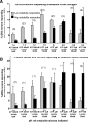
Graph of percentage of responding DRG neurons divided into high and low metabolite responders, respectively. A: graph is for all DRG neurons. B: graph includes only DRG neurons retrogradely labeled from hindlimb muscles. The numbers in parentheses indicate the number of wells analyzed. Each well in the top graph averaged 123 ± 8 neurons. Each well in the bottom graph averaged 28 ± 4 active DiI-labeled neurons.
FIG. 6.
FIG. 6.
Graph of calcium responses of DRG neurons retrogradely labeled from skeletal muscle to the application of the indicated metabolites for each bar. DRG neurons were divided into those that increased responses up to metabolites associated with pH 7.0, then decreased calcium responses to higher levels of metabolites (Low metabolite responders) and those that increased calcium responses to metabolites associated with pH levels <7.0 (High metabolite responders). Only neurons that showed a significant calcium increase above baseline were used in this analysis.
FIG. 7.
FIG. 7.
Effects of antagonists to acid-sensing ion channels (ASICs; A-317567: filled triangles) and transient receptor potential of the vanilloid type 1 (TRPV1; JYL-1433 and LJO-328: open circles and squares, respectively) at indicated doses on responses to increased metabolites as presented in previous graphs. Controls (filled diamonds) received only the metabolite increases indicated on the x-axis. For the antagonist applications, each well averaged 108 ± 5 neurons imaged for calcium increases. Statistical analyses used wells as the sample size.
FIG. 8.
FIG. 8.
Effects of the purinergic type 2X (P2X) antagonists trinitrophenyl-adenosine triphosphate (TNP-ATP) and pyridoxal-phosphate-6-azophenyl-2′,4′-disulfonate (PPADS) at various doses on responses of DRG neurons to increases in metabolites. Note that very low concentrations of TNP-ATP (10 nM) facilitated responses (inset graph), whereas concentrations of TNP-ATP that antagonize P2X5 (1 μM) reduced low metabolite responses, and concentrations that block all P2X receptors including P2X4 (200 μM) blocked nearly all responses to metabolites.
FIG. 9.
FIG. 9.
Percentage of DRG neurons responding with rapid calcium increase to application of 200 nM capsaicin. Black-filled bars are data from all DRG neurons. Gray-filled bars are from DRG neurons retrogradely labeled from hindlimb muscles. Unfilled bars further divide all DRG neurons (left) and muscle-labeled DRG neurons (right) into those responsive to low metabolites and high metabolites that also respond to capsaicin. Error bars are +SE. Statistical analysis was ANOVA followed by t-test, α <0.05.
FIG. 10.
FIG. 10.
Histograms of diameters of muscle-labeled DRG neurons responsive to increasing metabolites. Gray bars: diameter histogram generated from 184 active muscle-labeled DRG neurons and scaled to the number of metabolite-responsive neurons. Black bars: diameter histogram of 82 metabolite-responsive muscle-labeled DRG neurons. Open bars: diameter histogram generated from 90 capsaicin-responsive, muscle-labeled DRG neurons and scaled to the number of metabolite-responsive neurons. Note the overabundance of small diameters and decreased abundance of large diameters in the metabolite-responsive DRG neurons compared with all active muscle-labeled DRG neurons. Smoothed lines connecting the histogram bars were added to aid in observing distribution differences.

Comment in

Similar articles

Cited by

References

    1. Adreani CM, Hill JM, Kaufman MP. Responses of group III and IV muscle afferents to dynamic exercise. J Appl Physiol : 1811–1817, 1997. - PubMed
    1. Adreani CM, Kaufman MP. Effect of arterial occlusion on responses of group III and IV afferents to dynamic exercise. J Appl Physiol : 1827–1833, 1998. - PubMed
    1. Alam M, Smirk FH. Observations in man upon a blood pressure raising reflex arising from the voluntary muscles. J Physiol : 372–383, 1937. - PMC - PubMed
    1. Arendt-Nielsen L, Mense S, Graven-Nielsen T. Assessment of muscle pain and hyperalgesia. Experimental and clinical findings. Schmerz : 445–449, 2003. - PubMed
    1. Bangsbo J, Johansen L, Graham T, Saltin B. Lactate and H+ effluxes from human skeletal muscles during intense, dynamic exercise. J Physiol : 115–133, 1993. - PMC - PubMed

Publication types

MeSH terms

LinkOut - more resources