. 2008 Oct;128(10):2508-17.
doi: 10.1038/jid.2008.131. Epub 2008 May 29.

Inactivation of the vitamin D receptor enhances susceptibility of murine skin to UV-induced tumorigenesis

Affiliations

Inactivation of the vitamin D receptor enhances susceptibility of murine skin to UV-induced tumorigenesis

Tara I Ellison et al. J Invest Dermatol. 2008 Oct.

Abstract

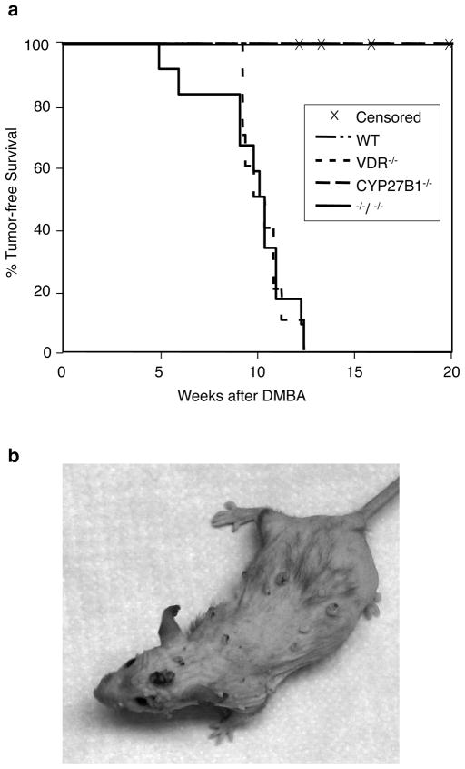
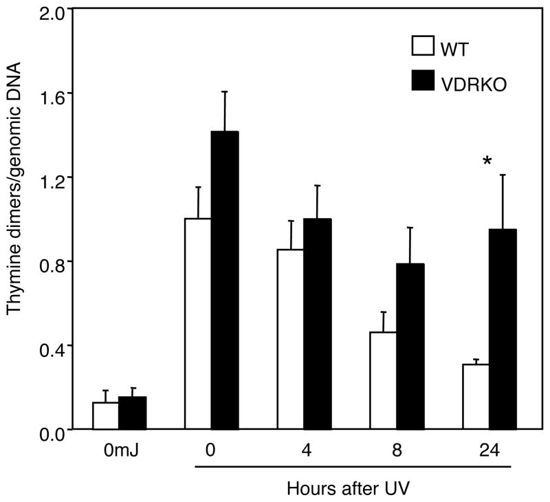
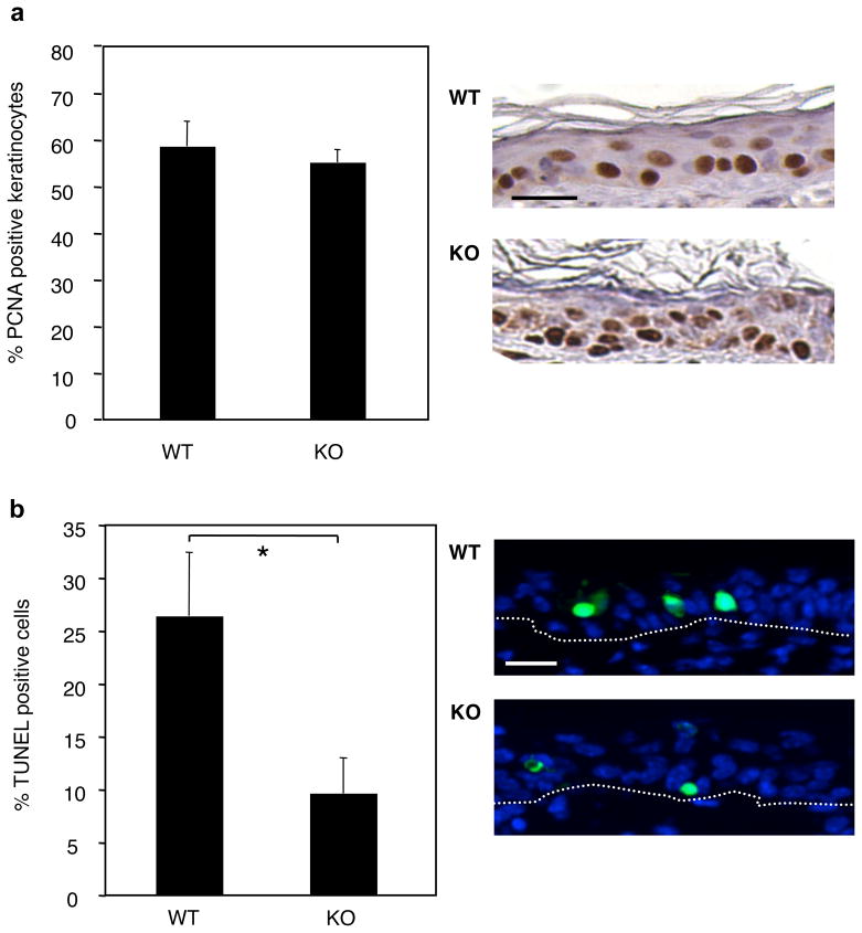
1,25-Dihydroxyvitamin D(3) (1,25(OH)(2)D(3)) is the biologically active ligand for the vitamin D receptor (VDR). VDR(-/-) mice have a hair follicle-cycling defect resulting in alopecia. However, mice lacking 25-hydroxyvitamin D(3) 1alpha-hydroxylase (CYP27B1(-/-)), and having no circulating 1,25(OH)(2)D(3), have normal follicular function. These mouse models indicate that VDR functions independently of 1,25(OH)(2)D(3) in regulating hair-follicle cycling. Here, we show that VDR(-/-) mice rapidly develop chemically induced skin tumors, whereas CYP27B1(-/-) and wild-type mice do not, indicating that VDR, and not the 1,25(OH)(2)D(3) ligand, is essential for protection against skin tumorigenesis. Because the majority of human skin cancer results from exposure to UV, the susceptibility of VDR(-/-) mice to this carcinogen was also evaluated. VDR(-/-) mice developed UV-induced tumors more rapidly and with greater penetrance than did VDR(+/+) mice. p53 protein levels were upregulated at similar rates in UV-treated keratinocytes of VDR(-/-) and VDR(+/+) mice. However, rates of thymine-dimer repair and UV-induced apoptosis were significantly lower in VDR(-/-) epidermis compared with the wild type epidermis. UV-induced epidermal thickening was also attenuated in VDR(-/-) skin, indicating that VDR plays a critical role in the repair and removal of severely damaged keratinocytes and adaptation of the skin to chronic UV exposure.


Conflict of interest statement

Conflict of Interest

The authors state no conflict of interest.

Figures

Figure 1
Figure 1. Protection against chemically-induced skin tumorigenesis is VDR-dependent and 1αOHase independent in mice
VDR+/+ (n=11), VDR+/− (n=6), and VDR−/− mice (n=10), CYP27B1+/+ (n=10), CYP27B1 +/− (n=11), and CYP27B1−/− mice (n=10), and VDR−/− CYP27B1−/− mice (n=13) were given two doses of DMBA by oral gavage at 5 and 6 weeks of age. Mice were monitored weekly by palpation and visual inspection for tumor development. a) Probability of tumor development was analyzed according to Kaplan-Meier. VDR+/+ and CYP27B1+/+ wildtype mice were grouped for statistical analysis. Mice were censored if they died or if they required euthanasia due to extreme sickness before developing a skin tumor. b) A representative image of a VDR−/− mouse bearing several DMBA-induced skin tumors.
Figure 2
Figure 2. VDR protects mice from UV-induced skin tumorigenesis
a) VDR+/+ (n=23) and VDR−/− (n=22) were shaved and treated with a depilatory lotion, then exposed to UV thrice weekly for 33 weeks as described in Materials and Methods. Mice were monitored weekly by palpation and visual inspection for tumor development. Probability of tumor development was analyzed according to Kaplan-Meier. Mice were censored if they died or if they required euthanasia due to extreme sickness before developing a skin tumor. b) Representative image of thickened but non-tumor bearing skin from a wildtype mouse after 26 weeks of regular UV exposure. Scale bar = 20 μm. c) Representative image of thickened but non-tumor bearing skin from a VDR−/− mouse after 29 weeks of regular UV exposure. Scale bar = 20 μm. Histological appearance of a representative benign papilloma (d and e), moderate papilloma (f and g), an invasive squamous cell carcinoma (SCC; h and i), and an atypical fibroxanthoma (AFX; j and k). Images were taken of hematoxylin and eosin stained paraffin-embedded sections (d, f, h, and j), and of sections immunohistochemically stained with an antibody against proliferating cell nuclear antigen (PCNA) to indicate proliferation (e, g, i, and k). PCNA positive cells appear brown. Scale bar (d–k) = 20 μm.
Figure 3
Figure 3. UV induces p53 protein levels and p53 phosphorylation equally in wildtype and VDR−/− keratinocytes
Keratinocytes were isolated from newborn wildtype and VDR−/− mice, allowed to grow for 24h, and irradiated with 50 mJ/cm2 of UVB through a thin film of PBS. Media were replaced and protein was harvested at the indicated time points. Protein expression was analyzed by western blot.
Figure 4
Figure 4. Incomplete repair of thymine dimers in VDR−/− epidermis compared to wildtype
Newborn mice were exposed to 120mJ/cm2 of UV and skin was collected at the indicated time points. Epidermal genomic DNA was isolated and thymine dimers were measured by southwestern slot blot. Thymine dimers were normalized to total genomic DNA with a radiolabeled mouse genomic DNA probe. Thymine dimers were measured in the epidermis of 2–3 individual mice for each time point, except for the unirradiated control. The data shown represents the mean from three independent measurements of thymine dimers relative to total genomic DNA from the epidermal samples. Error bars = SEM. *P<0.04
Figure 5
Figure 5. UVB-induced growth arrest is compromised in VDR−/− keratinocytes in vitro
Primary keratinocytes were isolated from newborn wildtype and VDR−/− mice, allowed to grow for 24h, and irradiated with the indicated amount of UVB through a thin film of PBS. Media were replaced and cell proliferation was measured at the indicated time points using the MTT assay as described in Materials and Methods. Cell growth of a) wildtype and b) VDR−/− keratinocytes is plotted over time.
Figure 6
Figure 6. In vivo apoptotic defects in VDR−/− keratinocytes following a single dose of UV
Wildtype (WT, n=4) and VDR−/− (KO, n=4) mice were exposed to 120mJ/cm2 of UV and a biopsy was collected 24 h later. a) Skin samples were stained for PCNA expression and counterstained with hematoxylin (positive cells are brown). PCNA positive keratinocytes in WT and KO skin were counted as a percentage of total number of keratinocytes. Error bars=SEM. Representative sections are shown on the right. Scale bar = 20 μm. b) Apoptotic cells were labeled by the TUNEL method and nuclei were counterstained with DAPI. Apoptosis rates were calculated as number of TUNEL positive cells as a percentage of total epidermal keratinocytes from multiple fields of TUNEL labeled paraffin-embedded skin sections, viewed at 100× magnification. Error bars=SD. Differences were analyzed by the two-tailed Student’s t test, * p = 0.005. Representative fields of TUNEL-labeled sections are on the right, showing TUNEL positive cells in green and nuclei in blue. Scale bar = 20 μm. Dotted lines indicate the epidermal/dermal junction.
Figure 7
Figure 7. Defective epidermal thickening of VDR−/− skin in response to chronic UV exposure
Wildtype (WT, n=4) and VDR−/− (KO, n=4) mice were exposed to six doses of UV (120mJ/cm2) during a 2-week period. Skin samples were collected and fixed 24 h after the final dose. a) Skin samples were stained for PCNA expression and counterstained with hematoxylin. PCNA positive keratinocytes in WT and KO skin were counted as a percentage of total number of keratinocytes. Wildtype epidermis was significantly more proliferative than VDR−/− epidermis following 6 exposures of UV, * p < 0.025. Error bars=SEM. Differences were analyzed by the two-tailed Student’s t test. b) Representative sections of wildtype and VDR−/− skin, stained for PCNA expression. Brackets indicate epidermal thickness. Scale bar = 40 μm. c) Epidermal thickness was calculated from 20–25 measurements from multiple fields of paraffin-embedded skin sections, viewed at 100× magnification. Error bars=SD. Differences in thickness were analyzed by the two-tailed Student’s t test. Wildtype epidermis was significantly thicker than VDR−/− epidermis following 6 exposures of UV, * p < 0.015.

Comment in

References

    1. Albert MR, Weinstock MA. Keratinocyte carcinoma. CA Cancer J Clin. 2003;53:292–302. - PubMed
    1. Barletta F, Freedman LP, Christakos S. Enhancement of VDR-mediated transcription by phosphorylation: correlation with increased interaction between the VDR and DRIP205, a subunit of the VDR-interacting protein coactivator complex. Mol Endocrinol. 2002;16:301–314. - PubMed
    1. Bikle DD. Vitamin D and skin cancer. J Nutr. 2004;134:3472S–3478S. - PubMed
    1. Bikle DD, Chang S, Crumrine D, Elalieh H, Man MQ, Choi EH, et al. 25 Hydroxyvitamin D 1 alpha-hydroxylase is required for optimal epidermal differentiation and permeability barrier homeostasis. J Invest Dermatol. 2004;122:984–992. - PubMed
    1. Bikle DD, Nemanic MK, Whitney JO, Elias PW. Neonatal human foreskin keratinocytes produce 1,25-dihydroxyvitamin D3. Biochemistry. 1986;25:1545–1548. - PubMed

Publication types

MeSH terms