

Plasmodium falciparum antigens on the surface of the gametocyte-infected erythrocyte
- PMID: 18509532
- PMCID: PMC2386550
- DOI: 10.1371/journal.pone.0002280
Plasmodium falciparum antigens on the surface of the gametocyte-infected erythrocyte
Abstract
Background: The asexual blood stages of the human malaria parasite Plasmodium falciparum produce highly immunogenic polymorphic antigens that are expressed on the surface of the host cell. In contrast, few studies have examined the surface of the gametocyte-infected erythrocyte.
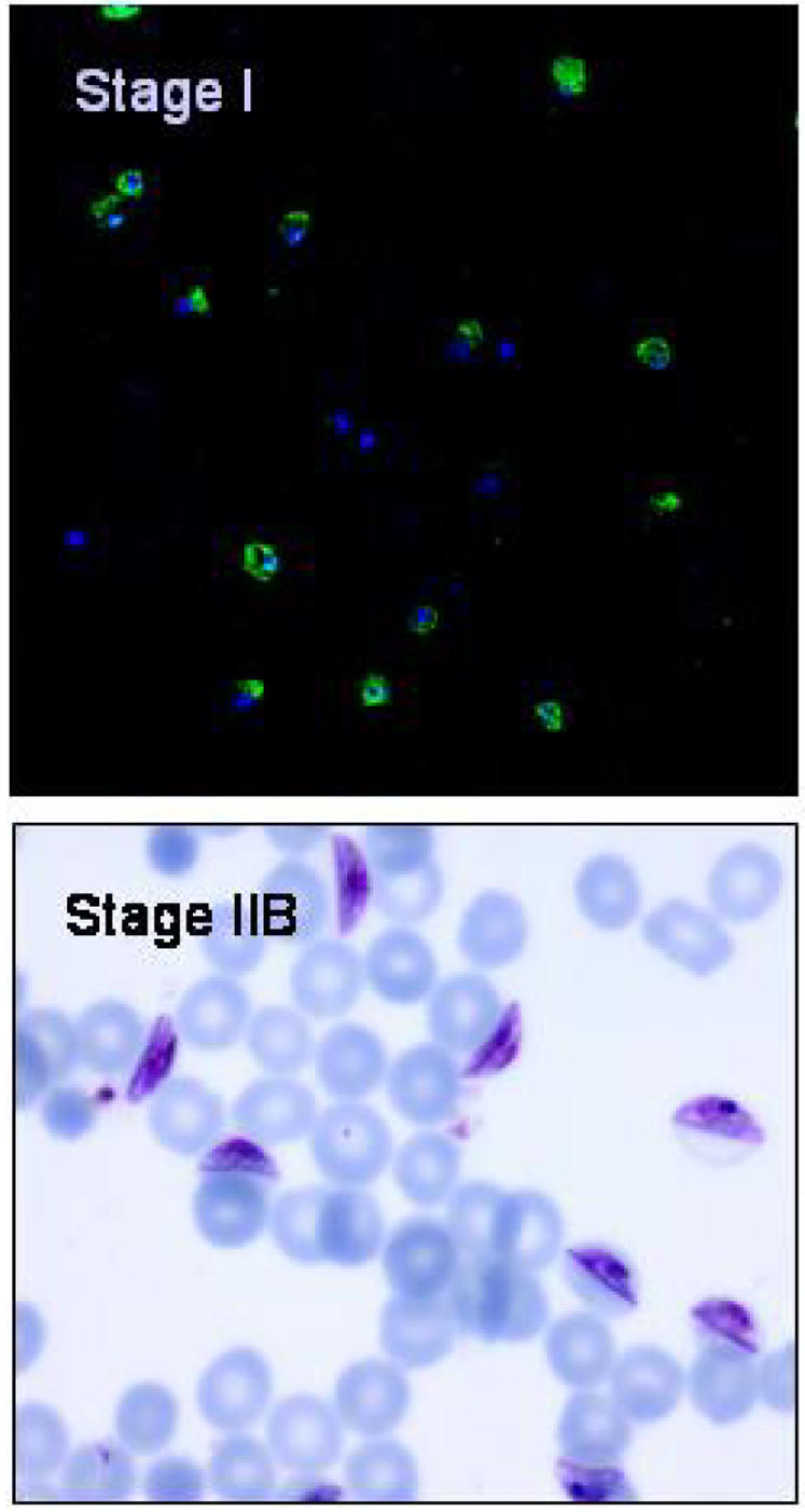
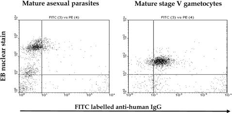
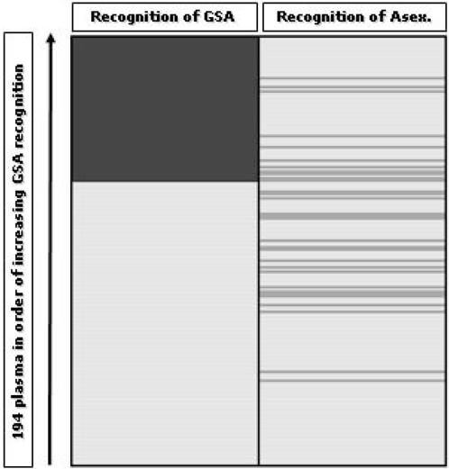
Methodology/principal findings: We used flow cytometry to detect antibodies recognising the surface of live cultured erythrocytes infected with gametocytes of P. falciparum strain 3D7 in the plasma of 200 Gambian children. The majority of children had been identified as carrying gametocytes after treatment for malaria, and each donated blood for mosquito-feeding experiments. None of the plasma recognised the surface of erythrocytes infected with developmental stages of gametocytes (I-IV), but 66 of 194 (34.0%) plasma contained IgG that recognised the surface of erythrocytes infected with mature (stage V) gametocytes. Thirty-four (17.0%) of 200 plasma tested recognised erythrocytes infected with trophozoites and schizonts, but there was no association with recognition of the surface of gametocyte-infected erythrocytes (odds ratio 1.08, 95% C.I. 0.434-2.57; P = 0.851). Plasma antibodies with the ability to recognise gametocyte surface antigens (GSA) were associated with the presence of antibodies that recognise the gamete antigen Pfs 230, but not Pfs48/45. Antibodies recognising GSA were associated with donors having lower gametocyte densities 4 weeks after antimalarial treatment.
Conclusions/significance: We provide evidence that GSA are distinct from antigens detected on the surface of asexual 3D7 parasites. Our findings suggest a novel strategy for the development of transmission-blocking vaccines.
Conflict of interest statement
Figures
References
-
- Epstein JE, Giersing B, Mullen G, Moorthy V, Richie TL. Malaria vaccines: are we getting closer? Curr Opin Mol Ther. 2007;9:12–24. - PubMed
-
- Brannan LR, Turner CM, Phillips RS. Malaria parasites undergo antigenic variation at high rates in vivo. Proc Biol Sci. 1994;256:71–75. - PubMed
-
- Baruch DI, Pasloske BL, Singh HB, Bi X, Ma XC, et al. Cloning the P. falciparum gene encoding PfEMP1, a malarial variant antigen and adherence receptor on the surface of parasitized human erythrocytes. Cell. 1995;82:77–87. - PubMed
Publication types
MeSH terms
Substances
Associated data
Grants and funding
LinkOut - more resources
Full Text Sources
