. 2008 May 30;133(5):789-800.
doi: 10.1016/j.cell.2008.04.030.

XPD helicase structures and activities: insights into the cancer and aging phenotypes from XPD mutations

Affiliations

XPD helicase structures and activities: insights into the cancer and aging phenotypes from XPD mutations

Li Fan et al. Cell. .

Abstract

Mutations in XPD helicase, required for nucleotide excision repair (NER) as part of the transcription/repair complex TFIIH, cause three distinct phenotypes: cancer-prone xeroderma pigmentosum (XP), or aging disorders Cockayne syndrome (CS), and trichothiodystrophy (TTD). To clarify molecular differences underlying these diseases, we determined crystal structures of the XPD catalytic core from Sulfolobus acidocaldarius and measured mutant enzyme activities. Substrate-binding grooves separate adjacent Rad51/RecA-like helicase domains (HD1, HD2) and an arch formed by 4FeS and Arch domains. XP mutations map along the HD1 ATP-binding edge and HD2 DNA-binding channel and impair helicase activity essential for NER. XP/CS mutations both impair helicase activity and likely affect HD2 functional movement. TTD mutants lose or retain helicase activity but map to sites in all four domains expected to cause framework defects impacting TFIIH integrity. These results provide a foundation for understanding disease consequences of mutations in XPD and related 4Fe-4S helicases including FancJ.


Figures
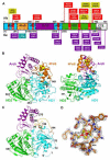
Figure 1
Figure 1. XPDcc conservation, functional motifs, mutation sites, domains, and structure
(A) XPDcc domains. Sequence comparison indicates that SaXPD contains the XPD conserved catalytic core, so the four XPDcc domains are shown schematically in boxes for HD1 (cyan), HD2 (green), 4FeS (orange), and Arch (purple) domains with conserved helicase motifs (red bars with white labels). Disease XP (red), XP/CS (yellow) and TTD (purple) mutation sites are labeled for human (Hs) and corresponding SaXPD (in parentheses) sites. Residue F136 corresponding to a FancJ mutation is highlighted by a blue-green flag. For the detailed sequence alignment with mutation sites, secondary structure, and domain fold see Supplemantary Data (Figure S1). The human enzyme has a partly ordered (grey) C-terminal extension (CTE) (as predicted by PONDR) that is a probable TFIIH p44 interface. (B) XPDcc fold and domains (ribbons). Helicase domains HD1 (cyan) and HD2 (green) form the ATP binding interface. Front view (left) shows the arch formed by the 4FeS (orange) and Arch (purple) domains, which are inserted into HD1. Side view (right) shows HD2 protruding from the flat box formed by HD1, 4FeS, and Arch as well as the HD2 helix-loop-helix insertion (green). Domain boundaries are indicated by residue numbers. (C) Apo XPDcc structure. Disordered regions (dashed lines with the boundaries indicated by residue numbers) show the 4Fe-4S cluster acts in ordering the 4FeS domain, the Arch interface, and parts of HD1. (D) Electron density for the 4Fe-4S cluster and key residues forming hydrogen bonds (green dashed lined) to the Cys ligands. Mutations at these sites cause TTD in XPD and Fanconi anemia in FancJ. Composite omit maps calculated from the model are displayed at 1 sigma level.
Figure 2
Figure 2. Structure-based model for DNA Interactions
(A) Conserved molecular surface (front view). Conserved residues are shown on the surface from deep blue (identical) to light blue (highly similar) to greenish light blue (similar) based on sequence alignment between HsXPD and SaXPD (Figure S1). (B) Electrostatic molecular surface of the arch region (back view). Electrostatic potential (blue positive to red negative) suggests a dsDNA binding site (arrow). (C) XPD-DNA binding model (DNA phosphate backbone as purple tube). ssDNA binding was located by superimposing known helicase-DNA complex structures (2P6R.pdb; Büttner et al., 2007) to the SaXPD structure over conserved HD2 helicase motifs IV and VI. The SaXPD dsDNA binding sites were located by a complete six-dimensional search. Helicase motifs are in red. (D) Electrostatic molecular surface for the DNA binding HD2 gateway. Electrostatic potential is calculated as in (B) and DNA (yellow tube) is modeled as in (C).
Figure 3
Figure 3. Structural roles of amino acid residues associated with disease-causing XPD mutations
Disease-causing mutations (Cα colored spheres) mapped in the SaXPD structure with hydrogen bonds (red dashed lines). (A) Mutation sites at the HD2 gateway DNA binding channel shown in Figures 2C & D. Solvent molecules including citrate (CIT), isopropyl alcohol (IPA), and glycerol (GOL) that mimic DNA backbone (purple tube, see Figure 2C), interact with charged residues K369, R373, and D529. (B) Mutation sites at the groove along HD2, Arch, and HD1 domains. (C) Mutation sites at the ATP binding groove between HD1 and HD2 domains. (D) Mutation sites at the edge of HD2 domain.
Figure 4
Figure 4. Effects of disease-causing XPD mutations
Catalytic activity and DNA-binding of SaXPD mutants, as percentage of wild-type activity. DNA-binding affinities were not determined for mutants R531W, C88S, and C102S. See Table 2 for rate and Kd measurements and Figure S5 for representative data.
Figure 5
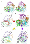
Figure 5. Structural placement of XPDcc disease-causing mutations
Mapping the three classes of mutations onto the SaXPD structure reveals patterns associated with each disease defect. (A) Stereo pair mapping the distribution of disease-causing mutations on a XPDcc Cα trace. Disease-causing mutation sites (Cα colored sphere): red (XP), greenish yellow (XP/CS), and purple (TTD). Residue F136 (a FancJ mutation) is also shown (cyan). (B) XPDcc fold and domain architecture (ribbons) with labeled disease-causing mutation sites as spheres colored as in (A). (C) XP mutations impact DNA and ATP binding regions. (D) XP/CS mutations impact HD1-HD2 conformational changes. (E) TTD mutations impact the overall framework stability.

References

    1. Bienstock RJ, Skorvaga M, Mandavilli BS, Van Houten B. Structural and functional characterization of the human DNA repair helicase XPD by comparative molecular modeling and site-directed mutagenesis of the bacterial repair protein UvrB. J. Biol. Chem. 2003;278:5309–5316. - PubMed
    1. Boal AK, Yavin E, Lukianova OA, O’Shea VL, David SS, Barton JK. DNA-bound redox activity of DNA repair glycosylases containing [4Fe-4S] clusters. Biochemistry. 2005;44:8397–8407. - PubMed
    1. Bootsma D, Hoeijmakers JH. DNA repair. Engagement with transcription. Nature. 1993;363:114–115. - PubMed
    1. Botta E, Nardo T, Lehmann AR, Egly JM, Pedrini AM, Stefanini M. Reduced level of the repair/transcription factor TFIIH in trichothiodystrophy. Hum Mol Genet. 2002;11:2919–2928. - PubMed
    1. Büttner K, Nehring S, Hopfner KP. Structural basis for DNA duplex separation by a superfamily-2 helicase. Nat. Struct. Mol. Biol. 2007;14:647–652. - PubMed

Publication types

MeSH terms