. 2008 Aug;212(2):337-47.
doi: 10.1016/j.expneurol.2008.04.009. Epub 2008 Apr 20.

Remote activation of microglia and pro-inflammatory cytokines predict the onset and severity of below-level neuropathic pain after spinal cord injury in rats

Affiliations

Remote activation of microglia and pro-inflammatory cytokines predict the onset and severity of below-level neuropathic pain after spinal cord injury in rats

Megan Ryan Detloff et al. Exp Neurol. 2008 Aug.

Abstract

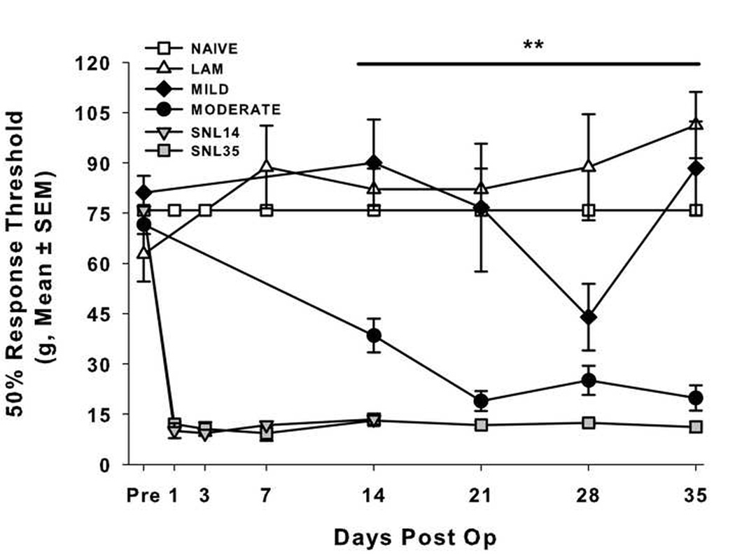
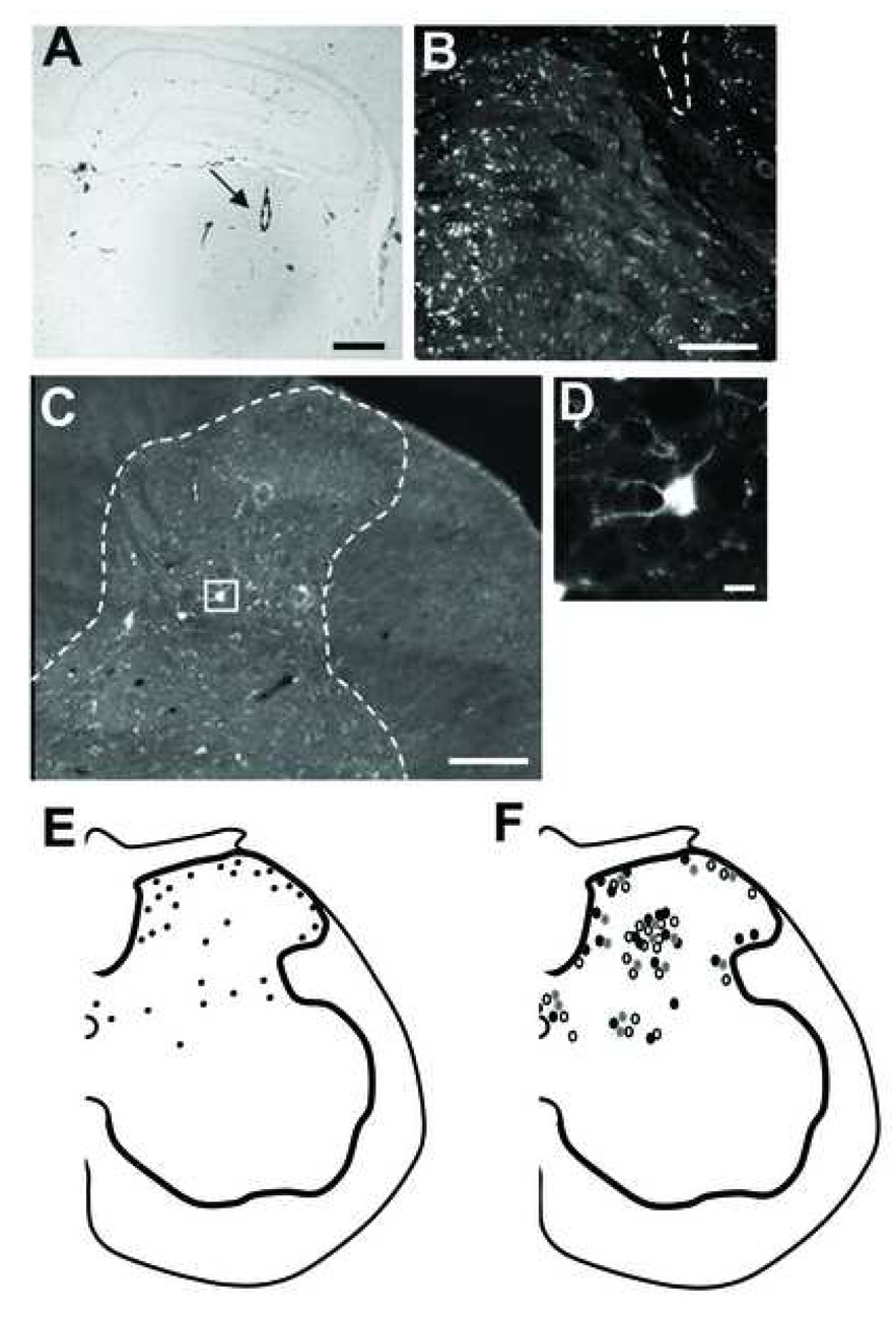
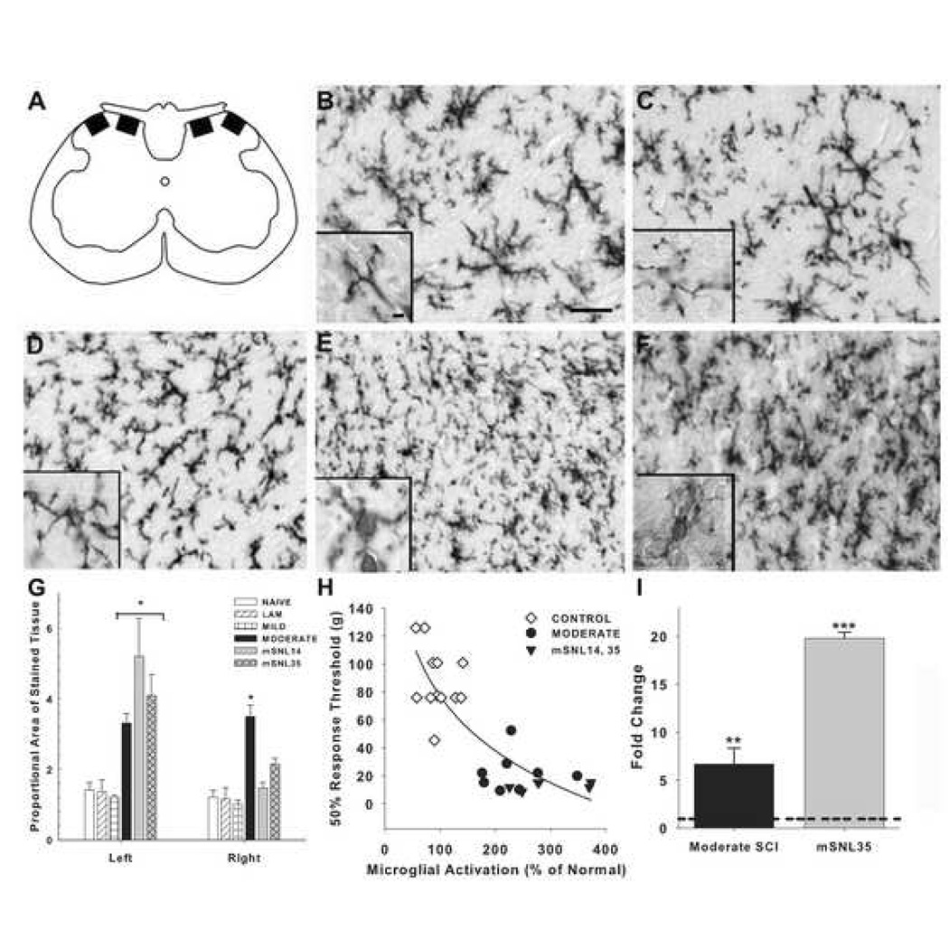
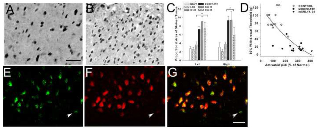
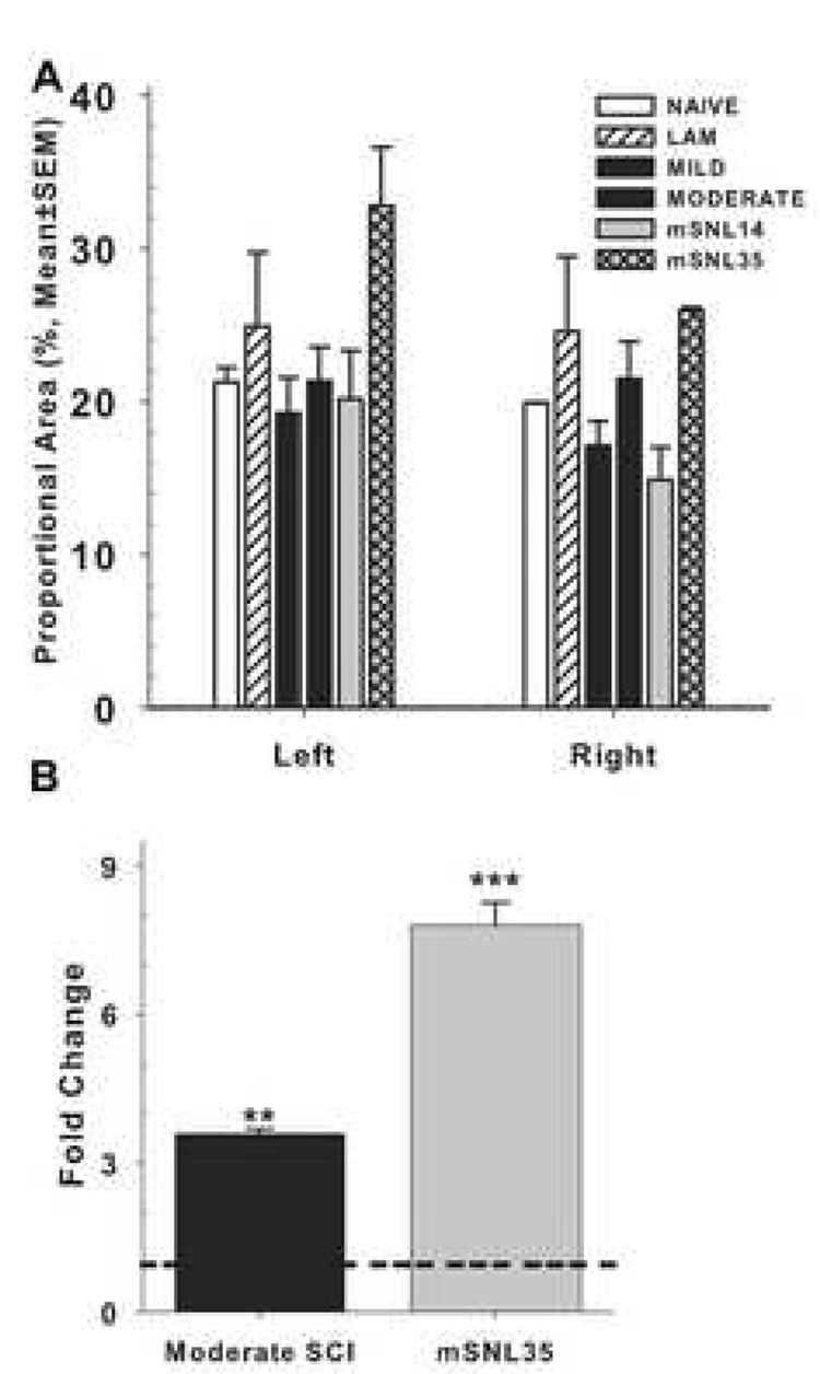
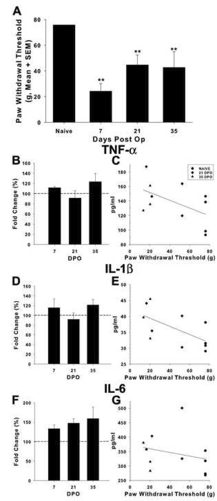
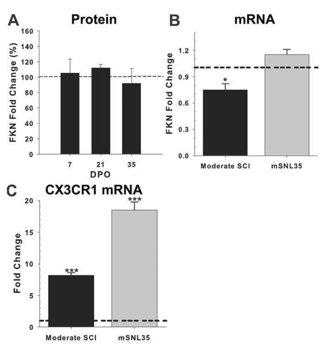
Spinal cord injury (SCI) impairs sensory systems causing chronic allodynia. Mechanisms underlying neuropathic pain have been more extensively studied following peripheral nerve injury (PNI) than after central trauma. Microglial activation, pro-inflammatory cytokine production and activation of p38 MAP kinase pathways may induce at-level allodynia following PNI. We investigated whether midthoracic SCI elicits similar behavioral and cellular responses below the level of injury (lumbar spinal cord; L5). Importantly, we show that anatomical connections between L5 and supraspinal centers remain intact after moderate SCI allowing direct comparison to a well-established model of peripheral nerve injury. We found that SCI elicits below-level allodynia of similar magnitude to at-level pain caused by a peripheral nerve injury. Moreover, the presence of robust microglial activation in L5 cord predicted allodynia in 86% of rats. Also increased phosphorylation of p38 MAP kinase occurred in the L5 dorsal horn of allodynic rats. For below-level allodynia after SCI, TNF-alpha and IL-1beta increased in the L5 dorsal horn by 7 dpo and returned to baseline by 35 dpo. Interestingly, IL-6 remains at normal levels early after SCI and increases at chronic time points. Increased levels of pro-inflammatory cytokines also occurred in the thalamus after SCI-induced allodynia. These data suggest that remote microglial activation is pivotal in the development and maintenance of below-level allodynia after SCI. Fractalkine, a known activator of microglia, and astrocytes were not primary modulators of below-level pain. Although the mechanisms of remote microglial activation are unknown, this response may be a viable target for limiting or preventing neuropathic pain after SCI in humans.


Figures

Figure 1
Figure 1
Pain-like sensory thresholds occurred after moderate SCI and modified SNL. Using 50% withdrawal to VFH stimulation, significantly lower sensory thresholds of the hindpaw occurred for moderate SCI and mSNL groups from 14 to 35 dpo. Naïve, laminectomy and mild SCI groups had normal sensory thresholds throughout the testing period. (Significance determined by repeated measures ANOVA; **p<.05 except at 28 dpo). Note that an equivalent severity of allodynia occurred between moderate SCI and mSNL, a well-established peripheral model of neuropathic pain.
Figure 2
Figure 2
Neuroanatomical substrates of the spinothalamic tract after moderate SCI. The ventroposterolateral (VPL) nucleus of the thalamus was injected with 2% fluorogold (FG) in rats with allodynia induced by moderate SCI (n=3). The FG injection was localized to the hindlimb region of the VPL visualized with brightfield (A) and fluorescence (B; dashed line indicates the injection track). FG-positive neurons were distributed in the L5 dorsal horn (C). Positive neuronal labeling included bright opacity of the cytoplasm and proximal dendrites (D; high power image of cell outlined by the square in C). The L5 spinothalamic neurons projecting to the VPL in a Naïve rat are distributed in the superficial and deep dorsal horn (E). The profiles reflect FG+ neurons in two sections 700 µm apart in the L5 dorsal horn. A similar distribution of spinothalamic neurons occurred after moderate SCI (F). Three SCI rats with allodynia are represented by different symbols for neuronal profiles in two sections 700 µm apart (F). Note that the majority of spared spinothalamic axons in allodynic rats project from the deep rather than superficial dorsal horn. [ Scale bars: A=1mm; B, C=300 µm; D=50 µm]
Figure 3
Figure 3
Immunolabeling and quantification of activated and resting microglia within the superficial dorsal horn. Basal levels of OX42 signal were observed within the L5 dorsal horn of Naïve (B) and Mild SCI (C) rats. Higher magnification displays the characteristic resting phenotype of microglia with slender processes and small cell body for naïve (inset of B) and mild SCI (inset C). After Moderate SCI (D) and spinal nerve ligation (mSNL14, E; mSNL35, F), microglia exhibited an activated phenotype with retracted, thickened processes and large cell bodies (high magnification insets D, E, F). Proportional area was measured within 2 regions of interest (0.026 mm2) aligned with anatomical landmarks along the medial and lateral portion of the dorsal horn as shown in A. The summed proportional areas for these regions of interest are represented in G. Microglial activation was significantly greater ipsilateral to mSNL (left) and bilaterally after Moderate SCI (right and left) compared to Mild SCI, naïve and laminectomy (LAM) controls (G). The degree of microglial activation predicted allodynia (H). Microglial activation of 200% or higher was associated with the development of mechanical allodynia (H; r2= 0.83; p<.01). Rats without microglial activation in the L5 dorsal horn displayed normal sensory thresholds (controls; Naïve, Laminectomy or Mild SCI). Using LCM/rtPCR detection of microglial marker CD11b (I) distinguished increased microglial message at 35 days after moderate SCI or mSNL compared to naïve (dashed line). [Significance in G: repeated measures ANOVA *p<.05 vs. Naïve, LAM and Mild SCI. Significance in I: one way ANOVA ** p<.01; *** p<.001]. All histological sections are of the medial box in the left dorsal horn. Scale Bar B–F = 200µm; Inset Scale bar = 10µm.
Figure 4
Figure 4
Immunolabeling and quantification of phosphorylated p38 in the L5 superficial dorsal horns. Representative section of the left L5 dorsal horns of Naïve (A) and Moderate SCI (B) at 35 dpo. Proportional area measurements within regions of interest depicted in figure 3A showed that rats with Moderate SCI and mSNL at short (14 day,) and long (35 day) survival had greater activated p38 protein compared to Naïve, Laminectomy (LAM) or Mild SCI groups (C). Phosphorylated p38 MAP kinase correlated to neuropathic pain (D, Pearson Coefficient = −0.78; p<.01; r2=0.634). Double-label immunofluorescence with p-p38 (E) and NeuN (F) antibodies, showed that p38 MAPK activation occurs predominantly in neurons (G; arrowhead denotes a p-p38+, NeuN- cell). (Significance in C: repeated measures ANOVA * p<.05 vs Naive, LAM and Mild SCI). Scale Bar A and B = 200µm. Scale Bar E, F, G = 20µm.
Figure 5
Figure 5
Quantification of reactive astrocytes at L5. Basal levels of GFAP signal were observed within the dorsal and ventral horn of L5 spinal cord regardless of injury. Proportional area of positively labeled tissue throughout the medial-lateral span of the superficial dorsal horn (A) showed no increase above naïve and laminectomy (LAM) controls after mild or moderate SCI or modified spinal nerve ligation despite a significant increase in GFAP mRNA in SCI and mSNL groups relative to naive(dashed line; B). (Significance in B: one way ANOVA **p<.01; ***p<.001).
Figure 6
Figure 6
Analysis of pro-inflammatory cytokine levels in the L5 dorsal spinal cord of rats with allodynia. Stimulation with VFH to the dorsal region of the L5 dermatome detected significantly lower sensory thresholds from 7 to 35 days after moderate SCI (A). Rats with pain-like behavior demonstrated significantly greater tumor necrosis factor-α (TNF-α, B) and interleukin (IL)-1β (D) protein early after injury, with levels returning to normal by 35 dpo. IL-6 protein (F) increased at later time points after SCI when pain-like behavior was fully established. Thus, TNF-α and IL-1β may play a role in the induction of pain-like behavior while IL-6 may be involved in pain maintenance. These increases in pro-inflammatory cytokines correlate to pain-like behavior as determined by a Spearmann Rank Test (p<.05; C, E, G). Greater cytokine proteins in the dorsal spinal cord predicted lower L5 sensory thresholds. Naïve expression levels are denoted as dashed lines (B, D, F). (Significance: one way ANOVA *p<.05; **p<.01 vs. Naïve).
Figure 7
Figure 7
Fractalkine (FKN) protein levels were unchanged in the L5 dorsal spinal cord of rats with SCI-induced allodynia over time (A). Interestingly, fractalkine mRNA did not increase above naïve levels after spinal nerve ligation and was significantly lower for moderate SCI at 35 dpo (B). The fractalkine receptor, CX3CR1, showed at least an eight-fold increase in message after moderate SCI and more than a 15-fold increase after mSNL at 35 dpo (C). Naïve expression levels are denoted as dashed lines. (Significance: one way ANOVA *p<.05; ** p<.01; ***p<.001 vs. naïve).

Comment in

Similar articles

Cited by

References

    1. International Association for the Study of Pain. Website 11-21-2005. 2008 Ref Type: Electronic Citation.
    1. Basso DM, Beattie MS, Bresnahan JC. A sensitive and reliable locomotor rating scale for open field testing in rats. J.Neurotrauma. 1995;12:1–21. - PubMed
    1. Behrmann DL, Bresnahan JC, Beattie MS, Shah BR. Spinal cord injury produced by consistent mechanical displacement of the cord in rats: Behavioral and histologic analysis. J.Neurotrauma. 1992;9:197–217. - PubMed
    1. Brenn D, Richter F, Schaible H-G. Sensitization of unmyelinated sensory fibers of the joint nerve to mechanical stimuli by interleukin-6 in the rat: An inflammatory mechanism of joint pain. Arthritis Rheum. 2007;56:351–359. - PubMed
    1. Bruce JC, Oatway MA, Weaver LC. Chronic pain after clip-compression injury of the rat spinal cord. Exp.Neurol. 2002;178:33–48. - PubMed

Publication types

MeSH terms

Substances