. 2008 Oct;38(12):1391-400.
doi: 10.1016/j.ijpara.2008.04.002. Epub 2008 Apr 25.

Bioluminescent imaging of Trypanosoma cruzi infection

Affiliations

Bioluminescent imaging of Trypanosoma cruzi infection

Kenneth V Hyland et al. Int J Parasitol. 2008 Oct.

Abstract

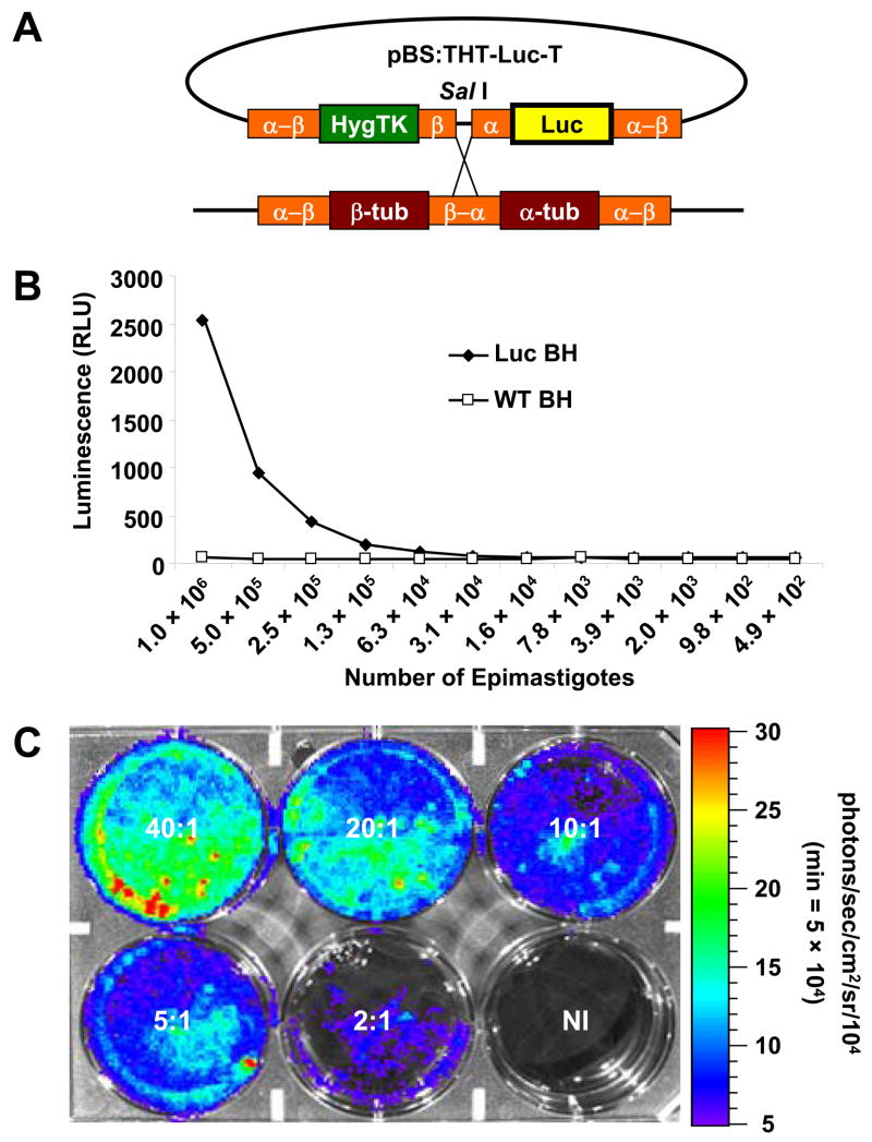
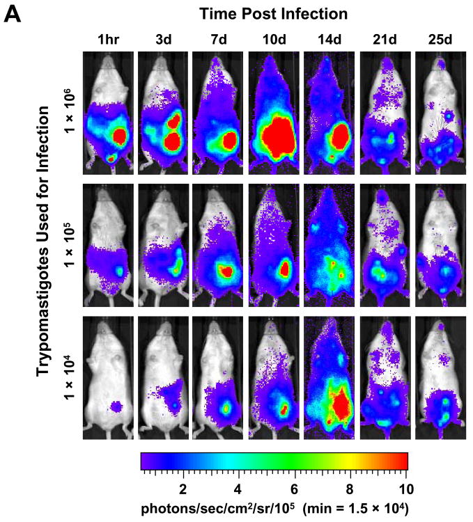
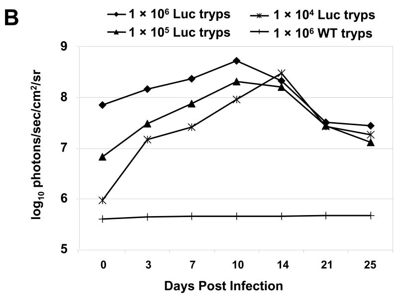
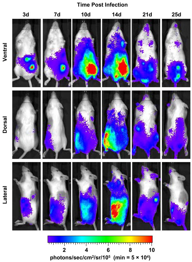
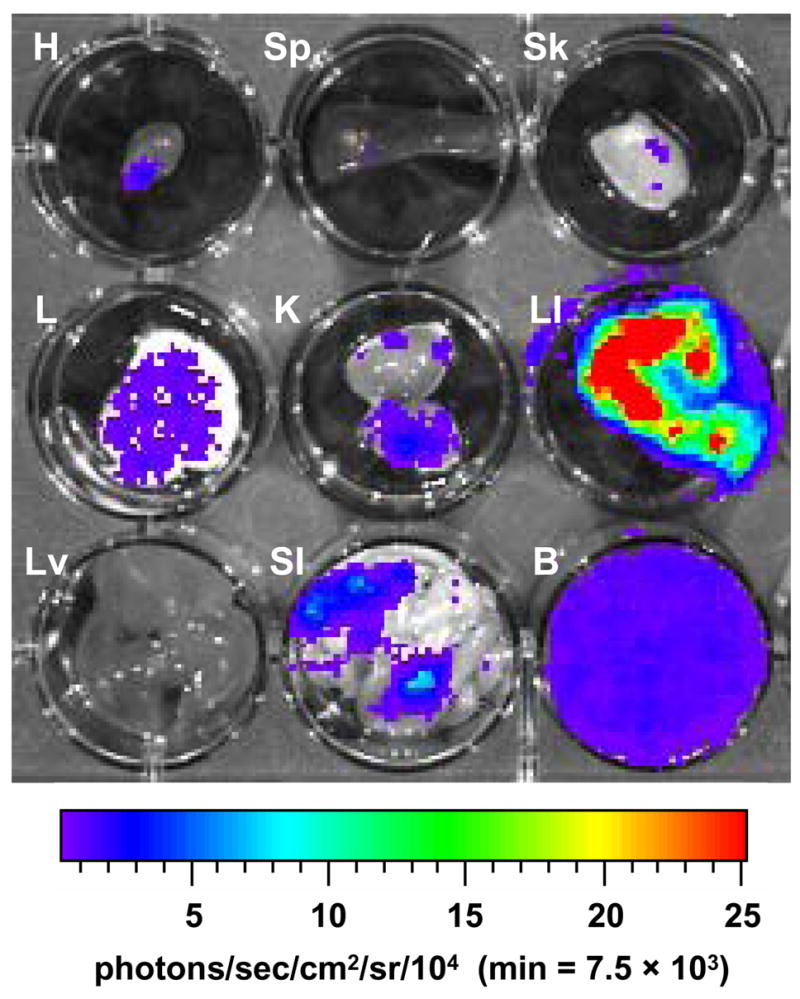
Chagas disease, caused by infection with the protozoan parasite Trypanosoma cruzi, is a major public health problem in Central and South America. The pathogenesis of Chagas disease is complex and the natural course of infection is not completely understood. The recent development of bioluminescence imaging technology has facilitated studies of a number of infectious and non-infectious diseases. We developed luminescent T. cruzi to facilitate similar studies of Chagas disease pathogenesis. Luminescent T. cruzi trypomastigotes and amastigotes were imaged in infections of rat myoblast cultures, which demonstrated a clear correlation of photon emission signal strength to the number of parasites used. This was also observed in mice infected with different numbers of luminescent parasites, where a stringent correlation of photon emission to parasite number was observed early at the site of inoculation, followed by dissemination of parasites to different sites over the course of a 25-day infection. Whole animal imaging from ventral, dorsal and lateral perspectives provided clear evidence of parasite dissemination. The tissue distribution of T. cruzi was further determined by imaging heart, spleen, skeletal muscle, lungs, kidneys, liver and intestines ex vivo. These results illustrate the natural dissemination of T. cruzi during infection and unveil a new tool for studying a number of aspects of Chagas disease, including rapid in vitro screening of potential therapeutical agents, roles of parasite and host factors in the outcome of infection, and analysis of differential tissue tropism in various parasite-host strain combinations.


Figures

Fig. 1
Fig. 1
Generation and analysis of luminescent Trypanosoma cruzi. (A) Construct and cloning strategy used to generate luminescent T. cruzi. pBS:THT-x-T (obtained from Wesley Van Voorhis, University of Washington), a plasmid with a pBluescript backbone and a hygromycin-resistance gene, was modified by insertion of the firefly luciferase gene. After linearization with SalI, the plasmid was transfected into T. cruzi epimastigotes and the Hyg and Luc genes integrated into the tubulin locus by homologous recombination. (B) Serial dilutions of antibiotic-resistant, T. cruzi epimastigote transfectants and wild-type epimastigotes were examined for luciferase activity. A black, 96-well plate containing 50 μL suspensions of parasites, mixed with 50 μL of Steady Glo reagent was read by a SpectraMax Gemini XS Microplate Spectrofluorometer and analyzed with SoftmaxPro 4.8 software. (C) Luminescent trypomastigotes and amastigotes were examined for luciferase activity in active, in vitro rat myoblast infections using parasite:myoblast ratios of 40:1, 20:1, 10:1, 5:1, 2:1 or uninfected (NI). Five days p.i., D-luciferin was added to each well and the 6-well plate was imaged with the Xenogen IVIS system after a 5-min incubation (see Materials and methods for detailed description). In the pseudocolor image the luciferase activity or photon intensity ranges from the lowest intensity (blue) to highest intensity (red). Abbreviations: RLU, relative light units; min, minimum.
Fig. 2
Fig. 2
Luminescent T. cruzi imaged at various times p.i. with an IVIS imaging system. (A) Trypomastigotes were isolated from in vitro myoblast infections and different amounts, as indicated, were injected i.p. into A/J mice. Mice were imaged ventrally starting 1 h after infection and monitored the days p.i. as shown. For all images shown, the color scale ranges from blue (just above background with a minimum set to 15,000 photons/sec/cm2/sr) to red (maximum of 1 × 10⁶ photons/sec/cm2/sr). (B) The course of whole-body parasite burden expressed in terms of the photonic signal resulting from infection of A/J mice with either 106, 105 or 104 luminescent or 106 wild-type T. cruzi trypomastigotes. The total light emission from the entire mouse body was measured and data points were generated from the analysis of at least two mice per infection condition. Day 0 p.i. corresponds to measurements acquired 1 h p.i. (see A). The signal shown for wild-type infection corresponds to background noise of the IVIS instrument. Abbreviation: min, minimum.
Fig. 2
Fig. 2
Luminescent T. cruzi imaged at various times p.i. with an IVIS imaging system. (A) Trypomastigotes were isolated from in vitro myoblast infections and different amounts, as indicated, were injected i.p. into A/J mice. Mice were imaged ventrally starting 1 h after infection and monitored the days p.i. as shown. For all images shown, the color scale ranges from blue (just above background with a minimum set to 15,000 photons/sec/cm2/sr) to red (maximum of 1 × 10⁶ photons/sec/cm2/sr). (B) The course of whole-body parasite burden expressed in terms of the photonic signal resulting from infection of A/J mice with either 106, 105 or 104 luminescent or 106 wild-type T. cruzi trypomastigotes. The total light emission from the entire mouse body was measured and data points were generated from the analysis of at least two mice per infection condition. Day 0 p.i. corresponds to measurements acquired 1 h p.i. (see A). The signal shown for wild-type infection corresponds to background noise of the IVIS instrument. Abbreviation: min, minimum.
Fig. 3
Fig. 3
Course of parasite dissemination in a mouse model of experimental Chagas heart disease. A/J mice were injected i.p. with 1 × 10⁴ luminescent Trypanosoma cruzi trypomastigotes and imaged either ventrally, dorsally or laterally over the course of infection prior to death typically observed by 30 days p.i.. For all images shown, the color scale ranges from blue (just above background with a minimum set to 50,000 photons/sec/cm2/sr) to red (maximum of 1 × 10⁶ photons/sec/cm2/sr). The minimum for this scale was adjusted to avoid signal saturation during the peak of signal intensity. The ventral, dorsal and lateral perspectives for each timepoint were taken from the same animal and all images are representative of at least two animals. Abbreviation: min, minimum.
Fig. 4
Fig. 4
Detection of luminescent T. cruzi in the internal organs of infected A/J mice. Mice were infected with 104 trypomastigotes and injected with D-luciferin substrate (as described in Materials and methods) prior to sacrifice and organ dissection. Twenty-five days p.i. luminescence was analyzed in heart (H), spleen (Sp), skeletal muscle (Sk), lung (L), kidney (K), large intestine (LI), liver (Lv), small intestine (SI) and whole blood (B). For all images shown, the color scale ranges from blue (just above background with a minimum set to 7,500 photons/sec/cm2/sr) to red (maximum of 2.5 × 10⁵ photons/sec/cm2/sr). The minimum and maximum for this scale was adjusted to enhance signal detection while avoiding saturation and is consistent for all organs imaged. Abbreviation: min, minimum.

Similar articles

Cited by

References

    1. Andrade LO, Machado CRS, Chiari E, Pena SDJ, Macedo AM. Trypanosoma cruzi: role of host genetic background in the differential tissue distribution of parasite clonal populations. Exp Parasitol. 2002;100:269–275. - PubMed
    1. Anez N, Carrasco H, Parada H, Crisante G, Rojas A, Fuenmayor C, Gonzalez N, Percoco G, Borges R, Guevara P, Ramirez JL. Myocardial parasite persistence in chronic chagasic patients. Am J Trop Med Hyg. 1999;60:726–732. - PubMed
    1. Ashutosh Gupta, Ramesh S, Sundar S, Goyal N. Use of Leishmania donovani field isolates expressing the luciferase reporter gene in in vitro drug screening. Antimicrob Agents Chemother. 2005;49:3776–3783. - PMC - PubMed
    1. Bellotti G, Bocchi EA, de Moraes AV, Higuchi ML, Barbero-Marcial M, Sosa E, Esteves-Filho A, Kalil R, Weiss R, Jatene A, Pileggi F. In vivo detection of Trypanosoma cruzi antigens in hearts of patients with chronic Chagas’ heart disease. Am Heart J. 1996;131:301–307. - PubMed
    1. Ben Younes-Chennoufi A, Hontebeyrie-Joskowicz M, Tricottet V, Eisen H, Reynes M, Said G. Persistence of Trypanosoma cruzi antigens in the inflammatory lesions of chronically infected mice. Trans R Soc Trop Med Hyg. 1988;82:77–83. - PubMed

Publication types