

Review
doi: 10.1016/j.biopsycho.2008.03.015.
Epub 2008 Apr 11.
Neurobiology of cognitive aging: insights from imaging genetics
Affiliations
- PMID: 18511173
- PMCID: PMC3127547
- DOI: 10.1016/j.biopsycho.2008.03.015
Item in Clipboard
Review
Neurobiology of cognitive aging: insights from imaging genetics
Biol Psychol.
2008 Sep.
Abstract
Over the last several years, neuroscientists have been increasingly using neuroimaging techniques to unravel the neurobiology underlying cognitive aging, and in more recent years to explore the role of genes on the variability of the aging process. One of the primary goals of this research is to identify proteins involved in cognitive aging with the hope that this would facilitate the development of novel treatments to combat cognitive impairment. Further, it is likely with early identification of susceptible individuals, early intervention through life-style changes and other methods could increase an individual's resilience to the effects of aging.
Figures
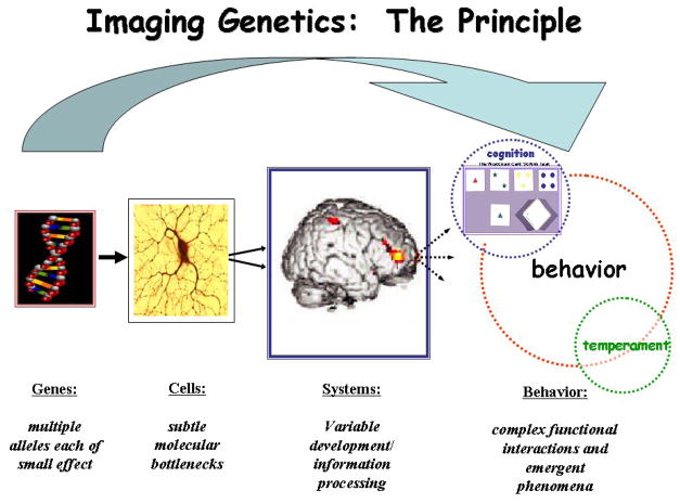
Imaging Genetics The biological impact of a variation in a gene on the brain traverses an increasingly divergent path from subtle molecular alterations at the cellular level to alterations in neural systems that eventually lead to variability in cognition and behavior. Imaging Genetics allows for the estimation of genetic effects at the level of neural systems or brain information processing, which represents a more proximate biological link to genes and serves as an obligatory intermediate of cognition, behavior and emergent phenomenon. Adapted from (Callicott & Weinberger, 2003) with permission.
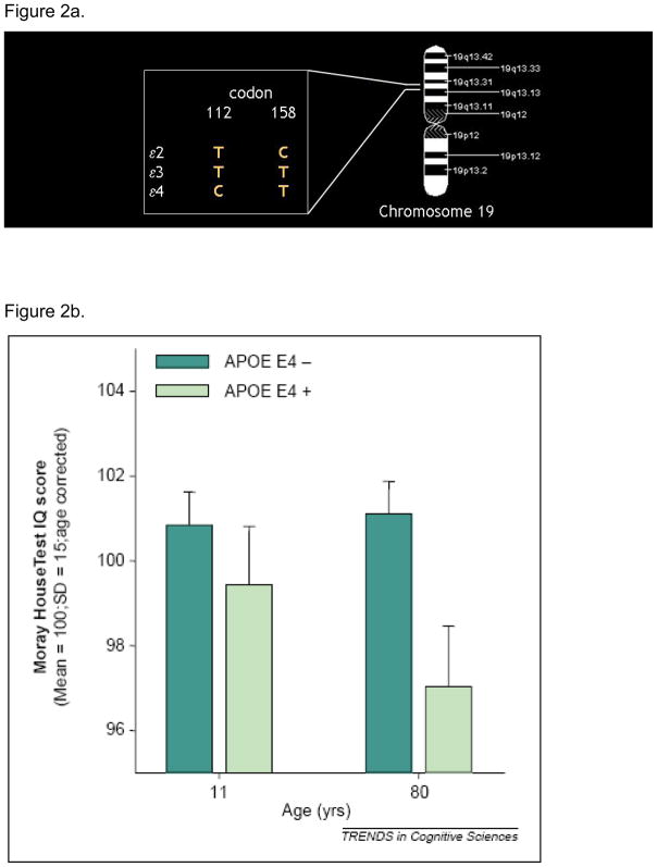
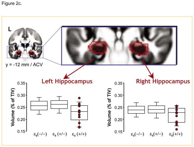
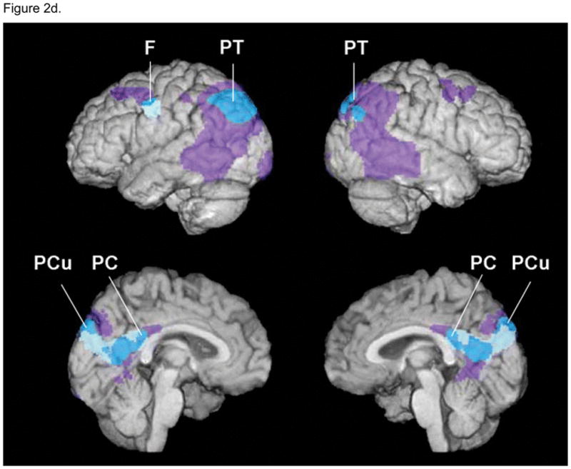
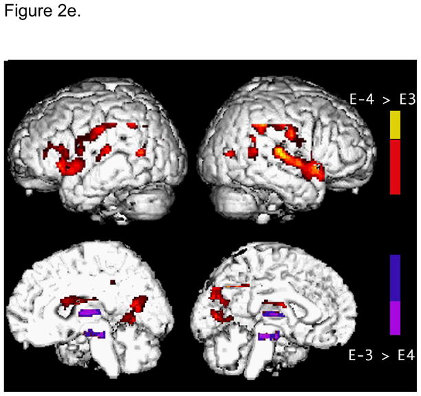
a: Gene for APOlipoprotein E: APOlipoprotein E (APOE) is a plasma glycoprotein involved in the transport of cholesterol and other lipids across the membrane of various cells. The gene for APOE is localized on chromosome 19 in a single locus with three alleles (e2, e3 and e4) responsible for the three APOE isoforms. The e4 allele results from a T - > C transition at codon 112 of the e3 allele. It is a well established risk factor for late onset (after 65 years) Alzheimer’s disease, with the risk increasing with the number of e4 allele one carries, or e4 gene dose effect (Corder et al., 1993). b. Effect of APOE on cognition: (Deary et al., 2002) report on the Moray House Test IQ score results from the Scottish Mental Survey. 466 subjects in this survey were tested twice, once at age 11 and again 70 years later. While there was no significant difference in IQ scores between subjects that possess an e4 allele and those that do not at age 11, there was a significant difference 70 years later at age 80, suggesting that possessing an e4 allele was associated with differences in normal cognitive aging. Reproduced from (Deary et al., 2002). c. Effect of APOE on brain structure: Using a large sample of about 750 subjects, (Lemaitre et al., 2005) show that in the absence of any difference in global brain compartment volume across the groups, healthy controls homozygous for the e4 allele had smaller hippocampal volumes than both heterozygotes and non-carriers. Reproduced from (Lemaitre et al., 2005). d. Effect of APOE on cerebral metabolism: Using FDG-PET, (Reiman et al., 2004) illustrate regions of the brain with abnormally low metabolism in young adult carriers of the APOE E4 allele in relation to those of patients with probable AD. A three dimensional (3D) surface-projection map of abnormally low glucose metabolism in young adult E4 carriers is superimposed on a map of abnormally low glucose metabolism in previously studied patients with probable AD. Purple areas represent brain areas in which glucose metabolism was abnormally low in patients with AD, muted blue areas represent brain areas in which glucose metabolism was abnormally low in E4 carriers, and bright blue areas represent areas in which glucose metabolism was abnormally low in both groups. Similar to patients with AD, young healthy adult E4 carriers had abnormally low glucose metabolism bilaterally in the posterior cingulate, and the parietal, temporal and prefrontal cortices. Reproduced from (Reiman et al., 2004). e. Effect of APOE on information processing during a memory task: During a paired associate learning task, for the same level of task performance, E4 carriers show greater activation in the left medial frontal, prefrontal and parietal regions than non-E4 carriers. This increased brain activity in healthy E4 carriers was interpreted as representing a compensatory process to maintain performance. Adapted from (Bookheimer et al., 2000).
a: Gene for APOlipoprotein E: APOlipoprotein E (APOE) is a plasma glycoprotein involved in the transport of cholesterol and other lipids across the membrane of various cells. The gene for APOE is localized on chromosome 19 in a single locus with three alleles (e2, e3 and e4) responsible for the three APOE isoforms. The e4 allele results from a T - > C transition at codon 112 of the e3 allele. It is a well established risk factor for late onset (after 65 years) Alzheimer’s disease, with the risk increasing with the number of e4 allele one carries, or e4 gene dose effect (Corder et al., 1993). b. Effect of APOE on cognition: (Deary et al., 2002) report on the Moray House Test IQ score results from the Scottish Mental Survey. 466 subjects in this survey were tested twice, once at age 11 and again 70 years later. While there was no significant difference in IQ scores between subjects that possess an e4 allele and those that do not at age 11, there was a significant difference 70 years later at age 80, suggesting that possessing an e4 allele was associated with differences in normal cognitive aging. Reproduced from (Deary et al., 2002). c. Effect of APOE on brain structure: Using a large sample of about 750 subjects, (Lemaitre et al., 2005) show that in the absence of any difference in global brain compartment volume across the groups, healthy controls homozygous for the e4 allele had smaller hippocampal volumes than both heterozygotes and non-carriers. Reproduced from (Lemaitre et al., 2005). d. Effect of APOE on cerebral metabolism: Using FDG-PET, (Reiman et al., 2004) illustrate regions of the brain with abnormally low metabolism in young adult carriers of the APOE E4 allele in relation to those of patients with probable AD. A three dimensional (3D) surface-projection map of abnormally low glucose metabolism in young adult E4 carriers is superimposed on a map of abnormally low glucose metabolism in previously studied patients with probable AD. Purple areas represent brain areas in which glucose metabolism was abnormally low in patients with AD, muted blue areas represent brain areas in which glucose metabolism was abnormally low in E4 carriers, and bright blue areas represent areas in which glucose metabolism was abnormally low in both groups. Similar to patients with AD, young healthy adult E4 carriers had abnormally low glucose metabolism bilaterally in the posterior cingulate, and the parietal, temporal and prefrontal cortices. Reproduced from (Reiman et al., 2004). e. Effect of APOE on information processing during a memory task: During a paired associate learning task, for the same level of task performance, E4 carriers show greater activation in the left medial frontal, prefrontal and parietal regions than non-E4 carriers. This increased brain activity in healthy E4 carriers was interpreted as representing a compensatory process to maintain performance. Adapted from (Bookheimer et al., 2000).
a: Gene for APOlipoprotein E: APOlipoprotein E (APOE) is a plasma glycoprotein involved in the transport of cholesterol and other lipids across the membrane of various cells. The gene for APOE is localized on chromosome 19 in a single locus with three alleles (e2, e3 and e4) responsible for the three APOE isoforms. The e4 allele results from a T - > C transition at codon 112 of the e3 allele. It is a well established risk factor for late onset (after 65 years) Alzheimer’s disease, with the risk increasing with the number of e4 allele one carries, or e4 gene dose effect (Corder et al., 1993). b. Effect of APOE on cognition: (Deary et al., 2002) report on the Moray House Test IQ score results from the Scottish Mental Survey. 466 subjects in this survey were tested twice, once at age 11 and again 70 years later. While there was no significant difference in IQ scores between subjects that possess an e4 allele and those that do not at age 11, there was a significant difference 70 years later at age 80, suggesting that possessing an e4 allele was associated with differences in normal cognitive aging. Reproduced from (Deary et al., 2002). c. Effect of APOE on brain structure: Using a large sample of about 750 subjects, (Lemaitre et al., 2005) show that in the absence of any difference in global brain compartment volume across the groups, healthy controls homozygous for the e4 allele had smaller hippocampal volumes than both heterozygotes and non-carriers. Reproduced from (Lemaitre et al., 2005). d. Effect of APOE on cerebral metabolism: Using FDG-PET, (Reiman et al., 2004) illustrate regions of the brain with abnormally low metabolism in young adult carriers of the APOE E4 allele in relation to those of patients with probable AD. A three dimensional (3D) surface-projection map of abnormally low glucose metabolism in young adult E4 carriers is superimposed on a map of abnormally low glucose metabolism in previously studied patients with probable AD. Purple areas represent brain areas in which glucose metabolism was abnormally low in patients with AD, muted blue areas represent brain areas in which glucose metabolism was abnormally low in E4 carriers, and bright blue areas represent areas in which glucose metabolism was abnormally low in both groups. Similar to patients with AD, young healthy adult E4 carriers had abnormally low glucose metabolism bilaterally in the posterior cingulate, and the parietal, temporal and prefrontal cortices. Reproduced from (Reiman et al., 2004). e. Effect of APOE on information processing during a memory task: During a paired associate learning task, for the same level of task performance, E4 carriers show greater activation in the left medial frontal, prefrontal and parietal regions than non-E4 carriers. This increased brain activity in healthy E4 carriers was interpreted as representing a compensatory process to maintain performance. Adapted from (Bookheimer et al., 2000).
a: Gene for APOlipoprotein E: APOlipoprotein E (APOE) is a plasma glycoprotein involved in the transport of cholesterol and other lipids across the membrane of various cells. The gene for APOE is localized on chromosome 19 in a single locus with three alleles (e2, e3 and e4) responsible for the three APOE isoforms. The e4 allele results from a T - > C transition at codon 112 of the e3 allele. It is a well established risk factor for late onset (after 65 years) Alzheimer’s disease, with the risk increasing with the number of e4 allele one carries, or e4 gene dose effect (Corder et al., 1993). b. Effect of APOE on cognition: (Deary et al., 2002) report on the Moray House Test IQ score results from the Scottish Mental Survey. 466 subjects in this survey were tested twice, once at age 11 and again 70 years later. While there was no significant difference in IQ scores between subjects that possess an e4 allele and those that do not at age 11, there was a significant difference 70 years later at age 80, suggesting that possessing an e4 allele was associated with differences in normal cognitive aging. Reproduced from (Deary et al., 2002). c. Effect of APOE on brain structure: Using a large sample of about 750 subjects, (Lemaitre et al., 2005) show that in the absence of any difference in global brain compartment volume across the groups, healthy controls homozygous for the e4 allele had smaller hippocampal volumes than both heterozygotes and non-carriers. Reproduced from (Lemaitre et al., 2005). d. Effect of APOE on cerebral metabolism: Using FDG-PET, (Reiman et al., 2004) illustrate regions of the brain with abnormally low metabolism in young adult carriers of the APOE E4 allele in relation to those of patients with probable AD. A three dimensional (3D) surface-projection map of abnormally low glucose metabolism in young adult E4 carriers is superimposed on a map of abnormally low glucose metabolism in previously studied patients with probable AD. Purple areas represent brain areas in which glucose metabolism was abnormally low in patients with AD, muted blue areas represent brain areas in which glucose metabolism was abnormally low in E4 carriers, and bright blue areas represent areas in which glucose metabolism was abnormally low in both groups. Similar to patients with AD, young healthy adult E4 carriers had abnormally low glucose metabolism bilaterally in the posterior cingulate, and the parietal, temporal and prefrontal cortices. Reproduced from (Reiman et al., 2004). e. Effect of APOE on information processing during a memory task: During a paired associate learning task, for the same level of task performance, E4 carriers show greater activation in the left medial frontal, prefrontal and parietal regions than non-E4 carriers. This increased brain activity in healthy E4 carriers was interpreted as representing a compensatory process to maintain performance. Adapted from (Bookheimer et al., 2000).
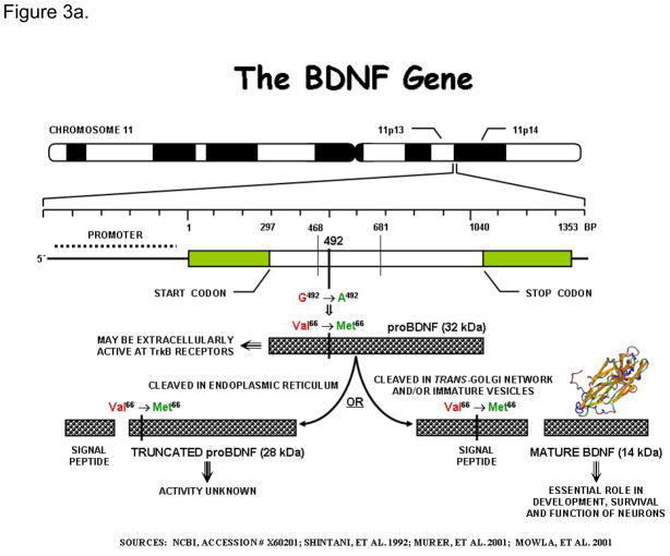
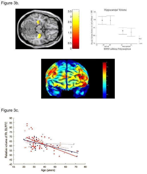
a. The gene for Brain Derived Neurotrophic Factor: The gene for BDNF is located on the short arm of chromosome 11. It encodes a precursor peptide (proBDNF), which is proteolytically cleaved to form the mature protein. In humans, a frequent single nucleotide polymorphism at nucleotide 196 (G/A) producing a non-conservative valine to methionine substitution at codon66 has been identified in this gene. This sequence variant, though located in the 5 pro-BDNF sequence, has been shown to affect intracellular processing and secrion of BDNF leading to impairments in hippocampal structure and function (Egan et al., 2003; Pezawas et al., 2004). The gene consists of at least nine exons, only one of which is translated. This translated exon is represented in the figure. b. Effect of BDNF val66met polymorphism on grey matter volume: Pezawas et al (Pezawas et al., 2004) using optimized VBM illustrate volume differences in BDNF met carriers relative to BDNF val/val individuals in the hippocampus (A) and prefrontal cortex (B). Consistent with the role of BDNF in cortical development and with the cellular and clinical effects of the BDNF val66met polymorphism, met carriers have relatively reduced gray matter volume in these brain regions. Reproduced from (Pezawas et al., 2004). c. BDNF val66met polymorphism modulates age related changes in the volume of the prefrontal cortex. Met-BDNF carriers show relatively more significant volume reduction with normal aging compared to individuals homozygous to Val-BDNF. Blue – male; Red – female; closed triangle – Met-BDNF carrier; open circle –homozygous Val-BDNF. Dotted lines represent regression lines for homozygous Val-BDNF individuals and solid lines represent regression lines for Met-BDNF carriers. Reproduced from (Nemoto et al., 2006).
a. The gene for Brain Derived Neurotrophic Factor: The gene for BDNF is located on the short arm of chromosome 11. It encodes a precursor peptide (proBDNF), which is proteolytically cleaved to form the mature protein. In humans, a frequent single nucleotide polymorphism at nucleotide 196 (G/A) producing a non-conservative valine to methionine substitution at codon66 has been identified in this gene. This sequence variant, though located in the 5 pro-BDNF sequence, has been shown to affect intracellular processing and secrion of BDNF leading to impairments in hippocampal structure and function (Egan et al., 2003; Pezawas et al., 2004). The gene consists of at least nine exons, only one of which is translated. This translated exon is represented in the figure. b. Effect of BDNF val66met polymorphism on grey matter volume: Pezawas et al (Pezawas et al., 2004) using optimized VBM illustrate volume differences in BDNF met carriers relative to BDNF val/val individuals in the hippocampus (A) and prefrontal cortex (B). Consistent with the role of BDNF in cortical development and with the cellular and clinical effects of the BDNF val66met polymorphism, met carriers have relatively reduced gray matter volume in these brain regions. Reproduced from (Pezawas et al., 2004). c. BDNF val66met polymorphism modulates age related changes in the volume of the prefrontal cortex. Met-BDNF carriers show relatively more significant volume reduction with normal aging compared to individuals homozygous to Val-BDNF. Blue – male; Red – female; closed triangle – Met-BDNF carrier; open circle –homozygous Val-BDNF. Dotted lines represent regression lines for homozygous Val-BDNF individuals and solid lines represent regression lines for Met-BDNF carriers. Reproduced from (Nemoto et al., 2006).
References
-
- Abe O, Aoki S, Hayashi N, Yamada H, Kunimatsu A, Mori H, et al. Normal aging in the central nervous system: quantitative MR diffusion-tensor analysis. Neurobiol Aging. 2002;23(3):433–441. - PubMed
-
- Akyol O, Yanik M, Elyas H, Namli M, Canatan H, Akin H, et al. Association between Ala-9Val polymorphism of Mn-SOD gene and schizophrenia. Progress in Neuro-Psychopharmacology and Biological Psychiatry. 2005;29(1):123–131. - PubMed
-
- Bartres-Faz D, Junque C, Clemente IC, Lopez-Alomar A, Valveny N, Lopez-Guillen A, et al. Angiotensin I converting enzyme polymorphism in humans with age-associated memory impairment: relationship with cognitive performance. Neurosci Lett. 2000;290(3):177–180. - PubMed
-
- Baune BT, Hohoff C, Berger K, Neumann A, Mortensen S, Roehrs T, et al. Association of the COMT val158met variant with antidepressant treatment response in major depression. Neuropsychopharmacology. 2008;33(4):924–932. - PubMed
Publication types
MeSH terms
Grants and funding
LinkOut - more resources
Full Text Sources
Medical
