. 2008 Aug;102(2):167-82.
doi: 10.1093/aob/mcn084. Epub 2008 May 29.

Embryological features of Tofieldia glutinosa and their bearing on the early diversification of monocotyledonous plants

Affiliations

Embryological features of Tofieldia glutinosa and their bearing on the early diversification of monocotyledonous plants

Samuel J Holloway et al. Ann Bot. 2008 Aug.

Abstract

Background and aims: Although much is known about the vegetative traits associated with early monocot evolution, less is known about the reproductive features of early monocotyledonous lineages. A study was made of the embryology of Tofieldia glutinosa, a member of an early divergent monocot clade (Tofieldiaceae), and aspects of its development were compared with the development of other early divergent monocots in order to gain insight into defining reproductive features of early monocots.

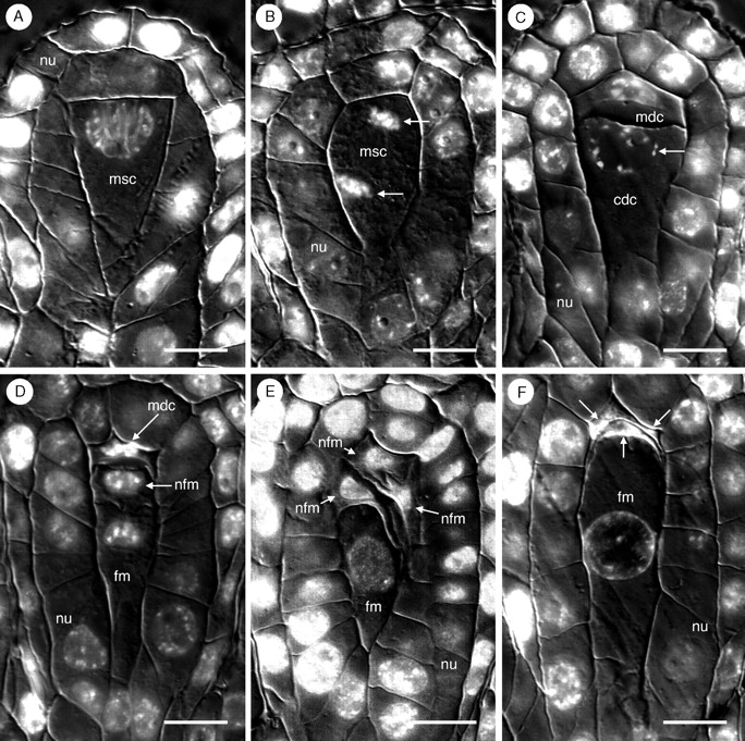
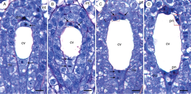
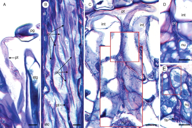
Methods: Field-collected developing gynoecial tissues of Tofieldia glutinosa were prepared for histological examination. Over 600 ovules were sectioned and studied using brightfield, differential interference contrast, and fluorescence microscopy. High-resolution digital imaging was used to document important stages of megasporogenesis, megagametogenesis and early endosperm development.

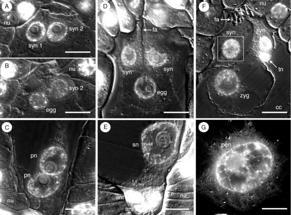
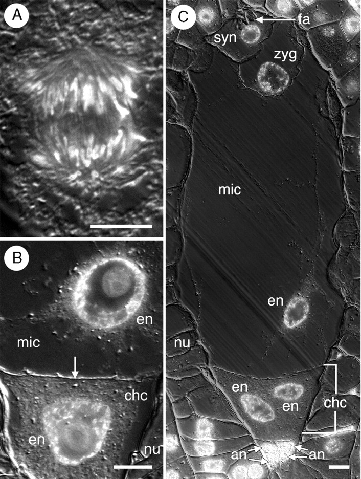
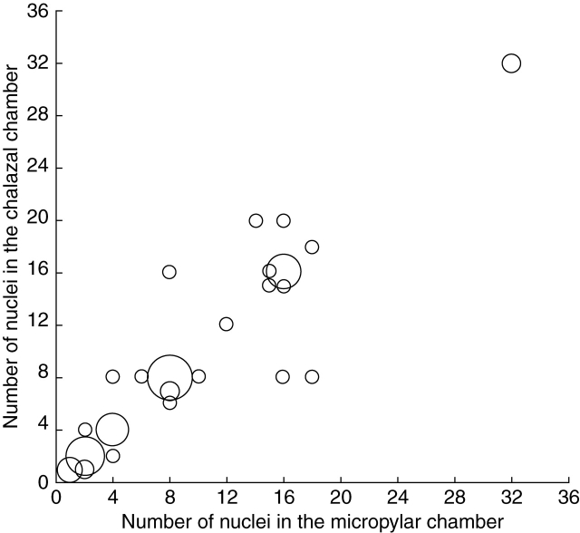
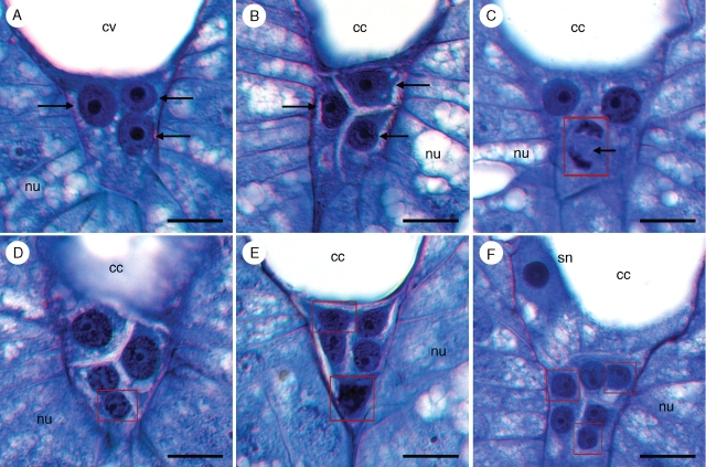
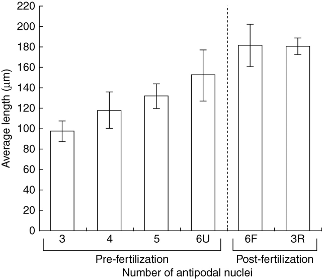
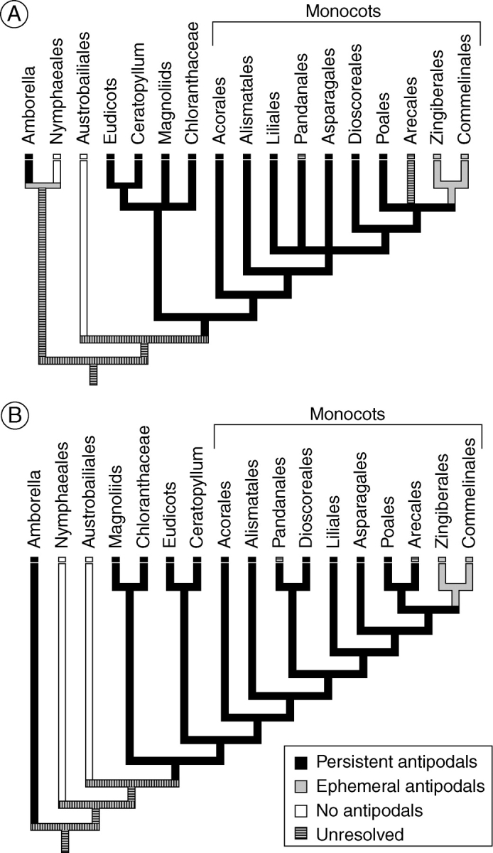
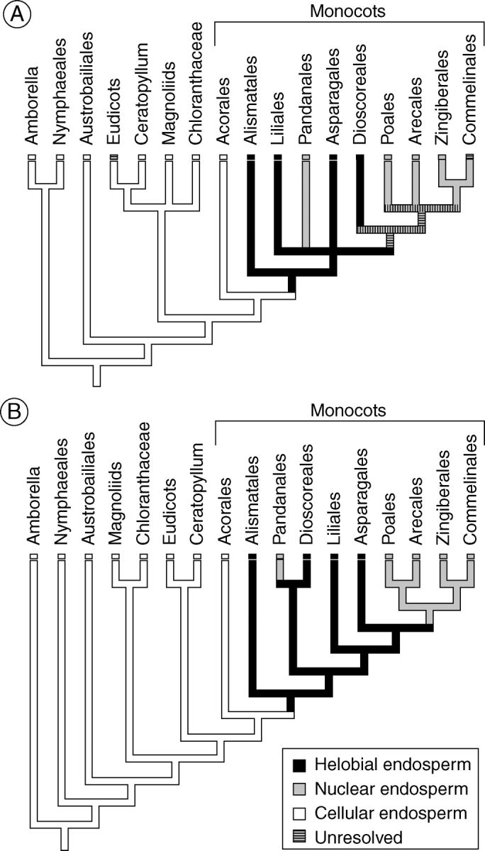
Key results: Development of the female gametophyte in T. glutinosa is of a modified Polygonum-type. At maturity the female gametophyte is seven-celled and 11-nucleate with a standard three-celled egg apparatus, a binucleate central cell (where ultimately, the two polar nuclei will fuse into a diploid secondary nucleus) and three binucleate antipodal cells. The antipodal nuclei persist past fertilization, and the process of double fertilization appears to yield a diploid zygote and triploid primary endosperm cell, as is characteristic of plants with Polygonum-type female gametophytes. Endosperm development is helobial, and free-nuclear growth initially proceeds at equal rates in both the micropylar and chalazal endosperm chambers.

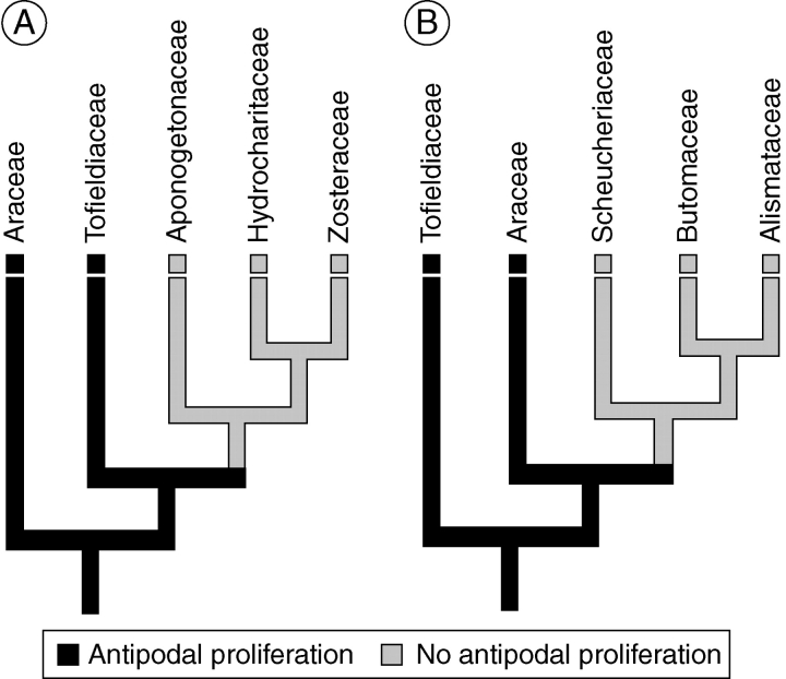
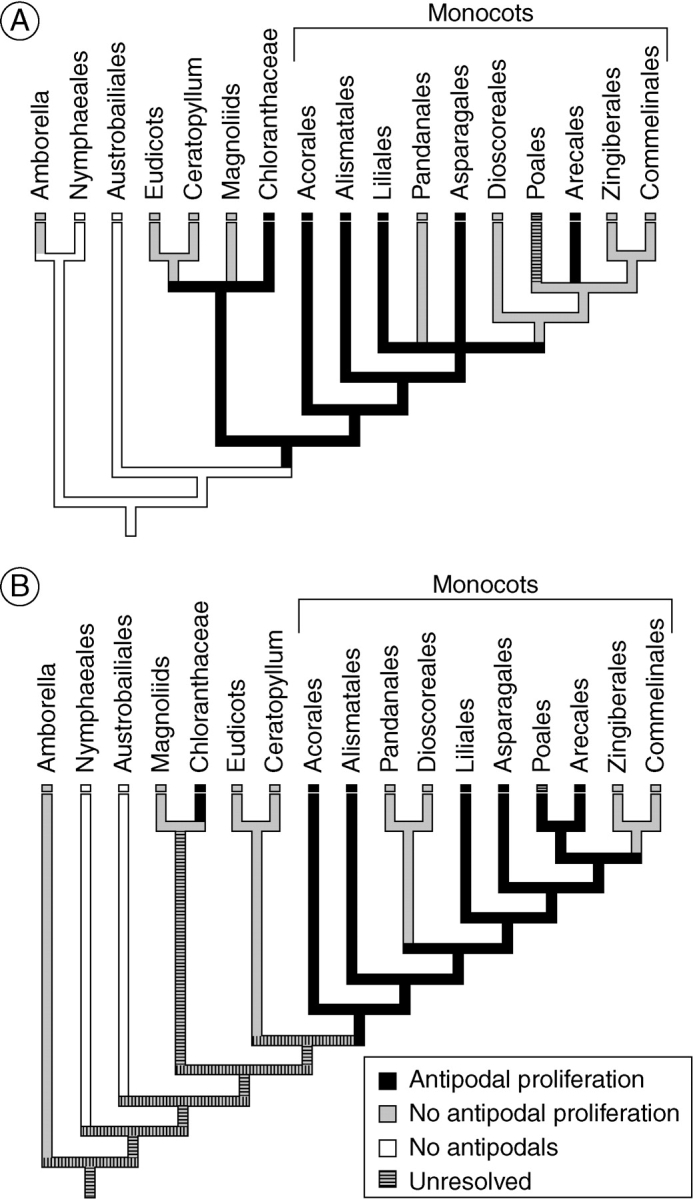
Conclusions: The analysis suggests that the shared common ancestor of monocots possessed persistent and proliferating antipodals similar to those found in T. glutinosa and other early-divergent monocots (e.g. Acorus and members of the Araceae). Helobial endosperm among monocots evolved once in the common ancestor of all monocots excluding Acorus. Thus, the analysis further suggests that helobial endosperm in monocots is homoplasious with those helobial endosperms that are present in water lilies and eudicot angiosperms.


Figures

F<sc>ig</sc>. 1.
Fig. 1.
Megasporogenesis in Tofieldia glutinosa produces a single functional megaspore. (A) Megasporocyte surrounded by two cell layers of nucellus. (B) First meiotic division of megasporocyte. Arrows indicate nuclei derived from meiosis I. (C) Dyad stage. The chalazal dyad cell, with nucleus in prophase (arrow) is significantly larger than the micropylar dyad cell. (D) Completion of meiosis II in the chalazal dyad to produce a functional megaspore. The micropylar dyad cell is arrested in prophase II. (E) Tetrad stage in which meiosis II was completed in both the micropylar and chalazal dyad cells. The chalazal-most cell is the functional megaspore. (F) Functional megaspore with three crushed and degenerated megaspores (arrows). All figures are oriented such that the micropylar pole is at the top and the chalazal pole is at the bottom. All sections were stained with the DNA fluorochrome DAPI and visualized with differential interference contrast (DIC) and fluorescence optics. Images are composites of two micrographs of the same section (one with fluorescence and one with DIC). cdc, Chalazal dyad cell; fm, functional megaspore; mdc, micropylar dyad cell; msc, megasporocyte; nfm, nonfunctional megaspore; nu, nucellus. Scale bars = 10 µm.
F<sc>ig</sc>. 2.
Fig. 2.
Syncytial development of the female gametophyte in Tofieldia glutinosa. (A) Two-nucleate stage with nuclei (arrows) located at each pole of the female gametophyte and separated by a large central vacuole. Degenerated megaspores are still visible, but not discernable from one another. (B) Transition to a four-nucleate female gametophyte. The two nuclei (arrows) at the micropylar pole are emerging from telophase while the two sets of chromosomes (arrows) at the chalazal pole are still in telophase. Note that the mitotic divisions are perpendicular to one another. (C) Four-nucleate (arrows) female gametophyte. (D) Eight-nucleate female gametophyte prior to cellularization and migration of polar nuclei. All sections were stained with toluidine blue. The red box indicates digital superposition of the nucleus from the adjacent histological section. cv, Central vacuole; dm, degenerate megaspores; int, integument; nu, nucellus; pn, polar nucleus. Scale bars = 10 µm.
F<sc>ig</sc>. 3.
Fig. 3.
Development of egg apparatus and central cell in Tofieldia glutinosa. (A) and (B) Serial sections of synergid cells and egg in recently cellularized female gametophyte (prior to fusion of the polar nuclei). At this stage, cells of the egg apparatus are confined to a narrow region between the central cell and the micropylar pole of the female gametophyte. syn 2 refers to the same synergid in (A) and (B). (C) Polar nuclei just prior to their fusion. (D) Mature egg apparatus containing an egg cell, two synergids and a rudimentary filiform apparatus. Cells of the egg apparatus are much larger at this stage than in previous stages. (E) Mature central cell containing a secondary nucleus, seen in close proximity to the antipodals. (F) Post-fertilization egg apparatus containing a zygote, two synergids, a pronounced filiform apparatus, and a tube nucleus (from a pollen tube). (G) Post-fertilization central cell containing a large primary endosperm nucleus. All sections were stained with the DNA fluorochrome DAPI and visualized with differential interference contrast (DIC) and fluorescence optics. Images are composites of two micrographs of the same section (one with fluorescence and one with DIC). The white box indicates digital superposition of the nucleus from the adjacent histological section. ant, Antipodal cell; cc, central cell; egg, egg cell; fa, filiform apparatus; nu, nucellus; pen, primary endosperm nucleus; pn, polar nucleus; sn, secondary nucleus; syn, synergid cell; tn, tube nucleus; zyg, zygote. Scale bars = 10 μm.
F<sc>ig</sc>. 4.
Fig. 4.
Pollen tube growth in Tofieldia glutinosa. (A) Pollen tube growing out of a pollen grain on the stigma. (B) Numerous pollen tubes grow through the stylar canal. (C) Pollen tube growth through the micropyle. This image is a composite of three serial histological sections of the same ovule. Red boxes indicate digital superposition of pollen tube and surrounding tissue from adjacent histological sections. (D) Penetration of the nucellus by a pollen tube. Pollen tube growth through the nucellus is intercellular. (E) Pollen tube growth through the nucellus and into the filiform apparatus of the female gametophyte. The red box indicates digital superposition of the pollen tube and surrounding tissue from the adjacent histological section. All sections were stained with toluidine blue. fa, Filiform apparatus; int, integument; nu, nucellus; pg, pollen grain; pt, pollen tube; st, style; stc, stylar canal; stg, stigma. Scale bars = 10 µm.
F<sc>ig</sc>. 5.
Fig. 5.
Post-fertilization egg apparatus in Tofieldia glutinosa. (A) Remnants of the pollen tube are visible at the micropylar pole of the female gametophyte. (B) A well-developed filiform apparatus inside the degenerated synergid cell. The tube nucleus is still visible at this stage. Sections were stained with toluidine blue and visualized with DIC optics. cc, Central cell; fa, filiform apparatus; nu, nucellus; syn, synergid cell; tn, pollen tube nucleus; zyg, zygote. Scale bars = 10 µm.
F<sc>ig</sc>. 6.
Fig. 6.
Double fertilization in Tofieldia glutinosa. (A) Fertilization of the egg nucleus. The sperm nucleus is still visible within the egg nucleus and the tube nucleus is present in the adjacent synergid. (B) Fertilization of the secondary nucleus by the second sperm nucleus. The sperm nucleus is still visible (with nucleolus) within the secondary nucleus. (C) Second fertilization event involving two unfused polar nuclei and a sperm nucleus. All sections were stained with the DNA fluorochrome DAPI and visualized with differential interference contrast (DIC) and fluorescence optics. Images are composites of two micrographs of the same section (one with fluorescence and one with DIC). egg, Egg cell; pn, polar nucleus; sn, secondary nucleus; sp, sperm nucleus; syn, synergid cell; tn, tube nucleus. Scale bars = 10 µm.
F<sc>ig</sc>. 7.
Fig. 7.
Helobial endosperm development in Tofieldia glutinosa. (A) The first mitotic division of the primary endosperm nucleus occurs in the bottom half of the central cell. (B) Two-celled, two-nucleate endosperm. A wall (arrow) forms between the two daughter nuclei of the primary endosperm nucleus partitioning the central cell into a large micropylar chamber and a smaller chalazal chamber. (C) Post-fertilization female gametophyte with binucleate chalazal endosperm chamber and binucleate (one nucleus is visible in this section) micropylar endosperm chamber. The zygote is visible at the micropylar pole. One synergid, the filiform apparatus, and four of six antipodal nuclei are also visible in this section. All sections were stained with DAPI and visualized with DIC and fluorescence optics. All images are composites of two micrographs of the same section (one utilizing fluorescence optics and one utilizing DIC optics). an, Antipodal nuclei; chc, chalazal chamber of endosperm; en, endosperm nucleus; fa, filiform apparatus; mic, micropylar chamber of endosperm; nu, nucellus; syn, synergid cell; zyg, zygote. Scale bars = 10 µm.
F<sc>ig</sc>. 8.
Fig. 8.
Relative rates of syncytial development in the micropylar and chalazal endosperm chambers in Tofieldia glutinosa. Endosperm nuclear proliferation occurs at roughly equal rates in each chamber. Endosperm beyond the 64-nucleate stage was not observed. The size of each data point is proportional to the number of times the specific endosperm configuration was observed.
F<sc>ig</sc>. 9.
Fig. 9.
Proliferation of antipodal nuclei in Tofieldia glutinosa. (A) Three antipodal nuclei (arrows) prior to cellularization of the female gametophyte. (B) Three uninucleate antipodal cells (arrows) following cellularization of the female gametophyte. (C) Antipodal nucleus in mitosis (arrow). Mitotic divisions of the antipodals are usually asynchronous. (D) Single binucleate antipodal and two uninucleate antipodals. (E) Two binucleate antipodals and a single uninucleate antipodal. (F) Three binucleate antipodal cells. The secondary nucleus is also visible in this section. All sections were stained with toluidine blue. Red boxes indicate digital superposition of nuclei from adjacent histological sections. cc, Central cell; cv, central vacuole; nu, nucellus; sn, secondary nucleus. Scale bars = 10 µm.
F<sc>ig</sc>. 10.
Fig. 10.
Female gametophyte length relative to the number of antipodal nuclei present in Tofieldia glutinosa. 6U refers to unfertilized female gametophytes with six antipodal nuclei. 6F refers to fertilized female gametophytes with six antipodal nuclei measured before division of the primary endosperm nucleus. 3R refers to fertilized female gametophytes in which the number of antipodal nuclei has been reduced from six to three.
F<sc>ig</sc>. 11.
Fig. 11.
Evolution of antipodal proliferation in Alismatales. (A) Phylogeny of Soltis et al. (2007). Antipodal proliferation has been reported in Hydrocharitaceae (Wylie, 1904), but more recent studies suggest that this is not actually the case (Johri et al., 1992). (B) Phylogeny of Graham et al. (2006). Parsimony analysis reveals that antipodal proliferation is the ancestral condition for Alismatales.
F<sc>ig</sc>. 12.
Fig. 12.
Evolution of antipodal proliferation in monocots. In all cases except for the Poales, the character state assigned to the group has been resolved as the ancestral condition for that group based on the present analysis. The ancestral condition within the Poales is unresolved, and is classified as polymorphic. (A) Phylogeny of Soltis et al. (2007). (B) Phylogeny of Graham et al. (2006). For both of these phylogenetic hypotheses of angiosperm and monocot relationships, parsimony analysis reveals that antipodal proliferation either evolved in or was present in the common ancestor of all monocots.
F<sc>ig</sc>. 13.
Fig. 13.
Evolution of persistence of antipodals in monocots. In all cases except for the Pandanales and the Arecales, the character state assigned to the group has been resolved as the ancestral condition for that group based on our analysis. The ancestral conditions within the Pandanales and the Arecales are unresolved, and are classified as polymorphic. (A) Phylogeny of Soltis et al. (2007). (B) Phylogeny of Graham et al. (2006). Parsimony analysis reveals that the common ancestor of all angiosperms exclusive of Amborella, Nymphaeales and Austrobaileyales had persistent antipodals. It is unclear whether the persistent antipodals in Amborella evolved independently or were inherited from the common ancestor of all angiosperms.
F<sc>ig</sc>. 14.
Fig. 14.
Evolution of helobial endosperm development in angiosperms. In all cases except for the Commelinales and eudicots, the character state assigned to the group has been resolved as the ancestral condition for that group based on the present analysis. The ancestral conditions within Commelinales and eudicots are unresolved, and are classified here as polymorphic. (A) Phylogeny of Soltis et al. (2007). (B) Phylogeny of Graham et al. (2006). Parsimony analysis reveals that helobial endosperm development evolved in the common ancestor of all monocots excluding the Acorales.

Similar articles

Cited by

References

    1. Anton AM, Cocucci AE. The grass megagametophyte and its possible phylogenetic implications. Plant Sytematics and Evolution. 1984;146:117–121.
    1. APG II. An update of the Angiosperm Phylogeny Group classification for the orders and families of flowering plants. Botanical Journal of the Linnean Society. 2003;141:399–436.
    1. Arber A. Monocotyledons: a morphological study. Cambridge: Cambridge University Press; 1925.
    1. Asplund I. Embryological studies in the genus Typha. Svensk Botanisk Tidskrift. 1972;66:1–17.
    1. Asplund I. Embryological studies in the genus Sparganium. Svensk Botanisk Tidskrift. 1973;67:177–200.

Publication types