. 2008 Jul 25;283(30):21145-52.
doi: 10.1074/jbc.M800610200. Epub 2008 May 29.

Substrate recognition mechanism of VAMP/synaptobrevin-cleaving clostridial neurotoxins

Affiliations

Substrate recognition mechanism of VAMP/synaptobrevin-cleaving clostridial neurotoxins

Stefan Sikorra et al. J Biol Chem. .

Abstract

Botulinum neurotoxins (BoNTs) and tetanus neurotoxin (TeNT) inhibit neurotransmitter release by proteolyzing a single peptide bond in one of the three soluble N-ethylmaleimide-sensitive factor attachment protein receptors SNAP-25, syntaxin, and vesicle-associated membrane protein (VAMP)/synaptobrevin. TeNT and BoNT/B, D, F, and G of the seven known BoNTs cleave the synaptic vesicle protein VAMP/synaptobrevin. Except for BoNT/B and TeNT, they cleave unique peptide bonds, and prior work suggested that different substrate segments are required for the interaction of each toxin. Although the mode of SNAP-25 cleavage by BoNT/A and E has recently been studied in detail, the mechanism of VAMP/synaptobrevin proteolysis is fragmentary. Here, we report the determination of all substrate residues that are involved in the interaction with BoNT/B, D, and F and TeNT by means of systematic mutagenesis of VAMP/synaptobrevin. For each of the toxins, three or more residues clustered at an N-terminal site remote from the respective scissile bond are identified that affect solely substrate binding. These exosites exhibit different sizes and distances to the scissile peptide bonds for each neurotoxin. Substrate segments C-terminal of the cleavage site (P4-P4') do not play a role in the catalytic process. Mutation of residues in the proximity of the scissile bond exclusively affects the turnover number; however, the importance of individual positions at the cleavage sites varied for each toxin. The data show that, similar to the SNAP-25 proteolyzing BoNT/A and E, VAMP/synaptobrevin-specific clostridial neurotoxins also initiate substrate interaction, employing an exosite located N-terminal of the scissile peptide bond.


Figures

FIGURE 1.
FIGURE 1.
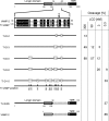
Determination of VAMP-2 regions that are essential for cleavage by BoNT/F and BoNT/D using various VAMP hybrids. Left, schematic representation of VAMP-2, TI-VAMP, and various TI-VAMP-VAMP-2 hybrids (TI-VH; numbers specify the number of VAMP-2 residues). F, D, B, and T together with numbers indicate the scissile bonds of respective BoNTs or TeNT. Numbers above (gray) and below (black) line drawings define border residues of VAMP-2 and TI-VAMP in the various hybrid proteins, respectively. Right, hybrid proteins were radiolabeled by in vitro transcription/translation and incubated for 1 h in the presence of 0.2 nm L chain of BoNT/D or BoNT/F. Samples were analyzed by SDS-PAGE and phosphorimaging. Values represent the percentage of cleavage versus VAMP-2-(1-96) ± S.D. of 2-8 independent experiments. Note that several TI-VHs were also analyzed as constructs lacking the longin domain (Δ Longin). n.a., not applicable; n.d., not determined.
FIGURE 2.
FIGURE 2.
Cleavage analysis of various VAMP-2 point mutants. Top, schematic representation of VAMP-2 and alignment of the amino acid region that was analyzed by systematic mutagenesis with the corresponding region of TI-VAMP. Identical residues are indicated by white letters on a black background. Conserved residues are shown in boxes shaded gray. Peptide bonds attacked by BoNT/F, D, B, or TeNT are marked F, D, B, and T. Bottom, in order to determine the effect on hydrolysis by CNT L chains, residues differing among VAMP-2 and TI-VAMP were individually replaced in VAMP-2 with the corresponding amino acids of TI-VAMP. Some of those were in addition replaced with alanine. All identical residues were substituted by alanine, and Ala-37, which corresponds to TI-VAMP Ala-131 was replaced with leucine. VAMP-2 mutants were radiolabeled by in vitro transcription/translation and incubated for 1 h in the presence of 0.2 nm LC/F, 0.2 nm LC/D, 20 nm LC/B, or 10 nm LC/T. Samples were analyzed by Tris/Tricine-PAGE using 15% gels. Columns represent percentages of cleavage versus the wild-type VAMP-2-(1-96). Data represent means ± S.D. of at least four independent experiments. The dotted lines specify thresholds of 10, 33, and 66% reduction of cleavability, and the color code applied to the columns is as follows: green, no or less than 10% reduction of cleavability; yellow, more than 10% reduction of cleavability; pink, more than 33% reduction of cleavability; red, more than 66% reduction of cleavability. Open column overlays specify the results of parallel mutations to alanine in those positions. Dark blue horizontal bars below individual charts denote putative exosites, light blue bars show the cleavage sites, and dark blue dots show interspersed anchor points (see “Discussion”).
FIGURE 3.
FIGURE 3.
Kinetic parameters of VAMP-2 cleavage by BoNT/F, BoNT/D, and TeNT. VAMP-2 or its mutants used at concentrations from 5 to 100 μm were incubated for 2 or 4 min in the presence of 0.08 nm LC/F, 0.08 nm LC/D, or 10 nm LC/T. Samples were analyzed by Tris/Tricine-PAGE using 15% gels. The percentage of hydrolyzed VAMP-2 was determined and used to calculate the initial velocity of substrate hydrolysis. Km and Vmax values were derived from Lineweaver-Burk plots (for further details, see “Experimental Procedures”). Data represent means ± S.D. of 2-4 independent experiments. The single-letter code below the lower columns was used to identify the mutants. Black columns, wild type (wt); white columns, exosite mutants; gray columns, cleavage site mutants.
FIGURE 4.
FIGURE 4.
Generation of BoNT/D-sensitive TI-VAMP variants. Left, alignment of a 32-amino acid segment encompassing the region of VAMP-2 supposed to be essential for proteolysis by BoNT/D with the corresponding region of TI-VAMP. Identical residues are indicated by white letters on a black background. Conserved residues are shown in boxes on a gray background. D, scissile peptide bond for BoNT/D. The numbers below specify mutated TI-VAMP residues. Right, mutated TI-VAMP variants were radiolabeled by in vitro transcription/translation and incubated for 1 h in the presence of 0.3-300 nm LC/D. The extent of cleavage was determined subsequent to SDS-PAGE (15% gels) and phosphorimaging. Values represent the average percentage of cleavage of 2-6 independent experiments. S.D. values did not exceed 30% of the measured values (e.g. TI-D-8; 9 ± 2.6%).

References

    1. Schiavo, G., Matteoli, M., and Montecucco, C. (2000) Physiol. Rev. 80 717-766 - PubMed
    1. Sollner, T., Bennett, M. K., Whiteheart, S. W., Scheller, R. H., and Rothman, J. E. (1993) Cell 75 409-418 - PubMed
    1. Schiavo, G., Rossetto, O., and Montecucco, C. (1994) Semin. Cell Biol. 5 221-229 - PubMed
    1. Niemann, H., Blasi, J., and Jahn, R. (1994) Trends Cell Biol. 4 179-185 - PubMed
    1. Advani, R. J., Bae, H. R., Bock, J. B., Chao, D. S., Doung, Y. C., Prekeris, R., Yoo, J. S., and Scheller, R. H. (1998) J. Biol. Chem. 273 10317-10324 - PubMed

Publication types

LinkOut - more resources