. 2008 Aug;26(8):2104-13.
doi: 10.1634/stemcells.2008-0115. Epub 2008 May 29.

Fate-mapping evidence that hepatic stellate cells are epithelial progenitors in adult mouse livers

Affiliations

Fate-mapping evidence that hepatic stellate cells are epithelial progenitors in adult mouse livers

Liu Yang et al. Stem Cells. 2008 Aug.

Abstract

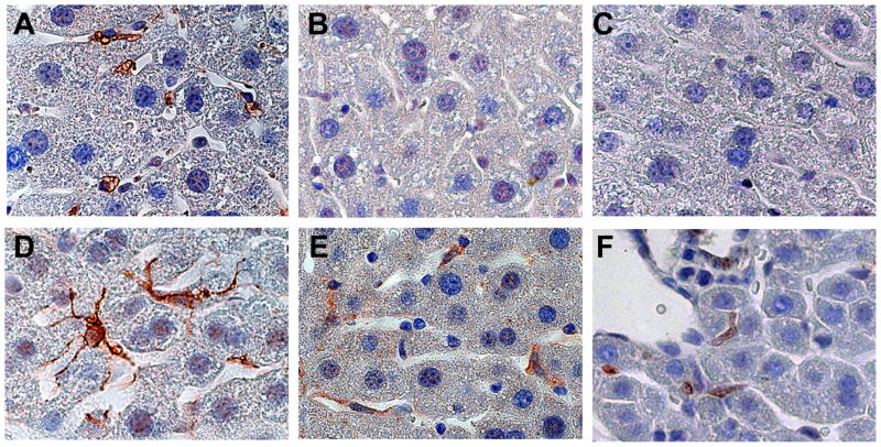
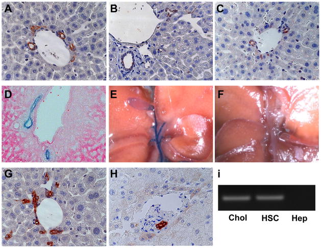
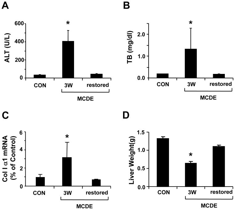
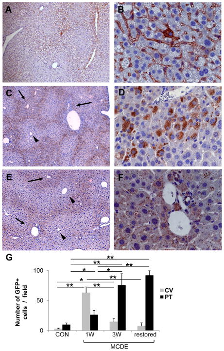
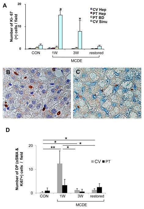
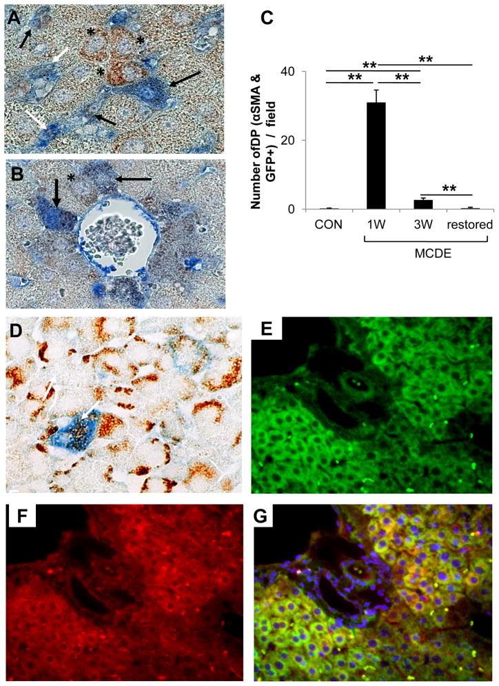
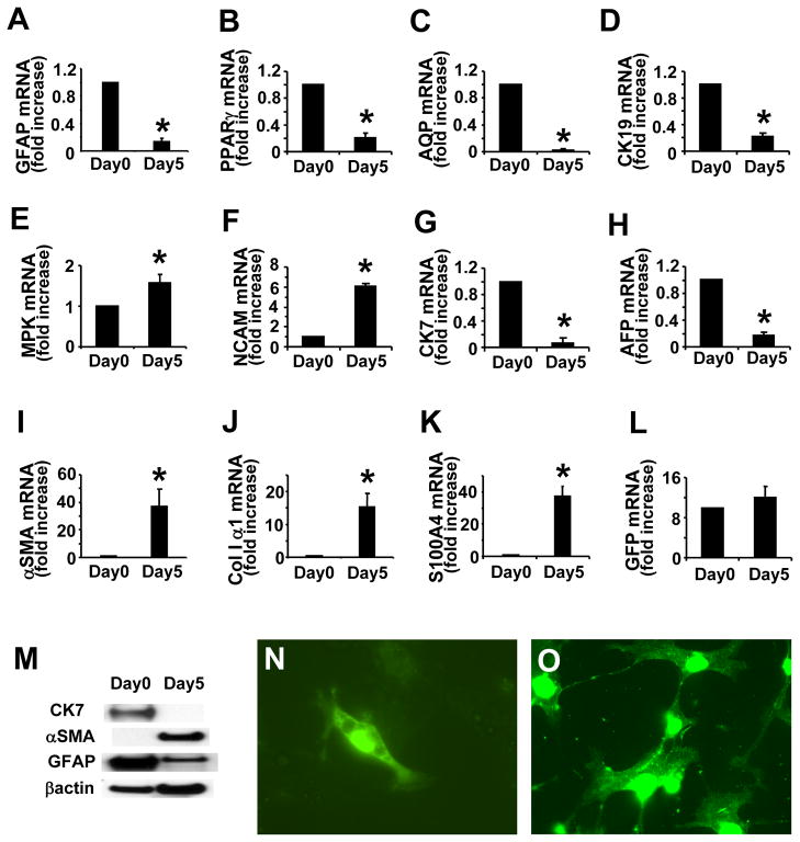
Liver injury activates quiescent hepatic stellate cells (Q-HSC) to proliferative myofibroblasts. Accumulation of myofibroblastic hepatic stellate cells (MF-HSC) sometimes causes cirrhosis and liver failure. However, MF-HSC also promote liver regeneration by producing growth factors for oval cells, bipotent progenitors of hepatocytes and cholangiocytes. Genes that are expressed by primary hepatic stellate cell (HSC) isolates overlap those expressed by oval cells, and hepatocytic and ductular cells emerge when HSC are cultured under certain conditions. We evaluated the hypothesis that HSC are a type of oval cell and, thus, capable of generating hepatocytes to regenerate injured livers. Because Q-HSC express glial fibrillary acidic protein (GFAP), we crossed mice in which GFAP promoter elements regulated Cre-recombinase with ROSA-loxP-stop-loxP-green fluorescent protein (GFP) mice to generate GFAP-Cre/GFP double-transgenic mice. These mice were fed methionine choline-deficient, ethionine-supplemented diets to activate and expand HSC and oval cell populations. GFP(+) progeny of GFAP-expressing precursors were characterized by immunohistochemistry. Basal expression of mesenchymal markers was negligible in GFAP(+)Q-HSC. When activated by liver injury or culture, HSC downregulated expression of GFAP but remained GFP(+); they became highly proliferative and began to coexpress markers of mesenchyme and oval cells. These transitional cells disappeared as GFP-expressing hepatocytes emerged, began to express albumin, and eventually repopulated large areas of the hepatic parenchyma. Ductular cells also expressed GFAP and GFP, but their proliferative activity did not increase in this model. These findings suggest that HSC are a type of oval cell that transitions through a mesenchymal phase before differentiating into hepatocytes during liver regeneration. Disclosure of potential conflicts of interest is found at the end of this article.


Figures

Figure 1
Figure 1. GFAP, Cre-recombinase and GFP expression in HSC of control and GFAP-Cre/reporter mice
Immunohistochemistry for GFAP (A,D), Cre-recombinase (B, E) and GFP (C,F) were performed in representative control (Ctr) (A–C) and GFAP-Cre/GFP (TG) mice (D–F). GFAP is expressed by hepatic stellate cells (HSC) in a representative Ctr mouse (A) and a representative GFAP-Cre/GFP (TG) mouse (D). HSC express Cre recombinase (E) and GFP (F) in TG mice, not in Ctr mice (B,C). (X100)
Figure 2
Figure 2. GFAP, Cre-recombinase and GFP expression in cholangiocytes of GFAP-Cre/reporter mice
GFAP (A), Cre-recombinase (B) and GFP (C) are expressed by bile ductular cells in portal triads of a representative GFAP-Cre/GFP (TG) mouse. Eosin-counterstained liver section from a representative GFAP-Cre/LacZ mouse showing LacZ(+) bile ductular cells (D). Extra-hepatic bile ducts, cystic duct and gallbladder exhibit β-galactosidase activity in a representative GFAP-Cre/LacZ mouse (E), but not in a Ctr mouse (F). Bile ductular cells in healthy adult mice (G) and a typical sample of non-diseased human liver (H) also express GFAP. Primary rat cholangiocytes (Chol) and HSC express GFAP mRNA, but primary hepatocytes (Hep) do not (I). (X63)
Figure 3
Figure 3. Effect of MCDE diet on serum liver enzymes levels, liver collagen I α1 mRNA and liver mass
Serum alanine aminotransferase (ALT) (A) and total bilirubin (TB) (B) levels were measured in controls (CON), mice fed MCDE diets for 3 weeks (MCDE 3W) and mice fed MCDE diets for 3 weeks and then returned to normal chow for 3 weeks before sacrifice (MCDE restored) (n=4 mice/group/time point). Collagen I α 1mRNA expression was evaluated in liver samples from the same mice using real time PCR (C). (D) Liver weight in the control and both MCDE diet fed groups (*P< 0.05 compared to control mice, n=4 mice/group/time point).
Figure 4
Figure 4. GFP expression in the livers of TG mice after injury
GFAP-Cre/GFP mice (n = 15) were fed MCDE-diets and survivors were sacrificed after 1 week (n = 4) or 3 weeks (n = 4). Other mice that survived 3 weeks of MCDE treatment (n = 4) were sacrificed after being switched back to normal diets for 3 weeks. GFP expression was assessed by immunohistochemistry. Representative results in mice that were sacrificed at the end of 1 week MCDE diet treatment (A, B), 3 weeks MCDE treatment (C, D), or 3 weeks after stopping a 3 week course of MDCE diets (E, F). Numbers of GFP-positive cells visualized at X40 were counted in different parts of the liver lobule (i.e., near central veins (CV) and portal tracts (PT)) (G). * < p 0.05, ** p < 0.005 (A, C, E original magnification X10; B, D, F X63). Arrows and arrowhead indicate portal triads and central veins, respectively.
Figure 5
Figure 5. Ki-67 expression in the livers of TG mice
GFAP-Cre/GFP mice (n = 15) were fed MCDE-diets and survivors were sacrificed as described above (n = 4/group/time point). Immunohistochemistry for Ki-67 or double-immunohistochemistry for Ki-67/αSMA was performed. Numbers of Ki-67-positive cells visualized at X63 (high power) were counted in different parts of the liver lobule (i.e., near central veins (CV) and portal tracts (PT)) and in different cell types (i.e., hepatocytes (Hep), bile ductular cells (BD), and perisinuosoidal lining cells (Sinu)). Results are graphed as numbers of positive cells/field in (A).* p < 0.05 Ki-67 expression (B) and Ki-67 and αSMA co-expression (C) in mice that were sacrificed at the end of 1 week MCDE treatment. Numbers of double positive (DP) cells for Ki-67and αSMA in different areas of the liver lobule (CV or PT) were counted at different time points and results are graphed in (D). * p < 0.05, ** p < 0.005 (A, C magnification X100)
Figure 6
Figure 6. Co-expression of GFP with αSMA, AE1/3 or albumin in the livers of TG mice after liver injury
GFAP-Cre/GFP mice (n = 15) were fed MCDE-diets and survivors were sacrificed as described above (n = 4/group/time point). Double immunohistochemistry for GFP and αSMA (A–B) or AE1/3 (D) in the 1 week MCDE diet group. Numbers of double positive (DP) cells for GFP and αSMA in different areas were counted at different time points and results are graphed in (C). * p < 0.05, ** p < 0.005. Co-staining for GFP (E), Albumin (F), and a merged photomicrograph demonstrating co-expression of these markers (G) in a representative mouse that was switched from MCDE-diet to normal chow for 3 weeks. (A, B, D X100; E–G original magnification X63)
Figure 7
Figure 7. GFP expression in HSC from GFAP-cre/GFP mice
Analysis of gene expression by freshly isolated and 5 day culture-activated primary HSC from adult mice. Realtime RT-PCR was used to evaluated GFAP (A), PPARγ (B), aquaporin-1 (AQP) (C), CK-19 (D), MPK (E), NCAM (F), CK-7 (G), AFP (H),α-SMA (I), collagen I α1 (J), S100A4 (K), and GFP (L) mRNA levels. Western blot analysis for CK-7, αSMA and GFAP (M). Cultures of HSC were stained with anti-GFP antibody on day 0 (N) and day 5 (O). (Original magnification X63, *p < 0.05 vs day 0)

References

    1. Kordes C, Sawitza I, Muller-Marbach A, et al. CD133+ hepatic stellate cells are progenitor cells. Biochem Biophys Res Commun. 2007;352:410–7. - PubMed
    1. Niki T, Pekny M, Hellemans K, et al. Class VI intermediate filament protein nestin is induced during activation of rat hepatic stellate cells. Hepatology. 1999;29:520–7. - PubMed
    1. Fujio K, Evarts RP, Hu Z, et al. Expression of stem cell factor and its receptor, c-kit, during liver regeneration from putative stem cells in adult rat. Lab Invest. 1994;70:511–6. - PubMed
    1. Hu Z, Evarts RP, Fujio K, et al. Expression of hepatocyte growth factor and c-met genes during hepatic differentiation and liver development in the rat. Am J Pathol. 1993;142:1823–30. - PMC - PubMed
    1. Asahina K, Sato H, Yamasaki C, et al. Pleiotrophin/heparin-binding growth-associated molecule as a mitogen of rat hepatocytes and its role in regeneration and development of liver. Am J Pathol. 2002;160:2191–205. - PMC - PubMed

Publication types

Substances