. 2008 Aug;26(8):2173-82.
doi: 10.1634/stemcells.2007-1104. Epub 2008 May 29.

Hypoxic preconditioning results in increased motility and improved therapeutic potential of human mesenchymal stem cells

Affiliations

Hypoxic preconditioning results in increased motility and improved therapeutic potential of human mesenchymal stem cells

Ivana Rosová et al. Stem Cells. 2008 Aug.

Abstract

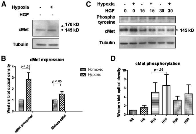
Mesenchymal stem cells (MSC) are adult multipotent cells found in bone marrow, adipose tissue, and other adult tissues. MSC have been shown to improve regeneration of injured tissues in vivo, but the mechanisms remain unclear. Typically, MSC are cultured under ambient, or normoxic, conditions (21% oxygen). However, the physiological niches for MSC in the bone marrow and other sites have much lower oxygen tension. When used as a therapeutic tool to repair tissue injuries, MSC cultured in standard conditions must adapt from 21% oxygen in culture to less than 1% oxygen in the ischemic tissue. We therefore examined the effects of preculturing human bone marrow-derived MSC in hypoxic conditions (1%-3% oxygen) to elucidate the best conditions that enhance their tissue regenerative potential. We demonstrated that MSC cultured in hypoxia activate the Akt signaling pathway while maintaining their viability and cell cycle rates. We also showed that MSC cultured in hypoxia induced expression of cMet, the major receptor for hepatocyte growth factor (HGF), and enhanced cMet signaling. MSC cultured in hypoxic conditions increased their migration rates. Since migration and HGF responsiveness are thought to be key mediators of MSC recruitment and/or activation in vivo, we next examined the tissue regenerative potential of MSC cultured under hypoxic conditions, using a murine hind limb ischemia model. We showed that local expression of HGF is increased in ischemic muscle in this model. Intra-arterial injection of MSC cultured in either normoxic or hypoxic conditions 24 hours after surgical induction of hind limb ischemia enhanced revascularization compared with saline controls. However, restoration of blood flow was observed significantly earlier in mice that had been injected with hypoxic preconditioned MSC. Collectively, these data suggest that preculturing MSC under hypoxic conditions prior to transplantation improves their tissue regenerative potential. Disclosure of potential conflicts of interest is found at the end of this article.


Conflict of interest statement

Disclosure of Potential Conflicts of Interest: The authors indicate no potential conflicts of interest.

Figures

Figure 1
Figure 1
Characterization of mesenchymal stem cells (MSC). Human MSC were cultured from bone marrow aspirates in complete medium. They were selected by adherence to plastic while hematopoietic contamination was excluded by fluorescent-conjugated antibody staining of the MSC for CD45 and CD34. The phenotypic markers CD105, CD90, and CD73 were assessed by flow cytometry. (A): Representative plots of the fluorescence-activated cell sorting analysis of unlabeled MSC. (B): Antibody-labeled samples. Assessment of the lack of CD34 and CD45 was performed with all cell cultures used for Western blot experiments, whereas three MSC cultures derived from three separate donors were analyzed for CD105, CD90, and CD73. When cultured in defined differentiation medium, MSC differentiated into the adipogenic lineage, shown here by oil red O stain (C), the osteogenic lineage, shown by alizarin red stain (D), and the chondrogenic lineage, shown by Alcian Blue stain (E). Abbreviations: APC, allophycocyanin; FL1, fluorescence channel 1, detecting FITC; FL2, fluorescence channel 2, detecting PE; FL4, fluorescence channel 4, detecting APC; PE, phycoerythrin.
Figure 2
Figure 2
Hypoxia and HGF treatment activate the Akt signaling pathway and maintain MSC viability. Bone marrow mesenchymal stem cells were incubated for 16 hours in serum-free medium exposed to hypoxic or normoxic conditions in the presence or absence of 25 ng/ml human recombinant HGF. MSC were tested for expression of Akt and its activation by Western blotting (A) and for viability by flow cytometry with Annexin V and propidium iodide staining (C). Western blot densitometry (B) showed a significant increase in Akt phosphorylation when cells were stimulated with hypoxia and HGF, whereas quantitation of flow cytometry data (D) demonstrated no significant difference between the different treatment groups, using the Student's t test. Shown are representative data of four (A) and five (C) separate experiments. Abbreviations: HGF, hepatocyte growth factor; MSC, mesenchymal stem cells.
Figure 3
Figure 3
HGF treatment stimulates Erk but not p38 or JNK signaling pathways. Mesenchymal stem cells were cultured in serum-free medium in hypoxia or normoxia in the presence or absence of 25 ng/ml HGF for 16 hours. Cell lysates were collected and separated on SDS-polyacrylamide gel electrophoresis gel and probed for Erk∼P, Erk (A), JNK∼P, JNK (B), and p38∼P, p38 (C) antibodies. Pictured are representative results of five (A) and four (B, C) separate experiments. Abbreviation: HGF, hepatocyte growth factor; JNK, Jun N-terminal Kinase.
Figure 4
Figure 4
Hypoxia and HGF treatment do not significantly change the proliferation status of mesenchymal stem cells (MSC). MSC were cultured in serum-free medium in hypoxic versus normoxic conditions in the presence or absence of 25 ng/ml HGF. Cells were collected, fixed, stained with PI, and analyzed with flow cytometry for cell cycle status. Fluorescence-activated cell sorting plots are representative results of five separate experiments (A). The bar graph shows quantitation of flow cytometry data (B). Differences were not significant, according to Student's t-test. Abbreviations: HGF, hepatocyte growth factor; PI, propidium iodide.
Figure 5
Figure 5
Hypoxia sensitizes human mesenchymal stem cells (MSC) by elevating the levels of HGF receptor cMet, thus increasing the ability of MSC to respond to HGF stimulation. MSC were cultured in serum-free medium for 16 hours in hypoxic or normoxic conditions and then collected to generate protein lysates, which were resolved on SDS-polyacrylamide gel electrophoresis (PAGE) gel and probed with cMet antibody (A). The bar graph represents Western blot densitometry results, demonstrating significant increases in the levels of mature cMet and its precursor in cells cultured in hypoxia (B). MSC were incubated in hypoxic or normoxic conditions in serum-free medium for 24 hours. Cultures were stimulated for 0, 15, or 30 minutes with 25 ng/ml HGF, and protein lysates were collected and immunoprecipitated with cMet antibody. Protein was resolved on SDS-PAGE gel and probed for anti-phosphotyrosine and cMet antibodies (C). The bar graph represents Western blot densitometry results of phosphorylated cMet (D). Shown are representative results of three (A, B) or four (C, D) separate experiments. Abbreviations: HGF, hepatocyte growth factor; kD, kilodaltons.
Figure 6
Figure 6
Hypoxia increases mesenchymal stem cell (MSC) migration and enhances functional recovery after hind limb ischemia. The growth area of MSC plated on tissue culture-treated plates was scratched with a sterile pipette tip to create a wound. The borderline of the scratch was immediately marked with a fine-tip marker, and the cultures were incubated in serum-free medium for 24 hr in hypoxia versus normoxia in the presence or absence of 25 ng/ml HGF. Photographs of cell migration were captured with bright-field microscopy at the endpoint. (A): Representative image of three separate experiments. The quantitation of migrated cells demonstrated significant fold increases in MSC motility in samples treated with HGF or combination of hypoxia and HGF, whereas cells cultured in hypoxia alone migrated the most (B). The bar graph (B) summarizes the results of three separate experiments. Hind limb ischemia and sham surgeries were performed on NOD/SCID mice (n = 4). Abductor muscles were harvested at 6, 12, 24, or 48 hr postsurgery, and muscle lysates were analyzed by enzyme-linked immunosorbent assay for the presence of HGF (C). The results show a slight increase in HGF levels in muscles of sham-operated animals starting at 12 hr postsurgery; however, the tissue ischemia caused a significant increase in the HGF levels in the injured muscle, which steadily increased up to 48 hr after surgery (two-way analysis of variance [ANOVA], p < .001). Alternatively, MSC cultured in complete medium were incubated overnight (16–24 hr) in hypoxic or normoxic conditions. Hind limb ischemia was performed on β-2-microglobulin knockout mice, which were then transplanted 1 day after surgery with hypoxic MSC, normoxic MSC, or PBS as a control. The functional recovery was measured by laser Doppler perfusion imaging immediately after the surgery and twice a week for 2 weeks after the transplantation and quantified by Moor LDI image software (D). The y-axis of the graph represents flux (measured in arbitrary units) ratio of injured to uninjured leg, and the three curves are average values for each treatment group (seven animals per group). The results demonstrate a significant improvement (ANOVA, p < .005) in mice transplanted with MSC versus saline control group, whereas the mice transplanted with hypoxic MSC improved significantly faster, as is shown at day 5 (*, p = .011; Student's t-test). Abbreviations: BM-MSC, bone marrow-derived mesenchymal stem cells; HGF, hepatocyte growth factor; hr, hours; PBS, phosphate-buffered saline.

References

    1. Prockop DJ. Marrow stromal cells as stem cells for nonhematopoietic tissues. Science. 1997;276:71–74. - PubMed
    1. Kinnaird T, Stabile E, Burnett MS, et al. Local delivery of marrow-derived stromal cells augments collateral perfusion through paracrine mechanisms. Circulation. 2004;109:1543–1549. - PubMed
    1. Al-Khaldi A, Al-Sabti H, Galipeau J, et al. Therapeutic angiogenesis using autologous bone marrow stromal cells: Improved blood flow in a chronic limb ischemia model. Ann Thorac Surg. 2003;75:204–209. - PubMed
    1. Iwase T, Nagaya N, Fujii T, et al. Comparison of angiogenic potency between mesenchymal stem cells and mononuclear cells in a rat model of hindlimb ischemia. Cardiovasc Res. 2005;66:543–551. - PubMed
    1. Moon MH, Kim SY, Kim YJ, et al. Human adipose tissue-derived mesenchymal stem cells improve postnatal neovascularization in a mouse model of hindlimb ischemia. Cell Physiol Biochem. 2006;17:279–290. - PubMed

Publication types