. 2008 Jul 9;27(13):1827-39.
doi: 10.1038/emboj.2008.108. Epub 2008 May 29.

The interaction network of the chaperonin CCT

Affiliations

The interaction network of the chaperonin CCT

Carien Dekker et al. EMBO J. .

Abstract

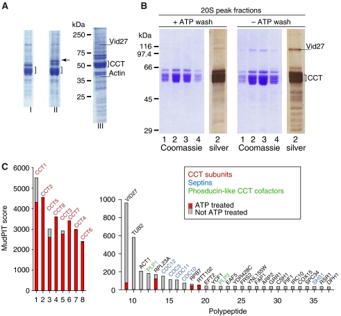
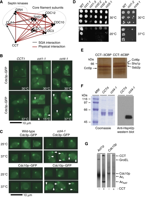
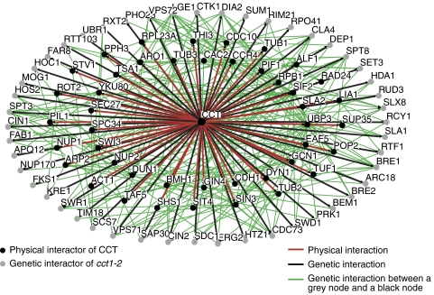
The eukaryotic cytosolic chaperonin containing TCP-1 (CCT) has an important function in maintaining cellular homoeostasis by assisting the folding of many proteins, including the cytoskeletal components actin and tubulin. Yet the nature of the proteins and cellular pathways dependent on CCT function has not been established globally. Here, we use proteomic and genomic approaches to define CCT interaction networks involving 136 proteins/genes that include links to the nuclear pore complex, chromatin remodelling, and protein degradation. Our study also identifies a third eukaryotic cytoskeletal system connected with CCT: the septin ring complex, which is essential for cytokinesis. CCT interactions with septins are ATP dependent, and disrupting the function of the chaperonin in yeast leads to loss of CCT-septin interaction and aberrant septin ring assembly. Our results therefore provide a rich framework for understanding the function of CCT in several essential cellular processes, including epigenetics and cell division.


Figures

Figure 1
Figure 1
Isolation of CCT substrate complexes. (A) Affinity purification of CCT–3CBP and CCT–6CBP on calmodulin resin. CaM-resin-purified CCT was analysed by 10% SDS–PAGE. (Lane I) CCT–3CBP complex: the eight CCT subunits migrating around the 60 kDa marker are indicated by the square bracket. (Lane II) CCT–6CBP complex: subunit Cct6p migrating at 70 kDa as a consequence of the inserted tag is indicated by an arrow. (Lane III) CCT–6CBP complex as used for in-house MALDI-MS and PMF-MS analysis. Molecular weight markers in kilodaltons are indicated on the left, Vid27p and actin are indicated as a reference (see also Supplementary Table S1). (B) Purification of CCT–3CBP in the presence and absence of ATP. CCT was incubated ±5 mM ATP at room temperature during the CaM affinity purification procedure. EDTA eluates of CCT were applied to 10–40% sucrose gradients. The panels +ATP wash and −ATP wash show four adjacent sucrose gradient fractions from each 20S CCT peak electrophoresed on 10% SDS gels followed by Coomassie (fractions 1–4) or silver staining (fraction 2 from each peak). (C) Histograms of the CCT–3CBP-bound polypeptides plotted against their MudPIT score (Tables I). Comparisons between the ATP-treated (red bars) and untreated CCT samples (gray bars) show the proteins that are removed by 5 mM ATP treatment during the CaM chromatography step.
Figure 2
Figure 2
Genetic and physical interactions of cct1-2 reveal numerous protein complexes functionally connected to the CCT chaperonin. Network diagrams incorporating either physical and genetic interactions (BD) or only genetic interactions (E, F) are shown. (A) Schematic method used to implicate CCT-binding proteins as putative substrates by integrating SGA and proteomic analyses. The presence of cct1-2 leads to misfolding of a substrate ‘X' that exhibits a known genetic interaction with gene ‘Y'. Therefore, the SGA hit connecting cct1-2 and gene ‘Y' actually reflects loss of function for a CCT substrate. Protein complexes or functional groupings are circled and labelled in each case and sorted into (B) cytoskeletal functions, (C) chromatin remodelling functions, (D) nuclear functions, and (E) other functional groups found within the SGA hits. (F) The remaining 15 SGA interactions with cct1-2 such that all 72 SGA interactions are represented in this figure. Genetic interactions are shown in black, physical interactions are shown in red and interactions within the protein complex groupings are shown in blue. SGA hits are represented by grey nodes and physical interactors are represented by black nodes.
Figure 3
Figure 3
Genomic and proteomic analyses identify a role for CCT in septin cytoskeleton function. (A) Physical and genetic interactions of yeast CCT with septin core components and septin kinases. (B) Localisation of GFP–Cdc3p in budded wild-type CCT1, cct1-1 cold-sensitive and cct4-1 heat-sensitive yeast cells. White arrows point to mis-shapen septin rings or additional patches of septin signal in cct mutant cells. CCT1 wild-type strain at 15°C which had normal septin localisation is not shown (see Table IIIA). (C) Localisation of GFP–Cdc3p and GFP–Cdc10p in wild-type and cct4-1 cells at permissive (25°C) and non-permissive temperatures (37°C). White arrows point to aberrant sites of septin localisation. (D) Allelic series of CCT cs and ts allele bearing strains to determine relative sensitivities of different alleles. CCT mutant cells were serially diluted, spotted onto rich growth media and grown at the temperatures indicated for 48 or 72 h. (E) Identification of Shs1p–Cct6p interaction. CCT–3CBP and CCT–6CBP were purified by CaM resin and sucrose gradient velocity sedimentation, after which the fraction of 18% sucrose normally containing individual CCT subunits was subjected to another CaM resin purification step. The eluates were run on an SDS gel and silver-stained and key bands were identified by PMF-MS as indicated. (F) Western blot: CaM resin-purified CCT from wild-type cells and mutant cct4-1 cells, blotted with anti-Hsp42p antibody, shows co-migration of Hsp42p with mutant CCT but not with wild-type CCT. (G) In vitro translation of Act1p and Cdc10p in E. coli-derived extract with (+) or without (−) added yeast CCT. AcI and AcNAT indicate the actin folding intermediate and native actin, respectively, according to Pappenberger et al (2006).
Figure 4
Figure 4
Integration of the CCT interactome with known interactions reveals putative substrate proteins. Network diagram showing integrated physical and genetic interactors of CCT. Those physical interactors of CCT that exhibit a genetic interaction with at least one genetic interactor of CCT (green lines) are shown. The outer ring (grey nodes) represents SGA hits and the two inner rings (black nodes) represent physical interactors of CCT (see text for description and Supplementary Table S7A).

References

    1. Adams A, Gottschling DE, Kaiser CA, Stearns T (1997) Methods in Yeast Genetics: a Cold Spring Harbor Laboratory Course Manual. Cold Spring Harbour, New York, USA: Cold Spring Harbour Laboratory Press
    1. Aloy P, Böttcher B, Ceulemans H, Leutwein C, Mellwig C, Fischer S, Gavin AC, Bork P, Superti-Furga G, Serrano L, Russell RB (2004) Structure-based assembly of protein complexes in yeast. Science 303: 2026–2029 - PubMed
    1. Amberg DC, Burke DJ, Strathern JN (2005) Methods in Yeast Genetics. Cold Spring Harbour, New York, USA: Cold Spring Harbour Laboratory Press
    1. Arava Y, Wang Y, Storey JD, Liu CL, Brown PO, Herschlag D (2003) Genome-wide analysis of mRNA translation profiles in Saccharomyces cerevisiae. Proc Natl Acad Sci USA 100: 3889–3894 - PMC - PubMed
    1. Bouhouche A, Benomar A, Bouslam N, Chkili T, Yahyaoui M (2006) Mutation in the epsilon subunit of the cytosolic chaperonin-containing t-complex peptide-1 (Cct5) gene causes autosomal recessive mutilating sensory neuropathy with spastic paraplegia. J Med Genet 43: 441–443 - PMC - PubMed

Publication types

MeSH terms