. 2008 Aug 1;509(4):382-99.
doi: 10.1002/cne.21771.

Plasticity of lumbosacral propriospinal neurons is associated with the development of autonomic dysreflexia after thoracic spinal cord transection

Affiliations

Plasticity of lumbosacral propriospinal neurons is associated with the development of autonomic dysreflexia after thoracic spinal cord transection

Shaoping Hou et al. J Comp Neurol. .

Abstract

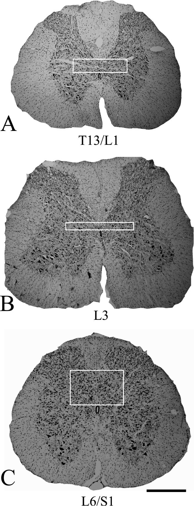
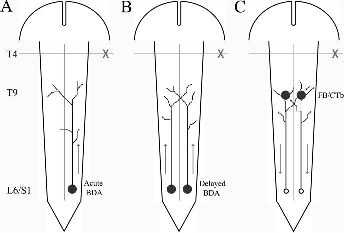
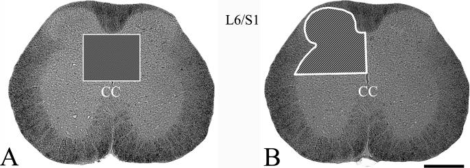
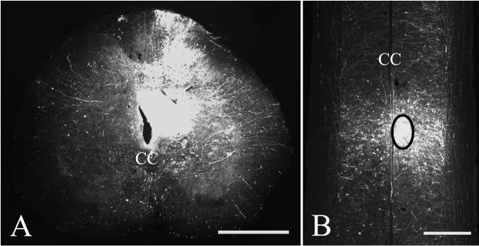
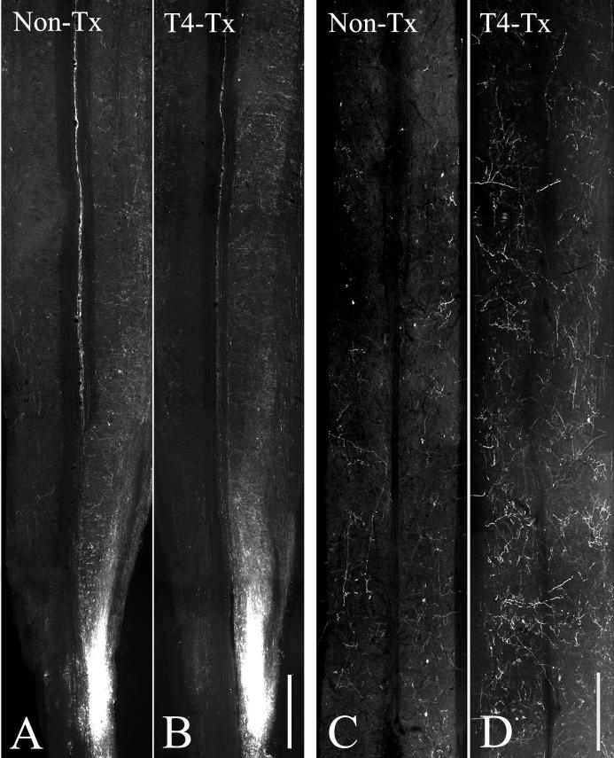
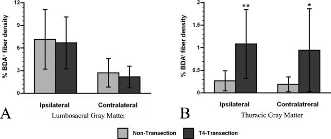
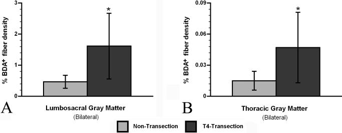
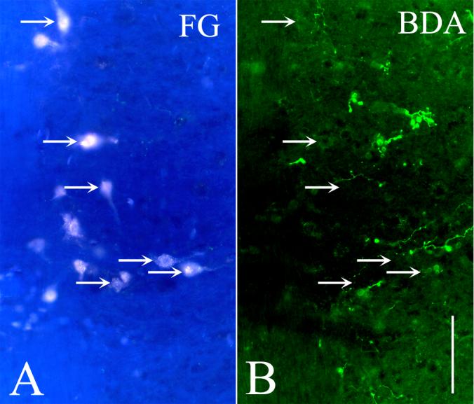
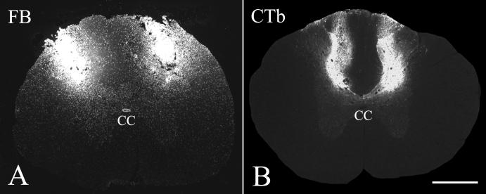
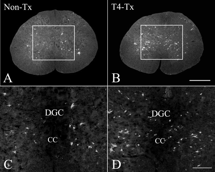
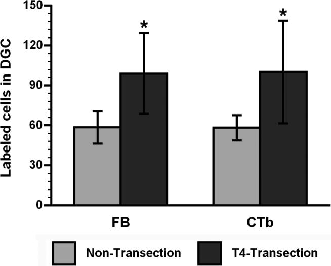
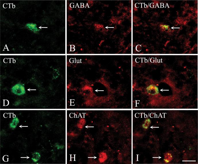
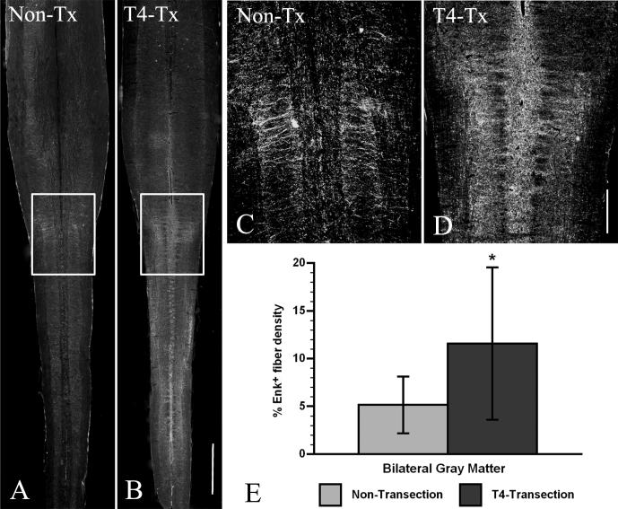
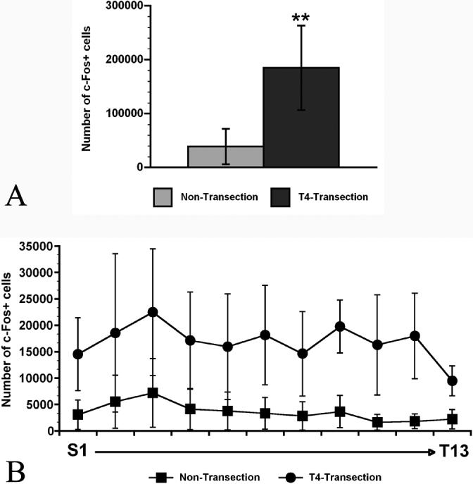
Complete thoracic (T) spinal cord injury (SCI) above the T6 level typically results in autonomic dysreflexia, an abnormal hypertensive condition commonly triggered by nociceptive stimuli below the level of SCI. Overexpression of nerve growth factor in the lumbosacral spinal cord induces profuse sprouting of nociceptive pelvic visceral afferent fibers that correlates with increased hypertension in response to noxious colorectal distension. After complete T4 SCI, we evaluated the plasticity of propriospinal neurons conveying visceral input rostrally to thoracic sympathetic preganglionic neurons. The anterograde tracer biotinylated dextran amine (BDA) was injected into the lumbosacral dorsal gray commissure (DGC) of injured/nontransected rats immediately after injury (acute) or 2 weeks later (delayed). At 1 or 2 weeks after delayed or acute injections, respectively, a higher density (P < 0.05) of BDA(+) fibers was found in thoracic dorsal gray matter of injured vs. nontransected spinal cords. For corroboration, fast blue (FB) or cholera toxin subunit beta (CTb) was injected into the T9 dorsal horns 2 weeks postinjury/nontransection. After 1 week transport, more retrogradely labeled (P < 0.05) DGC propriospinal neurons (T13-S1) were quantified in injured vs. nontransected cords. We also monitored immediate early gene c-fos expression following colorectal distension and found increased (P < 0.01) c-Fos(+) cell numbers throughout the DGC after injury. Collectively, these results imply that, in conjunction with local primary afferent fiber plasticity, injury-induced sprouting of DGC neurons may be a key constituent in relaying visceral sensory input to sympathetic preganglionic neurons that elicit autonomic dysreflexia after high thoracic SCI.


Figures

Figure 1
Figure 1
Nissl stained cross sections showing the dorsal gray commissure (DGC, boxed regions) at different spinal levels, spanning the regions that were examined (T13/L1-L6/S1). Scale bar = 500 μm.
Figure 2
Figure 2
Schematic diagrams illustrating the pathways under investigation and the tract tracer injection sites. (A) Acute (immediately after T4-transection/non-transection) unilateral biotinylated dextran amine (BDA) injection at the L6/S1 dorsal gray commissure (DGC) for anterograde labeling in ipsi-and contralateral gray matter, (B) delayed (2 weeks after T4-transection/non-transection) bilateral BDA injections for anterograde labeling in gray matter, bilaterally and (C) delayed (2 weeks after T4-transection/non-transection) fast blue (FB) or cholera toxin subunit beta (CTb) injections at T9 dorsal horns to retrogradely label lumbosacral DGC neurons. Arrows indicate direction of tracer transport.
Figure 3
Figure 3
Photomicrographs demonstrating the regions of interest (shaded insets) at L6/S1 level for stereological quantification of (A) retrogradely-labeled cells in the dorsal gray commissure (DGC) or (B) c-Fos-immunoreactive cells in the left dorsal gray matter. Scale bar = 500 μm. (CC central canal)
Figure 4
Figure 4
Representative photomicrographs demonstrate the distribution of unilateral BDA injection sites (right) in the L6/S1 dorsal gray commissure (DGC) in (A) coronal and (B) longitudinal, horizontal sections. The circumscribed area in B represents and example of the spatial extent of BDA diffusion used to calculate BDA injection volumes. Scale bars = 500 μm. (CC central canal)
Figure 5
Figure 5
BDA-labeled propriospinal pathways in (A, B) lumbosacral and (C, D) thoracic spinal cord segments 2 weeks after acute injection at L6/S1 in non-transected (Non-Tx) and T4-transected (T4-Tx) rats. Photomicrographs show apparent increased BDA+ fiber density in lumbosacral and thoracic gray matter in T4-transected compared to non-transected spinal cords. Scale bars = 1 mm (B), 100 μm (D).
Figure 6
Figure 6
Quantitative comparisons of BDA-labeled fiber density between non-transected and T4-transected spinal cords throughout the dorsal gray matter after unilateral acute injections. (A) In lumbosacral segments, BDA+ fiber density was not significantly different in either ipsi- or contralateral gray matter between injured and non-transected spinal cords. (B) On the contrary, in distal thoracic segments BDA+ fiber density in both ipsi- and contralateral gray matter was significantly greater in injured spinal cords compared to non-transected cords. Bars represent mean ± SD. * p < 0.05, ** p < 0.01
Figure 7
Figure 7
Quantitative comparisons of BDA-labeled fiber density between non-transected and T4-transected spinal cords throughout the dorsal gray matter after bilateral delayed injections. In both (A) lumbosacral and (B) distal thoracic segments, BDA+ fiber density was significantly greater in gray matter of injured spinal cords, bilaterally, compared to non-transected cords. Bars represent mean ± SD. * p < 0.05.
Figure 8
Figure 8
Peripheral i.p. injection of FluoroGold (FG) at 1 week post-BDA injection (acute) shows juxtaposition of (A) FG-labeled sympathetic preganglionic neurons (arrows) in the intermediolateral cell column of thoracolumbar segments with (B) BDA+ fiber projections. Scale bar = 100 μm.
Figure 9
Figure 9
Representative photomicrographs demonstrating retrograde tracer injection sites made 2 weeks after T4 transection/non-transection (delayed). (A) FB or (B) CTb were injected into dorsal horn gray matter at the T9 level, bilaterally, and allowed to transport for 1 week to retrogradely label lumbosacral dorsal gray commissure (DGC) neurons. Scale bar = 500 μm. (CC central canal)
Figure 10
Figure 10
Photomicrographs of L6/S1 cross sections showing FB retrogradely-labeled propriospinal neurons in the dorsal gray commissure (DGC). (A) Compared to non-transected cords (Non-Tx), more DGC neurons appeared labeled in (B) T4-transected (T4-Tx) spinal cords 1 week after FB injections at T9, bilaterally. C and D are higher magnifications of boxed regions in A and B, respectively. Scale bars = 500 μm (B); 200 μm (D). (DGC dorsal gray commissure, CC central canal)
Figure 11
Figure 11
Quantification of FB or CTb retrogradely-labeled dorsal gray commissure (DGC) neurons (T13-S1 segments) between non-transected and T4-transected spinal cords (see Figs. 1 and 3A for regions of interest). For each tracer, retrogradely-labeled DGC neurons were significantly greater in injured spinal cords compared to non-transected cords. Bars represent mean ± SD. * p < 0.05
Figure 12
Figure 12
Phenotypic characterization of CTb retrogradely-labeled dorsal gray commissure (DGC) neurons at L6/S1 level. Confocal photomicrographs show dual immunohistochemical staining (arrows) for CTb in conjunction with (A-C) γ-aminobutyric acid (GABA), (D-F) glutamate (Glut), or (G-I) choline acetyltransferase (ChAT) in coronal sections. Scale bar = 10 μm. Note: Magenta-green copy of images available as supplementary figure.
Figure 13
Figure 13
Photomicrographs demonstrate enkephalin (Enk)+ fiber plasticity 2 weeks after T4 transection in longitudinal, horizontal sections. Compared to non-transected (Non-Tx) spinal cords (A), immunohistochemical staining for Enk+ fibers showed greater density in T4-transected (T4-Tx) spinal cords (B) in the L6/S1 segments. C and D represent higher magnifications of boxed regions in A and B, respectively. (E) Densitometric analysis of Enk+ fiber density in the L6/S1 gray matter, bilaterally, showed significantly more immunoreactivity in T4-transected spinal cords compared to non-transected cords. Scale bars = 1 mm (B); 500 μm (D). Bars represent mean ± SD. * p < 0.05
Figure 14
Figure 14
Representative photomicrographs demonstrating c-Fos+ neurons in the T13/L1 (A, B) and L6/S1 (C, D) dorsal gray matter from both non-transected (Non-Tx) and T4-transected (T4-Tx) spinal cords following 2 hours of intermittent colorectal distension prior to perfusion and fixation. Compared to Non-Tx spinal cords, c-Fos+ cells were conspicuously increased throughout the dorsal horns and dorsal gray commissure 2 weeks after injury. Scale bar = 200 μm. (DGC dorsal gray commissure, CC central canal)
Figure 15
Figure 15
(A) Stereological quantification, in unilateral dorsal horns and dorsal gray commissure (DGC) from T13-S1 (see Fig. 3B for region of interest), showed that the total number of c-Fos+ cells was significantly increased in T4-transected spinal cords compared to non-transected cords. (B) Stereological comparisons across lumbosacral segments revealed that the number of c-Fos+ cells in T4-transected cords was significantly greater than non-transected cords at each level. Bars and symbols represent means ± SD. **p<0.01

References

    1. Abercrombie M, Johnson ML. Quantitative histology of Wallerian degeneration: I. Nuclear population in rabbit sciatic nerve. J Anat. 1946;80:37–50. - PubMed
    1. Al-Chaer ED, Lawand NB, Westlund KN, Willis WD. Pelvic visceral input into the nucleus gracilis is largely mediated by the postsynaptic dorsal column pathway. J Neurophysiol. 1996;76:2675–2690. - PubMed
    1. Anderson CR, Edwards SL. Intraperitoneal injections of Fluorogold reliably labels all sympathetic preganglionic neurons in the rat. J Neurosci Methods. 1994;53:137–141. - PubMed
    1. Bacon SJ, Smith AD. Preganglionic sympathetic neurones innervating the rat adrenal medulla: immunocytochemical evidence of synaptic input from nerve terminals containing substance P, GABA or 5-hydroxytryptamine. J Auton Nerv Syst. 1988;24:97–122. - PubMed
    1. Brown A, Ricci MJ, Weaver LC. NGF message and protein distribution in the injured rat spinal cord. Exp Neurol. 2004;188:115–127. - PubMed

Publication types

MeSH terms