

A comparison of five fMRI protocols for mapping speech comprehension systems
- PMID: 18513352
- PMCID: PMC2645716
- DOI: 10.1111/j.1528-1167.2008.01683.x
A comparison of five fMRI protocols for mapping speech comprehension systems
Abstract
Aims: Many fMRI protocols for localizing speech comprehension have been described, but there has been little quantitative comparison of these methods. We compared five such protocols in terms of areas activated, extent of activation, and lateralization.
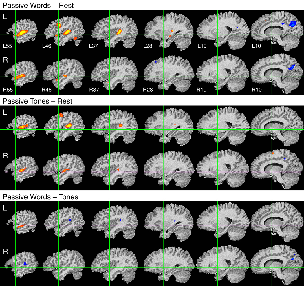
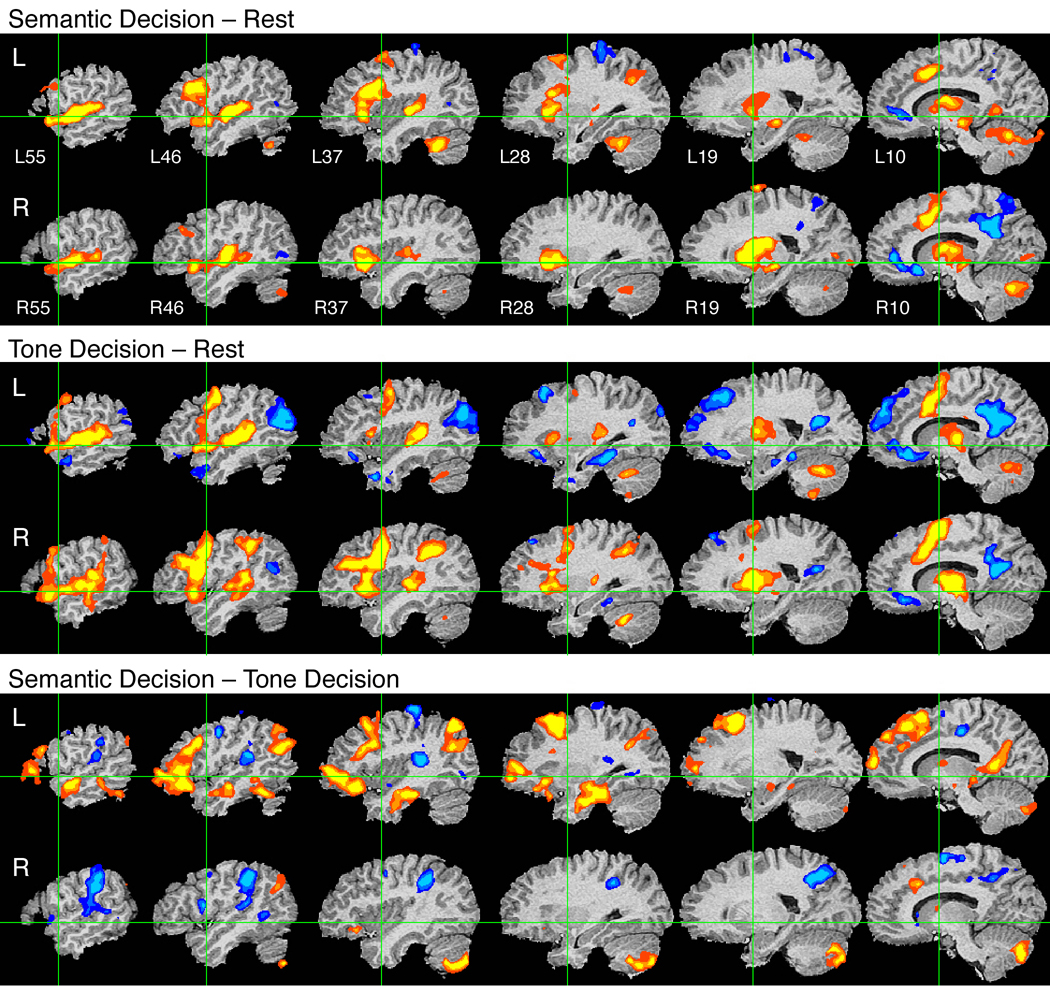
Methods: fMRI BOLD signals were measured in 26 healthy adults during passive listening and active tasks using words and tones. Contrasts were designed to identify speech perception and semantic processing systems. Activation extent and lateralization were quantified by counting activated voxels in each hemisphere for each participant.
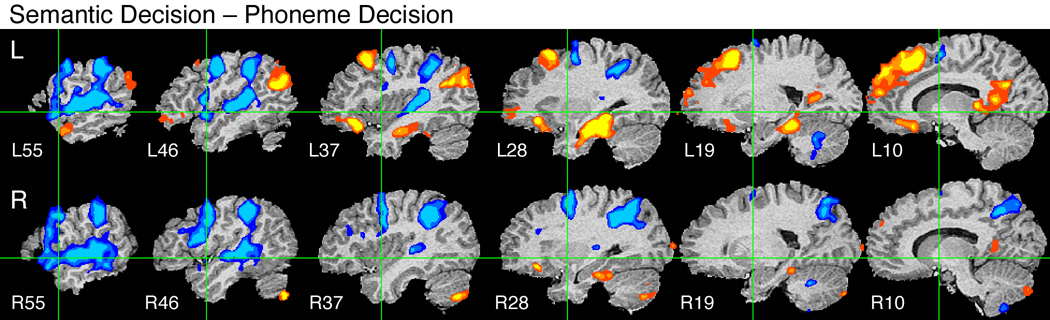
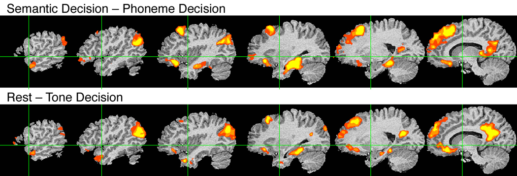
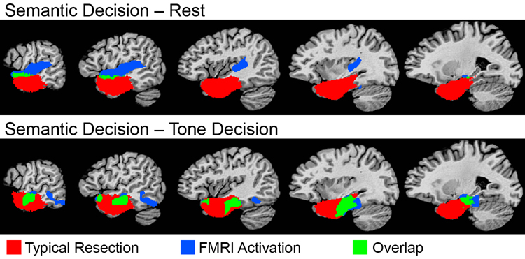
Results: Passive listening to words produced bilateral superior temporal activation. After controlling for prelinguistic auditory processing, only a small area in the left superior temporal sulcus responded selectively to speech. Active tasks engaged an extensive, bilateral attention, and executive processing network. Optimal results (consistent activation and strongly lateralized pattern) were obtained by contrasting an active semantic decision task with a tone decision task. There was striking similarity between the network of brain regions activated by the semantic task and the network of brain regions that showed task-induced deactivation, suggesting that semantic processing occurs during the resting state.
Conclusions: fMRI protocols for mapping speech comprehension systems differ dramatically in pattern, extent, and lateralization of activation. Brain regions involved in semantic processing were identified only when an active, nonlinguistic task was used as a baseline, supporting the notion that semantic processing occurs whenever attentional resources are not controlled. Identification of these lexical-semantic regions is particularly important for predicting language outcome in patients undergoing temporal lobe surgery.
Figures
References
-
- Adcock JE, Wise RG, Oxbury JM, Oxbury SM, Matthews PM. Quantitative fMRI assessment of the differences in lateralization of language-related brain activation in patients with temporal lobe epilepsy. Neuroimage. 2003;18:423–438. - PubMed
-
- Adler CM, Sax KW, Holland SK, Schmithorst V, Rosenberg L, Strakowski SM. Changes in neuronal activation with increasing attention demand in healthy volunteers: An fMRI study. Synapse. 2001;42:266–272. - PubMed
-
- Ahmad Z, Balsamo LM, Sachs BC, Xu B, Gaillard WD. Auditory comprehsnion of language in young children: Neural networks identified with fMRI. Neurology. 2003;60:1598–1605. - PubMed
-
- Alexander MP, Hiltbrunner B, Fischer RS. Distributed anatomy of transcortical sensory aphasia. Arch. Neurol. 1989;46:885–892. - PubMed
-
- Antrobus JS, Singer JL, Greenberg S. Studies in the stream of consciousness: Experimental enhancement and suppression of spontaneous cognitive processes. Perceptual and Motor Skills. 1966;23:399–417.
Publication types
MeSH terms
Substances
Grants and funding
LinkOut - more resources
Full Text Sources
