Comparative Study
. 2008;10(3):R63.
doi: 10.1186/ar2434. Epub 2008 May 30.

Cartilage degradation is fully reversible in the presence of aggrecanase but not matrix metalloproteinase activity

Affiliations
Comparative Study

Cartilage degradation is fully reversible in the presence of aggrecanase but not matrix metalloproteinase activity

Morten A Karsdal et al. Arthritis Res Ther. 2008.

Abstract

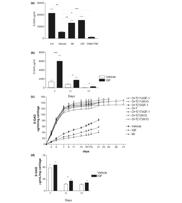
Introduction: Physiological and pathophysiological cartilage turnover may coexist in articular cartilage. The distinct enzymatic processes leading to irreversible cartilage damage, compared with those needed for continuous self-repair and regeneration, remain to be identified. We investigated the capacity of repair of chondrocytes by analyzing their ability to initiate an anabolic response subsequent to three different levels of catabolic stimulation.

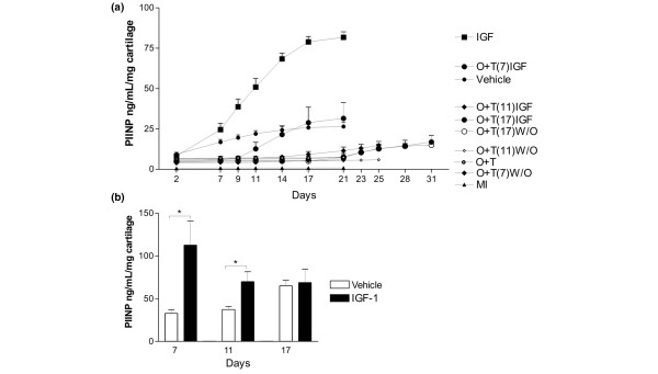
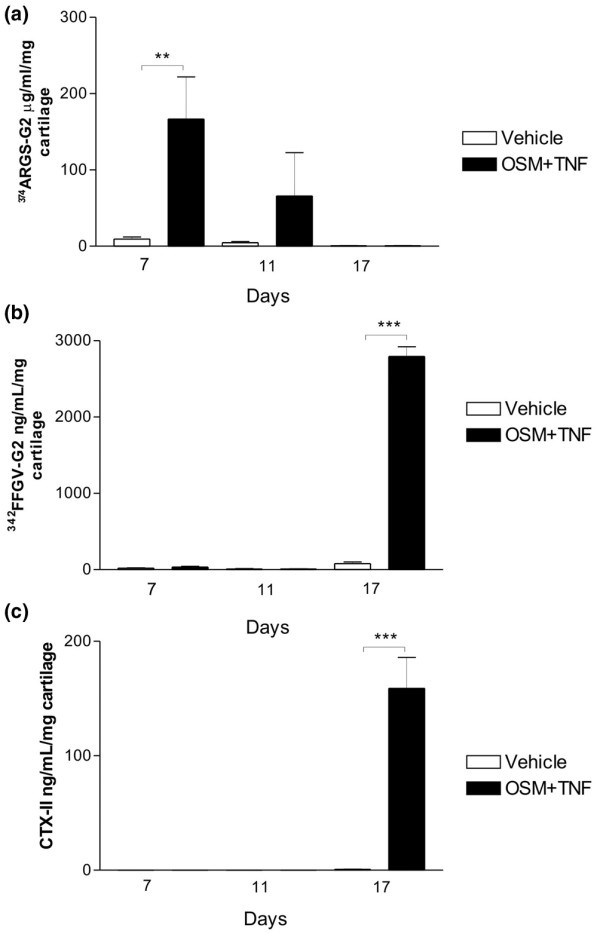
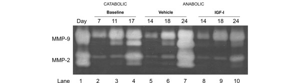
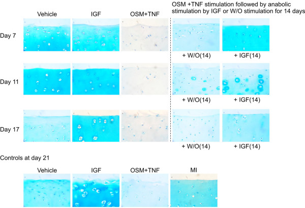
Methods: Cartilage degradation was induced by oncostatin M and tumour necrosis factor in articular cartilage explants for 7, 11, or 17 days. The catabolic period was followed by 2 weeks of anabolic stimulation (insulin growth factor-I). Cartilage formation was assessed by collagen type II formation (PIINP). Cartilage degradation was measured by matrix metalloproteinase (MMP) mediated type II collagen degradation (CTX-II), and MMP and aggrecanase mediated aggrecan degradation by detecting the 342FFGVG and 374ARGSV neoepitopes. Proteoglycan turnover, content, and localization were assessed by Alcian blue.

Results: Catabolic stimulation resulted in increased levels of cartilage degradation, with maximal levels of 374ARGSV (20-fold induction), CTX-II (150-fold induction), and 342FFGVG (30-fold induction) (P < 0.01). Highly distinct protease activities were found with aggrecanase-mediated aggrecan degradation at early stages, whereas MMP-mediated aggrecan and collagen degradation occurred during later stages. Anabolic treatment increased proteoglycan content at all time points (maximally, 250%; P < 0.001). By histology, we found a complete replenishment of glycosaminoglycan at early time points and pericellular localization at an intermediate time point. In contrast, only significantly increased collagen type II formation (200%; P < 0.01) was observed at early time points.

Conclusion: Cartilage degradation was completely reversible in the presence of high levels of aggrecanase-mediated aggrecan degradation. After induction of MMP-mediated aggrecan and collagen type II degradation, the chondrocytes had impaired repair capacity.


Figures

Figure 1
Figure 1
Quantification of aggrecan within the articular cartilage explants. The proteins of the cultured explants were extracted by liquid N2 pulverization. (a) Cartilage was extracted immediately after isolation (t = 0) or after culture for 21 days with vehicle, insulin growth factor (IGF), oncostatin M plus tumour necrosis factor (OSM + TNF), or metabolically inactive (MI) control for assessing passive physiochemical release. (b) Cartilage was extracted after the three different levels of cytokine treatment followed by an identical 14 days with either vehicle or IGF. IGF significantly stimulated proteoglycan content within the cartilage explants at all time points. (c) Quantification of sulphated glycosaminoglycan (S-GAG) from all treatments over the entire experimental period. S-GAG released from cartilage explants to the conditioned medium was quantified by the Alcian blue-binding assay. The curves represent the release at days when the conditioned medium was fully replaced, and the values were accumulated over the entire period. MI, metabolically inactive; O + T, oncostatin M plus tumour necrosis factor; W/O, without stimulation (vehicle control). (d) Quantification of S-GAG turnover 2 weeks after the catabolic induction. The aggrecan release in the identical 14-day period, with or without IGF stimulation following three different periods of catabolic stimulation, was measured by the Alcian blue-binding assay. The results show the accumulated release of S-GAG during the 2 weeks with anabolic stimulation (IGF) and without stimulation (vehicle). *P < 0.05, **P < 0.01, ***P < 0.001.
Figure 2
Figure 2
Quantification of pro-peptides of collagen type II. (a) Quantification of collagen type II synthesis from all treatments over the entire experimental period. Collagen type II synthesis in cartilage explants was measured by the concentration of N-terminal pro-peptides of type II collagen in the conditioned medium using the PIINP enzyme-linked immunosorbent assay (ELISA). The curves represent the release found at the specific day, where the conditioned medium was fully replaced, and the values were accumulated over the entire period. Vehicle control, metabolically inactive (MI), O + T, oncostatin M plus tumour necrosis factor. (b) Quantification of collagen type II formation 2 weeks after the catabolic induction. The collagen type II synthesis in the identical 14-day periods with or without IGF stimulation following the three different periods of catabolic stimulation was measured by the PIINP ELISA. The conditioned medium was fully replaced three times a week. The results show the accumulated release of collagen type II pro-peptide during the two weeks with anabolic stimulation (insulin growth factor, IGF) and without stimulation (vehicle). IGF-I significantly induced collagen type II formation at low and intermediate catabolic insult, but not at maximal insult. *P < 0.05. PIINP, N-terminal pro-peptide of pro-collagen type II.
Figure 3
Figure 3
Quantification of aggrecan and collagen degradation products at days 7, 11, and 17. Articular cartilage explants were cultured in the presence or absence of oncostatin M plus tumour necrosis factor (OSM + TNF). Conditioned medium was collected at days 7, 11, and 17. (a) Aggrecanase-mediated aggrecan degradation was measured by the 374ARGSV-G2 enzyme-linked immunosorbent assay (ELISA), (b) matrix metalloproteinase (MMP)-mediated aggrecan degradation was quantified by the 342FFGVG-G2 ELISA, and (c) MMP-mediated collagen type II degradation was quantified in the CTX-II ELISA. **P < 0.01, ***P < 0.001. CTX-II, crosslinked C-terminal neo-epitopes of type II collagen.
Figure 4
Figure 4
Gelatinase activity is attenuated but not abrogated during insulin growth factor (IGF) stimulation. Gelatinase activity in conditioned medium from bovine articular cartilage explants was investigated by zymography. Lane 1 shows standards for matrix metalloproteinase (MMP)-9 + MMP-2. Conditioned medium at the end of each catabolic culture period (7, 11, or 17 days) was used as a reference (lanes 2 to 4). Conditioned medium from cultures treated with IGF (lanes 8 to 10) or vehicle (lanes 5 to 7) 7 days after the catabolic period was analyzed. Compared with vehicle and baseline measurements, IGF only attenuated MMP production and activation.
Figure 5
Figure 5
Insulin growth factor (IGF) stimulates local replenishment of cartilage. Articular cartilage explants were cultured with either oncostatin M plus tumour necrosis factor (OSM + TNF) or vehicle for 7, 11, and 17 days. Subsequently, cartilage explants were paraffin-embedded and stained for aggrecan content as described in Materials and methods. Aggrecan is completely depleted from the tissue at 7, 11, and 17 days. Other cultures were treated with either OSM + TNF or vehicle for 7, 11, and 17 days followed by stimulation with either IGF or vehicle control for 14 days. Subsequently, cartilage explants were paraffin-embedded and stained for aggrecan content as described in Materials and methods. As a control experiment, articular cartilage explants were cultured for 21 days with vehicle, OSM + TNF, IGF, or metabolically inactive (MI) control for 21 days as controls (lower panel). W/O, without stimulation.

References

    1. Abramson SB, Attur M, Yazici Y. Prospects for disease modification in osteoarthritis. Nat Clin Pract Rheumatol. 2006;2:304–312. doi: 10.1038/ncprheum0193. - DOI - PubMed
    1. van Meurs JB, van Lent PL, Holthuysen AE, Singer II, Bayne EK, Berg WB Van den. Kinetics of aggrecanase- and metalloproteinase-induced neoepitopes in various stages of cartilage destruction in murine arthritis. Arthritis Rheum. 1999;42:1128–1139. doi: 10.1002/1529-0131(199906)42:6<1128::AID-ANR9>3.0.CO;2-2. - DOI - PubMed
    1. Karsdal MA, Henriksen K, Sørensen MG, Gram J, Schaller S, Dziegiel MH, Heegaard AM, Christophersen P, Martin TJ, Christiansen C, Bollerslev J. Acidification of the osteoclastic resorption compartment provides insight into the coupling of bone formation to bone resorption. Am J Pathol. 2005;166:467–476. - PMC - PubMed
    1. Behrens F, Kraft EL, Oegema TR., Jr Biochemical changes in articular cartilage after joint immobilization by casting or external fixation. J Orthop Res. 1989;7:335–343. doi: 10.1002/jor.1100070305. - DOI - PubMed
    1. Kiani C, Chen L, Wu YJ, Yee AJ, Yang BB. Structure and function of aggrecan. Cell Res. 2002;12:19–32. doi: 10.1038/sj.cr.7290106. - DOI - PubMed

Publication types