. 2008 May 30:3:24.
doi: 10.1186/1746-1596-3-24.

Intravascular lesions of the hand

Affiliations

Intravascular lesions of the hand

Liron Pantanowitz et al. Diagn Pathol. .

Abstract

Introduction: Intravascular lesions of the hand comprise reactive and neoplastic entities. The clinical diagnosis of such lesions is often difficult, and usually requires pathologic examination. We present the largest series to date of intravascular lesions affecting the hand.

Methods: A retrospective review of intravascular (arterial and venous) lesions involving the hand was conducted. Data regarding clinicopathologic findings were analyzed.

Results: We identified 10 patients with intravascular lesions of their hands including thromboemboli (n = 3), reactive intravascular conditions such as papillary endothelial hyperplasia or Masson's tumor (n = 2) and fasciitis (n = 1), as well as vascular neoplasms including pyogenic granuloma (n = 2) and angioleiomyoma (n = 2).

Conclusion: Blood vessel injury and/or venous thrombosis may predispose to several intravascular lesions of the hand. Recognition of reactive entities from neoplastic conditions is important.


Figures

Figure 1
Figure 1
Dry gangrene of an amputated hand due to infarction from septic emboli (case 9).
Figure 2
Figure 2
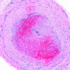
Organizing blood vessel thrombus from case 1 (H&E stain).
Figure 3
Figure 3
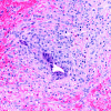
Organizing thrombus with central foreign material and an associated foreign body giant cell reaction seen at higher magnification (H&E stain).
Figure 4
Figure 4
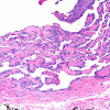
Intravascular papillary endothelial hyperplasia (H&E stain).
Figure 5
Figure 5
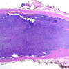
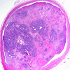
Intravascular fasciitis shown at low power magnification (H&E stain).
Figure 6
Figure 6
Intravascular fasciitis shown at high power magnification (H&E stain).
Figure 7
Figure 7
Intravenous pyogenic granuloma (H&E stain).
Figure 8
Figure 8
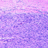
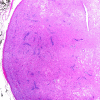
Angioleiomyoma shown at low power magnification (H&E stain).
Figure 9
Figure 9
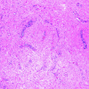
Angioleiomyoma shown at high power magnification (H&E stain).

Similar articles

Cited by

References

    1. Fleming AN, Smith PJ. Vascular cell tumors of the hand in children. Hand Clin. 2000;16:609–24. - PubMed
    1. Julliard FA, Baviera EE. Benign lesions of the hand simulating angiosarcoma: Masson's vegetating intravascular hemangioendothelioma (Article in French) Ann Chir Main. 1983;2:59–61. doi: 10.1016/S0753-9053(83)80082-9. - DOI - PubMed
    1. Anand A, Tsapakis EM, Narvani AA, Alhakim A, Cannon SR, Tsiridis E. "Pseudosarcoma" in a pregnant woman. World J Surg Oncol. 2007;18:5–7. - PMC - PubMed
    1. Kocer U, Aksoy HM, Tiftikcioglu YO, Karaaslan O. Intravenous pyogenic granuloma of the hand. Dermatol Surg. 2003;29:974–6. doi: 10.1046/j.1524-4725.2003.29263.x. - DOI - PubMed
    1. Hung CH, Kuo HW, Chiu YK, Huang PH. Intravascular pyogenic granuloma arising in an acquired arteriovenous malformation: report of a case and review of the literature. Dermatol Surg. 2004;30:1050–3. doi: 10.1111/j.1524-4725.2004.30316.x. - DOI - PubMed

LinkOut - more resources