. 2008 May 30:9:259.
doi: 10.1186/1471-2164-9-259.

Smoking-induced gene expression changes in the bronchial airway are reflected in nasal and buccal epithelium

Affiliations

Smoking-induced gene expression changes in the bronchial airway are reflected in nasal and buccal epithelium

Sriram Sridhar et al. BMC Genomics. .

Abstract

Background: Cigarette smoking is a leading cause of preventable death and a significant cause of lung cancer and chronic obstructive pulmonary disease. Prior studies have demonstrated that smoking creates a field of molecular injury throughout the airway epithelium exposed to cigarette smoke. We have previously characterized gene expression in the bronchial epithelium of never smokers and identified the gene expression changes that occur in the mainstem bronchus in response to smoking. In this study, we explored relationships in whole-genome gene expression between extrathorcic (buccal and nasal) and intrathoracic (bronchial) epithelium in healthy current and never smokers.

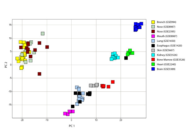
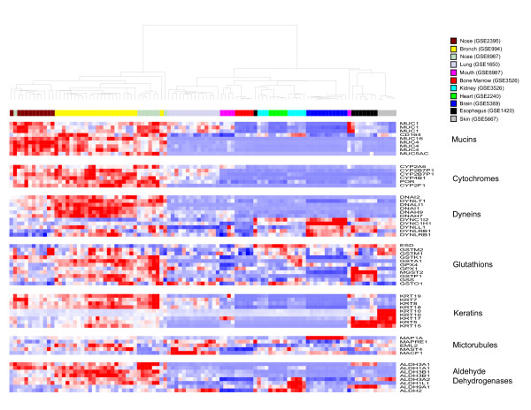
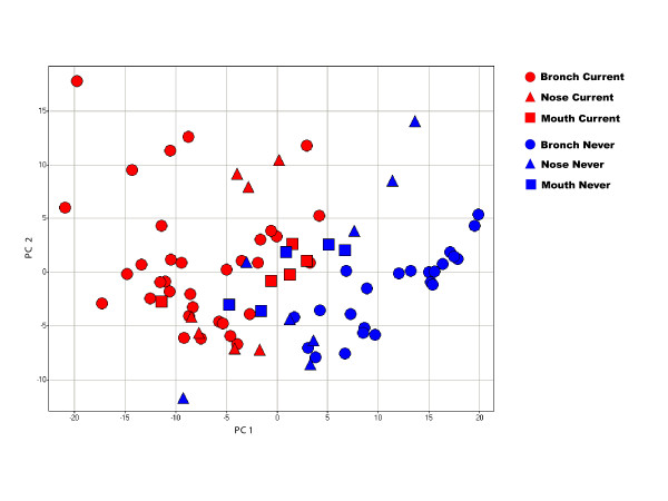
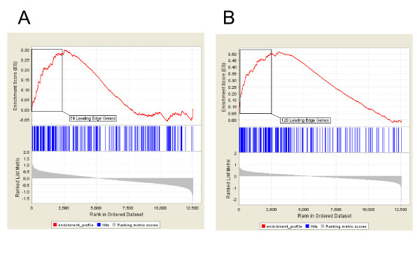
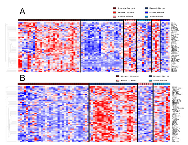
Results: Using genes that have been previously defined as being expressed in the bronchial airway of never smokers (the "normal airway transcriptome"), we found that bronchial and nasal epithelium from non-smokers were most similar in gene expression when compared to other epithelial and nonepithelial tissues, with several antioxidant, detoxification, and structural genes being highly expressed in both the bronchus and nose. Principle component analysis of previously defined smoking-induced genes from the bronchus suggested that smoking had a similar effect on gene expression in nasal epithelium. Gene set enrichment analysis demonstrated that this set of genes was also highly enriched among the genes most altered by smoking in both nasal and buccal epithelial samples. The expression of several detoxification genes was commonly altered by smoking in all three respiratory epithelial tissues, suggesting a common airway-wide response to tobacco exposure.

Conclusion: Our findings support a relationship between gene expression in extra- and intrathoracic airway epithelial cells and extend the concept of a smoking-induced field of injury to epithelial cells that line the mouth and nose. This relationship could potentially be utilized to develop a non-invasive biomarker for tobacco exposure as well as a non-invasive screening or diagnostic tool providing information about individual susceptibility to smoking-induced lung diseases.


Figures

Figure 1
Figure 1
Principal component analysis of 2382 genes from the normal bronchial airway transcriptome across 11 tissue datasets. Principal component analysis (PCA) of the expression of 2382 genes from the never-smoker bronchial-airway transcriptome [8] in samples from 11 different tissue datasets. Bronchial and nasal epithelial samples group together along the first and second principal components (representing 66% and 12% of the variance in gene expression respectively) based on the expression of these genes. Tissue types are color-coded.
Figure 2
Figure 2
Hierarchical clustering of 59 genes representing functional categories over-represented in the normal bronchial airway transcriptome. Supervised hierarchical clustering of the expression of 59 probesets representing genes in the mucin, dynein/microtubule, cytochrome P450, glutathione, aldehyde dehydrogenase, and keratin functional categories across samples from 11 tissue datasets. Nasal and bronchial epithelial samples cluster together based on similar expression patterns across these groups of genes (high expression = red, average expression = white, low expression = blue).
Figure 3
Figure 3
Principal component analysis of 361 differentially expressed bronchial epithelial genes across bronchial, nasal, and buccal mucosa samples. PCA of the expression of genes that are perturbed by smoking in the bronchial airway in samples of bronchial, nasal, and buccal epithelium from smokers and non-smokers. Bronchial and nasal epithelial samples separate according to smoking status, while the separation of buccal mucosa samples by smoking status is less pronounced. The first two principal components account for 27% and 9% of the total variance in gene expression respectively.
Figure 4
Figure 4
Enrichment of differentially expressed bronchial epithelial genes among smoking-induced genes in the buccal mucosa or nasal epithelium. Results from gene set enrichment analysis (GSEA) shows that 361 genes induced in bronchial epithelial cells from smokers are significantly skewed toward being among the genes most induced in buccal (A) or nasal (B) epithelial samples in response to smoking (p < 0.001). There are 74 genes that comprise the buccal "leading edge subset" and 120 genes that comprise the nasal "leading edge subset". These two analyses suggest that genes induced by smoking in the bronchus show a similar pattern of differential expression in buccal and nasal epithelium.
Figure 5
Figure 5
Hierarchical clustering of genes commonly perturbed by smoking across intra- and extrathoracic airway epithelium. A. Supervised hierarchical clustering of the expression of 45 genes induced by smoking in the bronchial airway that are present in both the nasal and buccal "leading edge subsets" in samples from smokers and non-smokers. These represent genes up-regulated by smoking in bronchial, nasal, and buccal epithelium. B. Supervised hierarchical clustering of the expression of 50 genes repressed by smoking in the bronchial airway that are present in the nasal "leading edge subset" in samples from smokers and nonsmokers. (High expression = red, average expression = white, low expression = blue).

References

    1. The Facts About Smoking and Health. WHO; 2006.
    1. Shields PG. Molecular epidemiology of lung cancer. Ann Oncol. 1999;10 Suppl 5:S7–11. doi: 10.1023/A:1008331616378. - DOI - PubMed
    1. Wistuba. Lam S, Behrens C, Virmani AK, Fong KM, LeRiche J, Samet JM, Srivastava S, Minna JD, Gazdar AF. Molecular damage in the bronchial epithelium of current and former smokers. J Natl Cancer Inst. 1997;89:1366–1373. doi: 10.1093/jnci/89.18.1366. - DOI - PMC - PubMed
    1. Powell CA, Klares S, O'Connor G, Brody JS. Loss of heterozygosity in epithelial cells obtained by bronchial brushing: clinical utility in lung cancer. Clin Cancer Res. 1999;5:2025–2034. - PubMed
    1. Franklin WA, Gazdar AF, Haney J, Wistuba. La Rosa FG, Kennedy T, Ritchey DM, Miller YE. Widely dispersed p53 mutation in respiratory epithelium. A novel mechanism for field carcinogenesis. J Clin Invest. 1997;100:2133–2137. doi: 10.1172/JCI119748. - DOI - PMC - PubMed

Publication types

MeSH terms