Case Reports
. 2008 Jun;82(6):1368-74.
doi: 10.1016/j.ajhg.2008.05.006.

Congenital joint dislocations caused by carbohydrate sulfotransferase 3 deficiency in recessive Larsen syndrome and humero-spinal dysostosis

Affiliations
Case Reports

Congenital joint dislocations caused by carbohydrate sulfotransferase 3 deficiency in recessive Larsen syndrome and humero-spinal dysostosis

Pia Hermanns et al. Am J Hum Genet. 2008 Jun.

Erratum in

  • Am J Hum Genet. 2008 Aug;83(2):293

Abstract

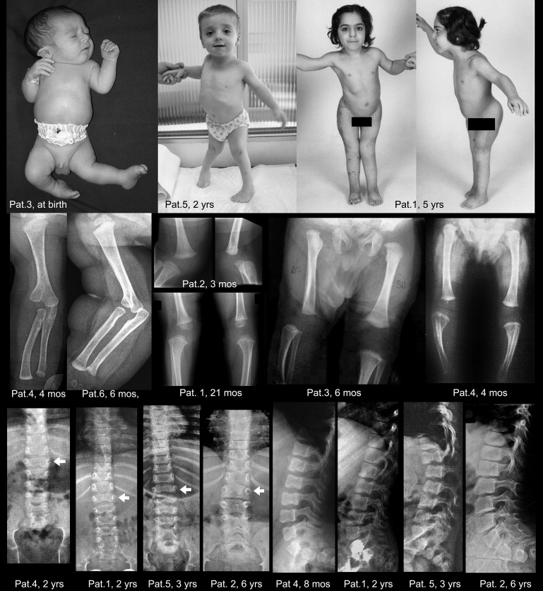
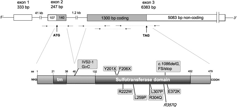
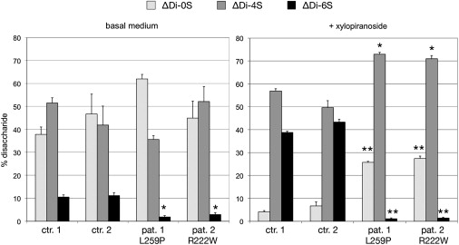
Deficiency of carbohydrate sulfotransferase 3 (CHST3; also known as chondroitin-6-sulfotransferase) has been reported in a single kindred so far and in association with a phenotype of severe chondrodysplasia with progressive spinal involvement. We report eight CHST3 mutations in six unrelated individuals who presented at birth with congenital joint dislocations. These patients had been given a diagnosis of either Larsen syndrome (three individuals) or humero-spinal dysostosis (three individuals), and their clinical features included congenital dislocation of the knees, elbow joint dysplasia with subluxation and limited extension, hip dysplasia or dislocation, clubfoot, short stature, and kyphoscoliosis developing in late childhood. Analysis of chondroitin sulfate proteoglycans in dermal fibroblasts showed markedly decreased 6-O-sulfation but enhanced 4-O-sulfation, confirming functional impairment of CHST3 and distinguishing them from diastrophic dysplasia sulphate transporter (DTDST)-deficient cells. These observations provide a molecular basis for recessive Larsen syndrome and indicate that recessive Larsen syndrome, humero-spinal dysostosis, and spondyloepiphyseal dysplasia Omani type form a phenotypic spectrum.


Figures

Figure 1
Figure 1
Synopsis of Clinical Aspect and Main Radiographic Features The first row shows congenital dislocation of the left knee and clubfoot in patient 3, diagnosed with Larsen syndrome, at a few days of age (the appearance is identical to that of the HSD patient reported by Hall11). At birth, the left leg was extended in such a way that the foot rested against the thoracic wall. The central photograph shows marked valgus deformity of the knees and bilateral clubfoot in patient 5 at age 2 yr. The right panels show patient 1, a girl, at age 5 yr, after several surgical procedures had been carried out to stabilize the dislocated knees and the dysplastic hips. Note also the inability to extend the elbows completely. The second row shows the bifid humerus and radial dislocation, as well as the knee dislocation or subluxation with delayed development of the knee epiphyses (patient identification and ages are within the panels). The third row shows the diagnostically useful spinal changes: In the AP projection, there is marked widening of the interpedicular space from T12 to L1 (the arrows mark the vertebral body of L1); the interpedicular space then decreases progressively from L3 to L5. In the lateral projection, notching or scalloping of the upper and lower endplates of the lumbar vertebral bodies can be seen; these changes have been emphasized in the original descriptions of humero-spinal dysostosis. The changes are more pronounced in the younger patients.
Figure 2
Figure 2
Scheme of the CHST3 Gene and Protein The upper part is a schematic representation of the CHST3 exon-intron structure and of the coding regions. Immediately below, the small horizontal arrows indicate the position of primer couples used to amplify genomic DNA. The lower part is a schematic representation of the CHST3 protein with its transmembrane domain and the intraluminal sulfotransferase domain as inferred from data on the Expasy site. Above the protein are mutations leading to premature stop codons as well as the putative splicing mutation (see Table 1 and Supplemental Material and Methods, available online); below the proteins are amino acid substitutions, including the R304Q substitution reported by Thiele et al., and the known polymorphism R357Q.
Figure 3
Figure 3
Disaccharide Composition of Chondroitin Sulfate Synthesized by Fibroblast Cultures Fibroblasts from two controls and from patients 1 and 2 were labeled simultaneously with the radiolabeled glycosaminoglycan sugar precursor, [6-3H]glucosamine, and the chondroitin sulfate synthesized was harvested, purified, and cleaved into its constituent disaccharides, ΔDi-0S, ΔDi-4S and ΔDi-6S. These were separated by high-performance liquid chromatography (HPLC) and measured (see Supplemental Material and Methods). In patients' cells, the proportion of ΔDi-6S (black bars) is markedly reduced at basal condition; under stimulation of GAG synthesis with beta-D-xylopiranoside, the difference between controls and patients increases further. The bars show the percentage of each of the three disaccharides (and the standard error of the mean) in each experiment. The statistical significance of the difference between the value of each disaccharide in the patient cell lines and the mean of the same disaccharide in the two control lines was calculated with Student's t test; p < 0.01; ∗∗ p < 0.001.

References

    1. Raman R., Sasisekharan V., Sasisekharan R. Structural insights into biological roles of protein-glycosaminoglycan interactions. Chem. Biol. 2005;12:267–277. - PubMed
    1. Thiele H., Sakano M., Kitagawa H., Sugahara K., Rajab A., Hohne W., Ritter H., Leschik G., Nurnberg P., Mundlos S. Loss of chondroitin 6-O-sulfotransferase-1 function results in severe human chondrodysplasia with progressive spinal involvement. Proc. Natl. Acad. Sci. USA. 2004;101:10155–10160. - PMC - PubMed
    1. Rajab A., Kunze J., Mundlos S. Spondyloepiphyseal dysplasia Omani type: A new recessive type of SED with progressive spinal involvement. Am. J. Med. Genet. A. 2004;126:413–419. - PubMed
    1. Larsen L.J., Schottstaedt E.R., Bost F.C. Multiple congenital dislocations associated with characteristic facial abnormality. J. Pediatr. 1950;37:574–581. - PubMed
    1. Latta R.J., Graham C.B., Aase J., Scham S.M., Smith D.W. Larsen's syndrome: A skeletal dysplasia with multiple joint dislocations and unusual facies. J. Pediatr. 1971;78:291–298. - PubMed

Publication types

MeSH terms

Substances