Case Reports
. 2008 Jun;82(6):1290-305.
doi: 10.1016/j.ajhg.2008.05.001.

Spondylocheiro dysplastic form of the Ehlers-Danlos syndrome--an autosomal-recessive entity caused by mutations in the zinc transporter gene SLC39A13

Affiliations
Case Reports

Spondylocheiro dysplastic form of the Ehlers-Danlos syndrome--an autosomal-recessive entity caused by mutations in the zinc transporter gene SLC39A13

Cecilia Giunta et al. Am J Hum Genet. 2008 Jun.

Abstract

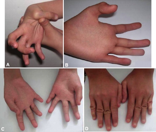
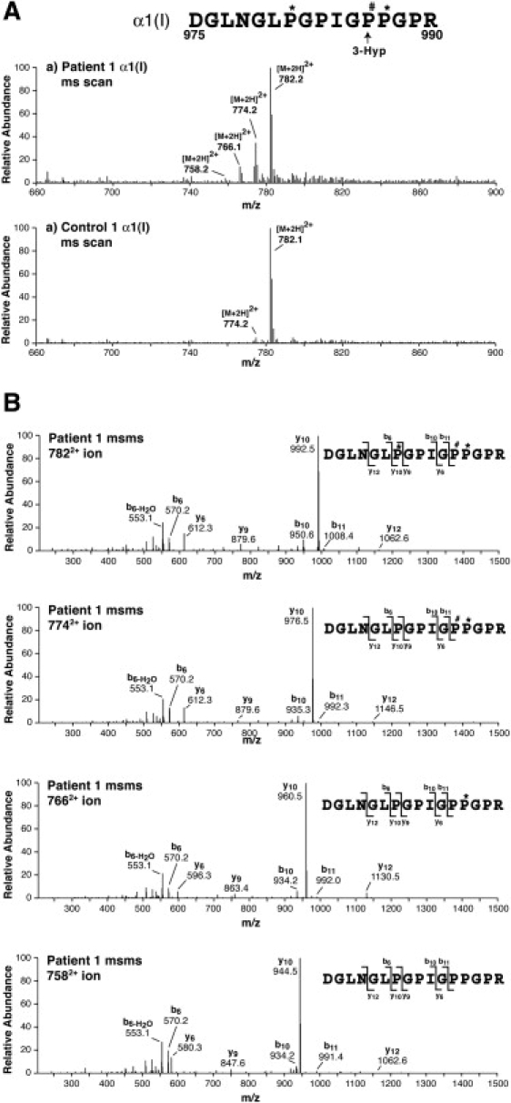
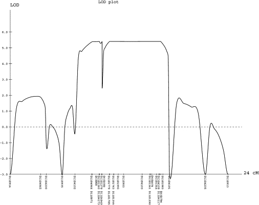
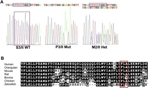
We present clinical, radiological, biochemical, and genetic findings on six patients from two consanguineous families that show EDS-like features and radiological findings of a mild skeletal dysplasia. The EDS-like findings comprise hyperelastic, thin, and bruisable skin, hypermobility of the small joints with a tendency to contractures, protuberant eyes with bluish sclerae, hands with finely wrinkled palms, atrophy of the thenar muscles, and tapering fingers. The skeletal dysplasia comprises platyspondyly with moderate short stature, osteopenia, and widened metaphyses. Patients have an increased ratio of total urinary pyridinolines, lysyl pyridinoline/hydroxylysyl pyridinoline (LP/HP), of approximately 1 as opposed to approximately 6 in EDS VI or approximately 0.2 in controls. Lysyl and prolyl residues of collagens were underhydroxylated despite normal lysyl hydroxylase and prolyl 4-hydroxylase activities; underhydroxylation was a generalized process as shown by mass spectrometry of the alpha1(I)- and alpha2(I)-chain-derived peptides of collagen type I and involved at least collagen types I and II. A genome-wide SNP scan and sequence analyses identified in all patients a homozygous c.483_491 del9 SLC39A13 mutation that encodes for a membrane-bound zinc transporter SLC39A13. We hypothesize that an increased Zn(2+) content inside the endoplasmic reticulum competes with Fe(2+), a cofactor that is necessary for hydroxylation of lysyl and prolyl residues, and thus explains the biochemical findings. These data suggest an entity that we have designated "spondylocheiro dysplastic form of EDS (SCD-EDS)" to indicate a generalized skeletal dysplasia involving mainly the spine (spondylo) and striking clinical abnormalities of the hands (cheiro) in addition to the EDS-like features.


Figures

Figure 1
Figure 1
Pedigrees and Haplotype Analysis Family I and family II are shown in (A) and (B), respectively. Affected individuals are depicted by black symbols. Genotypes for 39 microsatellite-marker loci spanning the linkage interval on chromosome 11p11.2-p13 are shown. The haplotypes are displayed by vertical bars. The disease-associated haplotype is denoted by a red box around the vertical bars. Recombinations in both families define a candidate region flanked by markers D11S1779 and D11S4191.
Figure 2
Figure 2
Hands with Characteristic Changes (A) P3/II at age 10.1 years. The thumb is hypermobile and can easily be adducted to the forearm; the fingers are tapering and display flexion contractures. (B) P3/II at age 10.1 years. The palm is excessively wrinkled, the thenar is hypotrophic, the fingers are tapering, and the endphalange of the thumb contracted. (C) P6/III at age 12.5 years. The palms are wrinkled, the thenar and hypothenar muscles are hypotrophic, the fingers are tapering, and there is partial syndactyly of fingers 2 and 3, and 3 and 4. (D) P6/III at age 12.5 years. The dorsal aspect of the hands show tapering fingers, broadened regions over the interphalangeal joints with abundant skin, and flexion contractures, especially of the fifth fingers.
Figure 3
Figure 3
Radiographs of the Low Thoracic and Lumbar Spines, the Hands, the Pelvis, and the Knees (A) P2/I at age 3.5 years. Note mild to moderate flattening, osteopenia, and irregular endplates of the vertebral bodies. (B) P1/I at age 11.5 years. There are similar findings to (A) with flattening, irregular endplates and osteopenia of the vertebral bodies. (C) P5/III at age 28 years. Again, platyspondyly, osteopenia, and irregular endplates of the vertebral bodies with depressions resembling notochordal remnants are seen. (D) P1/I at age 11.5 years. Note the mildly short metacarpals and phalanges with widening of the ends and relative narrowing of the diaphyses. The epiphyses of the short tubular bones are flat. Osteopenia is not present. (E) P2/I hand at age 3.5 years. Skeletal features of this younger child are similar but less pronounced as compared to (A); alterations of shape and flat epiphyses of the short tubular bones are noted. (F) Pelvis of P3/II at age 10 years. Small ilia, mild flattening of the proximal epiphyses, and short and wide femoral necks are seen. (G) Knee of P5/III at age 28 years. The knees show normal-sized epiphyses and a normal metaphyseal contour on the long tubular bones, but the intercondylar fossa of the distal femur is shallow in this adult patient.
Figure 4
Figure 4
Decreased Lysyl Hydroxylation of the Collagen α1(I) and α2(I) Chains Revealed by TMS (A) SDS-PAGE resolution of collagen α1(I)- and α2(I)-chains of collagen type I from pepsin-treated culture medium. The α1(I) chains were excised from the gel and subjected to in-gel digestion with trypsin. Lanes 1–4 refer to samples obtained by fibroblasts from P1/I, P3/II, and controls 1 and 2. Please note that the electrophoretic mobility of the α(I) chains from the patients' samples is barely different from that of the controls. (B) SDS-PAGE resolution of CNBr-peptides prepared from collagen type I from culture medium. Peptide α2(I)CB4 was excised from the gel and subjected to in-gel digestion by trypsin. Lanes 1, 2, and 3 were from patients P1/I, P3/II, and a control, respectively. (C) Mass-spectral scans across the regions of the LCMS profiles from the α1(I) chains of one patient and a control that capture the post-translational variants of the tryptic peptide. The lowest panel shows an MS/MS spectrum from the 11492+ precursor ion, which establishes the identity of the peptide sequence including placement of hydroxyl groups on the three prolyl residues and the single lysyl residue. Similar MS/MS spectra showed that the 11412+ ion lacked the hydroxyl group (16 mass units) of the lysyl residue 684 in the two patients. (D) Mass-spectral scans across regions of the LCMS profiles from peptide α2(I)CB4 of one patient and a control that capture posttranslational variants of the tryptic peptide shown. The lowest panel shows the MS/MS spectrum from the 12182+ precursor ion, which establishes the identity of the peptide including the placement of hydroxyl groups on three prolines and the single lysine. Similar MS/MS spectra showed that the 12102+ ion lacked the hydroxyl (16 mass units) on Lys219 in the two patients.
Figure 5
Figure 5
Decreased Prolyl 3-Hydroxylation and Prolyl 4-Hydroxylation Revealed by TMS Mass spectra are shown for the tryptic peptide containing Pro986, the site of 3-Hyp formation in the α1(I) chain from one patient and one control fibroblast cultures run on SDS-PAGE (shown in Figure 4A). (A) Mass-spectral scans encompassing all variants of the tryptic peptide detected in the LC profiles (∼28 min elution time). Four posttranslational variants are revealed in the patient's collagen. (B) MS/MS spectra of the four peptide variants differing by 16 mass units identify the placement of hydroxyl groups. Underhydroxylation at the 3-Hyp position and the two 4-Hyp positions can be quantified from the identified structures and their relative abundance as shown in Figure 4.
Figure 6
Figure 6
Results of Linkage Analysis The parametric multipoint LOD score analysis for 39 microsatellite markers spanning the linkage interval on chromosome 11p11.2-q13 in families I and II is shown. Because of space constraints, only 31 markers are shown. Genetic distances, in cM, based on the deCODE map and the LOD scores are shown on the x axis and on the y axis, respectively.
Figure 7
Figure 7
Mutation Identification and Comparative Analysis of the ZIP13 Protein (A) Electropherograms indicating the wild-type (S3/II), the homozygous (P3/II), and the heterozygous (M2/II) mutation sequences. The 9 bp deletion in exon 4 of SLC39A13 (c.483_491 del9; p.F162_164 del) is boxed. (B) ClustalW alignment and BOXSHADE analysis of the protein sequence of the transmembrane domain III of ZIP13 around the mutation (the deleted amino acids FLA are boxed). The protein sequence is highly conserved across vertebrates, from primates through to birds and primitive fish.

References

    1. Beighton P., De Paepe A., Steinmann B., Tsipouras P., Wenstrup R.J. Ehlers-Danlos syndromes: Revised nosology, Villefranche, 1997. Am. J. Med. Genet. 1998;77:31–37. - PubMed
    1. Steinmann B., Royce P.M., Superti-Furga A. The Ehlers-Danlos syndrome. In: Royce P.M., Steinmann B., editors. Connective Tissue and its Heritable Disorders. Second Edition. Wiley-Liss; New York: 2002. pp. 431–523.
    1. Kraenzlin, M.E., Kraenzlin, C.A., Meier, C., Giunta, C., and Steinmann, B. Evaluation of an automated HPLC system for measurement of urinary collagen crosslinks: Effect of age, menopause and metabolic bone diseases. Clin. Chem., in press. - PubMed
    1. Steinmann B., Eyre D.R., Shao P. Urinary pyridinoline cross-links in Ehlers-Danlos syndrome type VI. Am. J. Hum. Genet. 1995;57:1505–1508. - PMC - PubMed
    1. Giunta C., Randolph A., Al-Gazali L., Brunner H.G., Kraenzlin M.E., Steinmann B. The Nevo syndrome is allelic to the kyphoscoliotic type of the Ehlers-Danlos syndrome (EDS VIA) Am. J. Med. Genet. 2005;133:158–164. - PubMed

Publication types

MeSH terms