. 2008 Jul 15;319(2):223-33.
doi: 10.1016/j.ydbio.2008.04.014. Epub 2008 Apr 23.

Patterning of the heart field in the chick

Affiliations

Patterning of the heart field in the chick

Radwan Abu-Issa et al. Dev Biol. .

Abstract

In human development, it is postulated based on histological sections, that the cardiogenic mesoderm rotates 180 degrees with the pericardial cavity. This is also thought to be the case in mouse development where gene expression data suggests that the progenitors of the right ventricle and outflow tract invert their position with respect to the progenitors of the atria and left ventricle. However, the inversion in both cases is inferred and has never been shown directly. We have used 3D reconstructions and cell tracing in chick embryos to show that the cardiogenic mesoderm is organized such that the lateralmost cells are incorporated into the cardiac inflow (atria and left ventricle) while medially placed cells are incorporated into the cardiac outflow (right ventricle and outflow tract). This happens because the cardiogenic mesoderm is inverted. The inversion is concomitant with movement of the anterior intestinal portal which rolls caudally to form the foregut pocket. The bilateral cranial cardiogenic fields fold medially and ventrally and fuse. After heart looping the seam made by ventral fusion will become the greater curvature of the heart loop. The caudal border of the cardiogenic mesoderm which ends up dorsally coincides with the inner curvature. Physical ablation of selected areas of the cardiogenic mesoderm based on this new fate map confirmed these results and, in addition, showed that the right and left atria arise from the right and left heart fields. The inversion and the new fate map account for several unexplained observations and provide a unified concept of heart fields and heart tube formation for avians and mammals.


Figures

Figure 1
Figure 1
Nkx2.5 expression pattern in transverse sections of stage HH8 chick embryo. The sections are oriented with dorsal up and ventral down. The sections span from the buccopharyngeal membrane cranially (A) to the first somite caudally (H). (B) Higher magnification of the box in (A) showing Nkx2.5 expression in the ventral endoderm (En) and ventral ectoderm (Ec) at the midline and the mesoderm laterally. (C) Mesoderm expressing Nkx2.5 in the dorsomedial wall of the coelom (coelom indicated by arrow in D-G). The Nkx2.5 expressing mesodermal cells extend medially but are separated by a gap at the midline. (D) Higher magnification of the box in (C). Nkx2.5 expression in the splanchnic mesoderm continues partially into the paraxial mesoderm (arrowhead). (E, F) Strong expression of Nkx2.5 in the splanchnic mesoderm at the anterior intestinal portal (arrowhead). Nkx2.5 expressing cells are located mediodorsal to the coelom in (E) and mediodorsally and medioventrally in (F). (G) Just cranial to somite 1, the splanchnic mesoderm can be seen as a small area of cells expressing Nkx2.5 that are positioned ventral to the coelom (arrow). (H) Nkx2.5 and the coelom both disappear at the first somite.
Figure 2
Figure 2
Three different orientations of the 3D reconstructions of stage HH8 heart field. Each is shown embedded in three representative histological sections. Below each panel is an artist's rendering of the 3D image. The histological sections are at the level of the buccopharyngeal membrane, anterior intestinal portal, and first somite from cranial to caudal. The colors represent the 44 sections incorporated into the 3D reconstruction. (A) Ventral view shows the proximity of the unfused cardiogenic mesoderm across the midline at an intermediate level within the wings of the heart field. The length of the left-right axis cranially and caudally is also shown. (A′) Dorsal view shows the 120-130° inversion of the cardiogenic mesoderm which coincides with the anterior intestinal portal. (A″) Lateral view shows the dorsoventral axis of the cardiogenic mesoderm, and the craniocaudal axis.
Figure 3
Figure 3
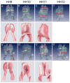
3D reconstruction of the cardiogenic mesoderm at stages HH9-12. Below the 3D images for stages HH9-11 is an artist's rendering of the 3D image. (A-D) Ventral view. (A′-D′) Dorsal view. (A, A′) Stage HH9 cardiogenic mesoderm from right and left approximate at the midline but are not fused. They form two bulges (arrows) separated by the ventral mesocardium. (B, B′) Stage HH10, the cardiogenic mesoderm fuses ventrally at the midline and forms a myocardial trough which is open dorsally (arrow). (C, C′) At stage HH11, the myocardial trough begins looping but is still open dorsally. The caudal portion of the myocardial trough just cranial to the anterior intestinal portal is closing (arrow). (D, D′) Stage HH12 shows a looped myocardial tube with dorsal fusion in progress (bracket). Star (*) indicates the position of the mediolateral inversion, which moves caudally with the anterior intestinal portal during development.
Figure 4
Figure 4
Stage 7/8 chick embryo with 3+ somites labeled at the cranial end of the heart field with DiI/rhodamine on the right and DiO/fluorescein on the left. A-F show the embryo at the 0 timepoint and at 3 hr intervals thereafter. The green dye contained a higher concentration of DMSO and diffused more widely while the red dye was more limited and better represents the cranial heart field cells. However, both dyes show labeling in the outer curvature with red limited to the outer curvature. G-I show a caudocranial series of sections of the same embryo. In the most caudal section nearest to the anterior intestinal portal (G) the labeling is in the ventral midline. In more cranial sections the label can be seen on the right as the outer curvature develops and the heart loops to the right side.
Figure 5
Figure 5
Double labeling of the cardiogenic mesoderm of a stage HH8 chick embryo. A) Position of the DiI (red) and DiO (green) labels in the cardiogenic mesoderm based on the 3D reconstructions. The 3D reconstruction of the same stage is overlaid on the embryo. (B-H) Ventral views of the same embryo at successive times from 0-28 hrs after labeling. (B-D) From 0 time to 6 hours, the two labeled populations (green medially and red laterally) are at the caudal edge of the anterior intestinal portal (white arrowhead). (C-F) The red labeled cells move toward the midline, inverting their position completely with the green labeled cells. (D-E) The red labeled cells also begin to move caudally along with the anterior intestinal portal away from the green cells, which are stationary (D). (E-G) From 9 to 28 hours, the heart tube forms and loops. The red labeled cells located in the heart tube continue their movement caudally along the anterior intestinal portal giving the impression that the green population is moving cranially. Red labeled cells are incorporated into the inflow (I) myocardium. The green population approaches the outflow (O) tract and is incorporated into it. (H) Higher magnification of the heart in G. (I -J) Transverse cryosections 5 hours after double labeling of a stage HH8 embryo. (J) Higher magnification of the labeled cells in (I). Note that both the red and green labeled cells are in the cardiogenic mesoderm.
Figure 6
Figure 6
Mapping the cardiogenic mesoderm in cultured stage HH8-9 embryos. 3-D reconstructions are overlaid on embryos of the same stages in the far left panels (A,F,K). (A-E) Labeling of the craniolateral cardiogenic mesoderm. The same embryo is shown from a ventral view from the time of labeling (0hr) to 45 hr after the label was placed. The red labeled cells appeared to move toward the midline and were incorporated into the myocardium of the outer curvature of the outflow tract (O). I, inflow tract. (F-J) Labeling the mid-wing region of the cardiogenic mesoderm. The same embryo is seen from the ventral side at the times indicated after injection of the label. At 0 time, the labeled cells (red) were at the center of the inversion point of the heart field. The labeled cells appeared to move toward the midline and split into two populations. The more lateral population appeared to move cranially to the outer curvature of the outflow (O) myocardium while the more medial population appeared to move caudally to the outer curvature of the inflow (I) myocardium. (K-O) Double labeling of the cranial and caudal extremities of the cardiogenic mesoderm. Whole embryos are seen from the ventral side at the times indicated. At 0 time, the two labeled populations (green cranially and red caudally) in the medial part of the cardiogenic mesoderm. The labels both move toward the outflow tract with the cranial labeled cells (green) populating the outer curvature while the caudal labeled cells (red) populate the inner curvature. Panel O is a section through the outflow tract of the same embryo showing red (black arrowhead) and green (white arrowhead) labeled cells on opposite sides of the outflow myocardium.
Figure 7
Figure 7
Labeling in ovo to observe location of labeled cells after addition of cardiogenic mesoderm to the outflow myocardium and the atrium. A) The position of the labels in the cardiogenic mesoderm shown in a dorsal view of the 3D reconstruction of an HH11 embryo. Double labeling with crystals of DiI (red) and DiO (green). The green label is implanted into inverting region while the red label is in the lateral leg of the cardiogenic mesoderm, of a stage HH11 embryo. The labeling was followed for 45 hrs. B) Position of the crystals immediately after they were placed in the embryo. C,D) Labeling in the outflow (O, green) and inflow (I, red) after 45 hrs. D) shows higher magnification of the boxed region in C. E) Embryo labeled at stage 8 with Neurotrace green placed in the caudal lateral cardiogenic mesoderm. White arrowhead indicates somite 1. F,G) show the same embryo at 45 hrs after the labeling. G shows an enlargement of the box in F. Green cells can be seen in the wall of the left side of the atrium.
Figure 8
Figure 8
Ablations of selected regions of the heart field and the resulting heart phenotype. The far left panels show 3D reconstructions overlaid on the embryos to indicate what part of the heart field was ablated. (A-A″) Unilateral right ablation of the medial leg and region of inversion (square, A and A′) at stage HH9 caused reduction of the right ventricle (RV) and outflow tract (OFT) myocardium which resulted in double outlet right ventricle (A″). Arrowhead indicates the ventricular septum; LV, left ventricle. (B-B″) Ablation of the right leg of cardiogenic mesoderm at stage HH10 (circled, B and B′) resulted in absence of the right atrium (LA) and reduction of the left ventricle while the left atrium (LA) is developing well (B″). (C-C″) Ablation of the left leg of cardiogenic mesoderm (circled, C and C′) resulted in absence of the left atrium and reduction of the ventricles while the right atrium (RA) is developing well (D″).
Figure 9
Figure 9
Model of cardiogenic mesoderm and heart tube formation. Between stages HH3-7, cells in the epiblast ingress through the primitive streak and become localized in the anterolateral lateral plate mesoderm. In the epiblast and primitive streak the cells are located in the same craniocaudal position as they will be in the looped heart tube. During gastrulation however there is a 90° shift such that inflow precursors (blue) are located laterally in the cardiogenic mesoderm and outflow precursors (red) are located medially in the flat cardiogenic mesoderm at stage HH7. Formation of the foregut pocket by the anterior intestinal portal inverts the cardiogenic mesoderm 120-130° which returns the position of the inflow and outflow progenitors to the original craniocaudal orientation. The cranial parts of the heart fields are brought to the ventral midline where they will form the outer curvature after fusion at stage HH10. Note that even at stage HH11 when the heart tube has started to loop, there is still a cardiogenic mesoderm that will be added to both the inflow and outflow myocardium.

References

    1. Alsan BH, Schultheiss TM. Regulation of avian cardiogenesis by Fgf8 signaling. Development. 2002;129:1935–1943. - PubMed
    1. Barron M, Gao M, Lough J. Requirement for BMP and FGF signaling during cardiogenic induction in non-precardiac mesoderm is specific, transient, and cooperative. Dev Dyn. 2000;218:383–393. - PubMed
    1. Bruneau BG, Logan M, Davis N, Levi T, Tabin CJ, Seidman JG, Seidman CE. Chamber-specific cardiac expression of Tbx5 and heart defects in Holt-Oram syndrome. Dev Biol. 1999;211:100–108. - PubMed
    1. Buckingham M, Meilhac S, Zaffran S. Building the mammalian heart from two sources of myocardial cells. Nat Rev Genet. 2005;6:826–35. - PubMed
    1. Cai CL, Shi Y, Pfaff S, Chen J, Evans SM. Isl1 identifies a cardiac progenitor population that proliferates prior to differentiation and contributes a majority of cells to the heart. Dev Cell. 2003;5:878–889. - PMC - PubMed

Publication types

LinkOut - more resources