. 2008 Jun 27;380(1):181-92.
doi: 10.1016/j.jmb.2008.04.073. Epub 2008 May 7.

Structures and functional implications of an AMP-binding cystathionine beta-synthase domain protein from a hyperthermophilic archaeon

Affiliations

Structures and functional implications of an AMP-binding cystathionine beta-synthase domain protein from a hyperthermophilic archaeon

Neil P King et al. J Mol Biol. .

Abstract

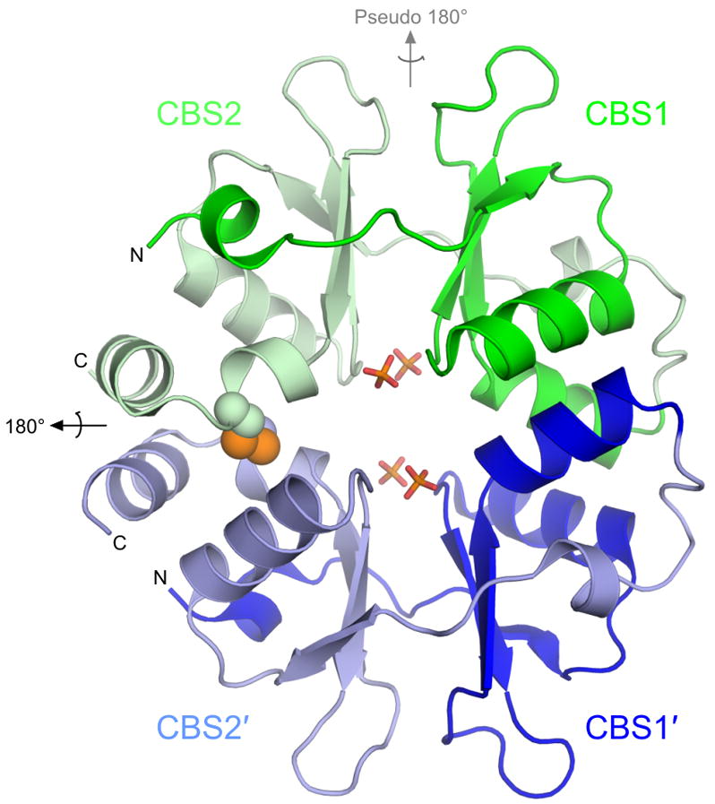
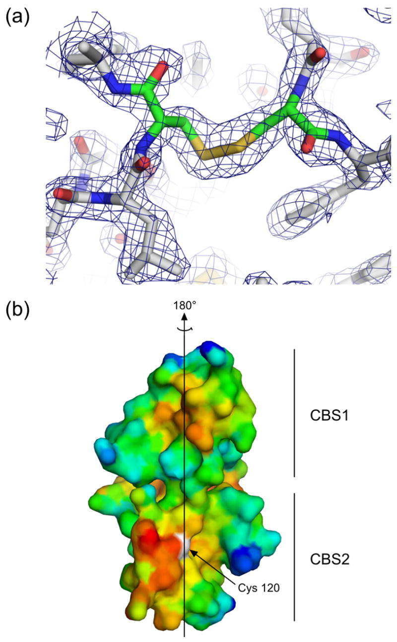
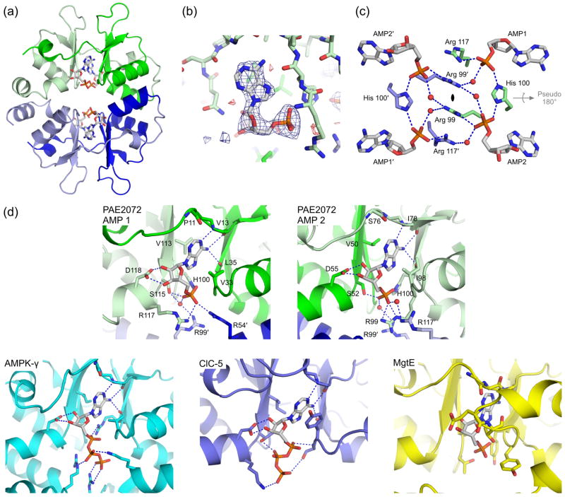
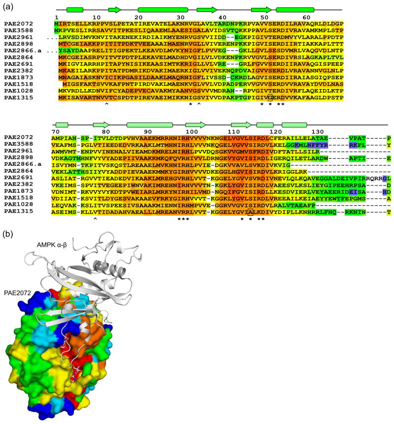
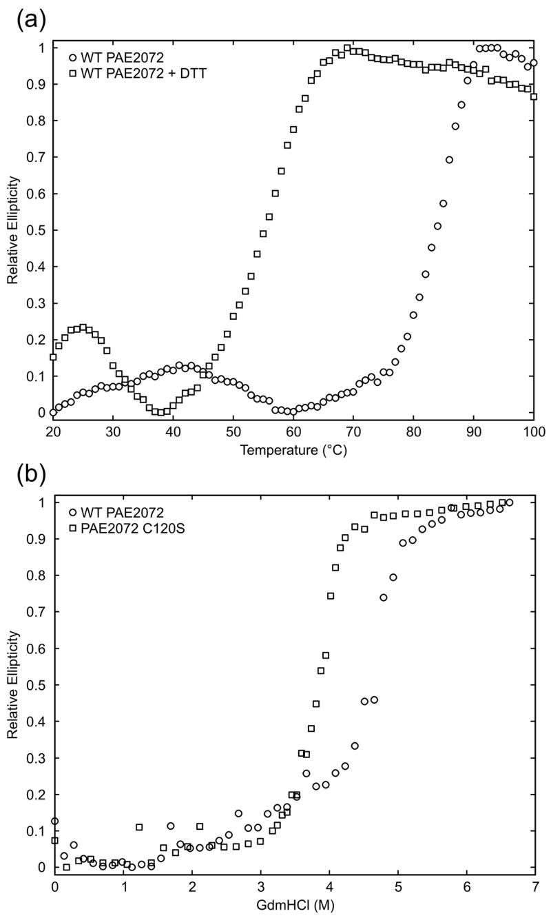
Cystathionine beta-synthase domains are found in a myriad of proteins from organisms across the tree of life and have been hypothesized to function as regulatory modules that sense the energy charge of cells. Here we characterize the structure and stability of PAE2072, a dimeric tandem cystathionine beta-synthase domain protein from the hyperthermophilic crenarchaeon Pyrobaculum aerophilum. Crystal structures of the protein in unliganded and AMP-bound forms, determined at resolutions of 2.10 and 2.35 A, respectively, reveal remarkable conservation of key functional features seen in the gamma subunit of the eukaryotic AMP-activated protein kinase. The structures also confirm the presence of a suspected intermolecular disulfide bond between the two subunits that is shown to stabilize the protein. Our AMP-bound structure represents a first step in investigating the function of a large class of uncharacterized prokaryotic proteins. In addition, this work extends previous studies that have suggested that, in certain thermophilic microbes, disulfide bonds play a key role in stabilizing intracellular proteins and protein-protein complexes.


Figures

Figure 1
Figure 1. Overall structure of PAE2072
The unliganded PAE2072 dimer is shown in cartoon representation, with each CBS domain colored individually. The intermolecular disulfide bond between cysteines 120 and 120′ is shown as spheres, and the sulfate ions that occupy the same positions as the phosphate groups of AMP in the AMP-bound structure are shown in stick representation. The dimeric two-fold axis is indicated in black, and the pseudo two-fold axis in gray. The core β-α-β-β-α fold conserved in CBS domain proteins is easily identifiable; an additional short helix punctuates the extended N-terminus of each CBS domain.
Figure 2
Figure 2. The dimeric interface
(a) Residues in the area of the disulfide bond in the unliganded structure are shown in stick representation, and electron density from a simulated annealing 2Fo-Fc omit map in which residues Cys120 and Cys120′ were omitted is contoured at 1.2 σ, demonstrating clear density for the disulfide bond. Similar density was seen for the disulfides in an omit map made using the coordinates and structure factors from the AMP-bound structure. (b) A surface representation of the dimeric interface of one subunit of PAE2072 is shown, colored according to hydrophobicity. Red indicates regions of higher hydrophobicity, while blue indicates more polar areas. The dimeric twofold axis is shown, and a prominent hydrophobic patch can be seen in each CBS domain. The sulfur atom of cysteine 120, representing the site of the intermolecular disulfide bond, is colored white.
Figure 3
Figure 3. Ligand binding by PAE2072
(a) A view of one of the two crystallographically independent dimers in the AMP-bound structure shows the positions of the four bound AMP molecules. Note the lack of any significant conformational change from the unliganded structure in Figure 1. (b) Difference density from a simulated annealing Fo-Fc omit map, contoured at 3.0 σ, reveals readily interpretable density for AMP. The coordinates from the final model are shown in stick representation. (c) Three residues from each subunit (Arg99, His100, and Arg117) are involved in coordinating multiple AMP molecules, which may suggest cooperative binding. Several well-ordered water molecules form bridging interactions, and may indicate space where additional phosphate groups, such as those on ATP, could be accommodated. The dimeric two-fold axis is indicated in the center of the figure and runs perpendicular to the page; the pseudo two-fold is shown in gray. (d) The mechanism of adenine nucleotide binding is strongly conserved in the CBS domains of PAE2072 (both binding sites are shown), mammalian AMPK (PDB code 2V92), and human ClC-5 (PDB code 2J9L), but not in bacterial MgtE (PDB code 2YVY). The four central beta strands of one subunit of PAE2072 were aligned with those of the tandem CBS domains of MgtE (RMSD = 0.81 Å over 20 Cα atoms) to model the potential position of a bound AMP molecule. Strong steric clashes between the protein and the modeled AMP can be seen, and no basic residues are in position to coordinate the phosphate residues. It therefore seems unlikely that MgtE binds adenine nucleotides in a manner similar to that observed in the other CBS domains shown here.
Figure 4
Figure 4. Sequence-structure analysis of Pyrobaculum CBS domain proteins
(a) A multiple sequence alignment of 12 standalone CBS domain proteins from P. aerophilum, generated using the alignment program T-COFFEE, revealed nearly universal conservation of the residues involved in adenylate binding in PAE2072. Residues with side chains (*) or backbone atoms (^) that contact AMP are indicated. Two substitutions likely to result in the loss of a contact are boxed. The sequences of two regions other than the termini were found to be highly variable in this family of proteins (residues 40–46 and 71–84 in PAE2072). The first 10 residues of PAE2961 and 27 residues of PAE2866.a are represented by ellipses for clarity. (b) The regions of high sequence variability in Pyrobaculum CBS domain proteins map to a single surface patch on PAE2072 which coincides precisely with the site of interaction between the γ subunit of AMPK and the α and β subunits. The four central beta strands of PAE2072 were aligned with those of the γ subunit of S. pombe AMPK (PDB ID 2OOX; RMSD = 0.88 over 20 Cα atoms) to model PAE2072 in the place of the γ subunit. The surface of PAE2072 is shown and colored according to sequence conservation; red indicates highly conserved positions, while blue indicates highly variable positions. The α and β subunits of AMPK are shown in cartoon representation and can be seen to dock onto the surface patch defined by the highly variable segments (blue and cyan). The AMP molecules bound to PAE2072 are shown in sticks to emphasize their surface accessibility, which is presumably important for the mechanism of regulation by PAE2072.
Figure 5
Figure 5. Contribution of the disulfide bond to protein stability
(a) Thermal melts of PAE2072 in the presence and absence of DTT indicate a dramatic loss of protein stability upon reduction of the disulfide bond. (b) Chemical denaturation of wild-type PAE2072 and a C120S mutant also reveal a stabilizing role for the disulfide bond as evidenced by the lower midpoint of denaturation for the mutant lacking the disulfide.

References

    1. Gilbert HF. Molecular and cellular aspects of thiol-disulfide exchange. Adv Enzymol Relat Areas Mol Biol. 1990;63:69–172. - PubMed
    1. Derman AI, Beckwith J. Escherichia coli alkaline phosphatase fails to acquire disulfide bonds when retained in the cytoplasm. J Bacteriol. 1991;173:7719–22. - PMC - PubMed
    1. Prinz WA, Aslund F, Holmgren A, Beckwith J. The role of the thioredoxin and glutaredoxin pathways in reducing protein disulfide bonds in the Escherichia coli cytoplasm. J Biol Chem. 1997;272:15661–7. - PubMed
    1. Kadokura H, Katzen F, Beckwith J. Protein disulfide bond formation in prokaryotes. Annu Rev Biochem. 2003;72:111–35. - PubMed
    1. Chiu HJ, Johnson E, Schroder I, Rees DC. Crystal structures of a novel ferric reductase from the hyperthermophilic archaeon Archaeoglobus fulgidus and its complex with NADP+ Structure. 2001;9:311–9. - PubMed

Publication types

LinkOut - more resources