. 2008 Jun;82(6):1249-61.
doi: 10.1016/j.ajhg.2008.04.020.

Hyperglycosylation and reduced GABA currents of mutated GABRB3 polypeptide in remitting childhood absence epilepsy

Affiliations

Hyperglycosylation and reduced GABA currents of mutated GABRB3 polypeptide in remitting childhood absence epilepsy

Miyabi Tanaka et al. Am J Hum Genet. 2008 Jun.

Abstract

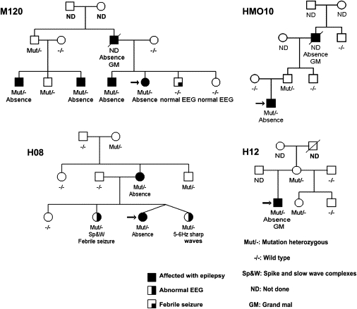
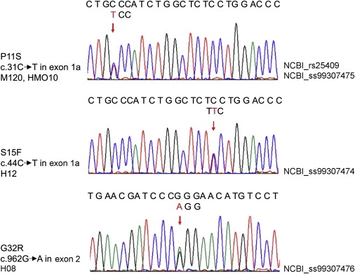
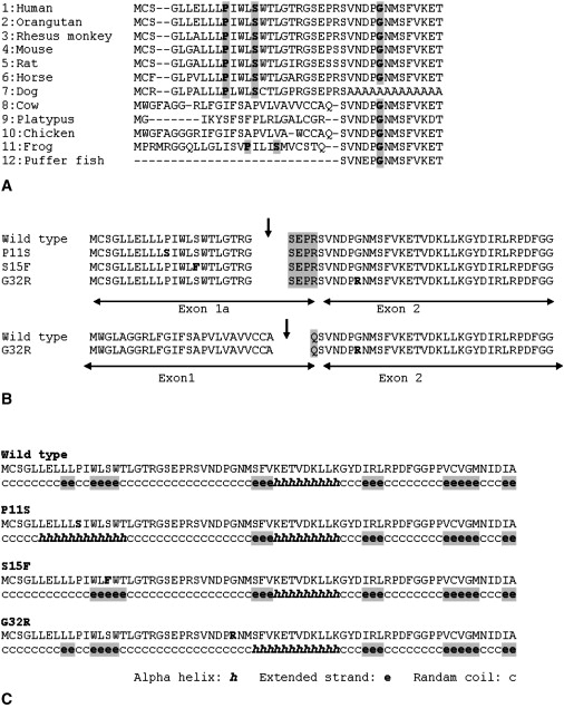
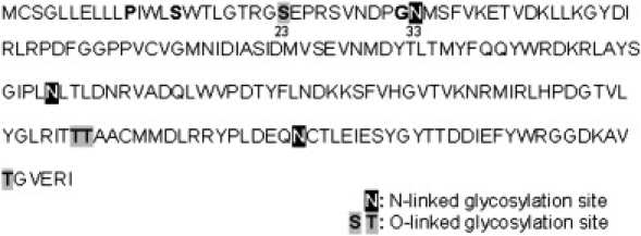
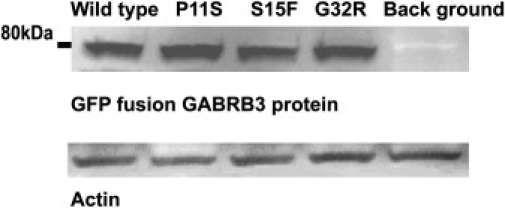
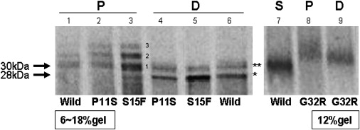
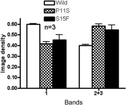
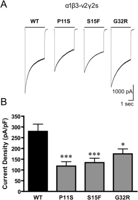
Childhood absence epilepsy (CAE) accounts for 10% to 12% of epilepsy in children under 16 years of age. We screened for mutations in the GABA(A) receptor (GABAR) beta 3 subunit gene (GABRB3) in 48 probands and families with remitting CAE. We found that four out of 48 families (8%) had mutations in GABRB3. One heterozygous missense mutation (P11S) in exon 1a segregated with four CAE-affected persons in one multiplex, two-generation Mexican family. P11S was also found in a singleton from Mexico. Another heterozygous missense mutation (S15F) was present in a singleton from Honduras. An exon 2 heterozygous missense mutation (G32R) was present in two CAE-affected persons and two persons affected with EEG-recorded spike and/or sharp wave in a two-generation Honduran family. All mutations were absent in 630 controls. We studied functions and possible pathogenicity by expressing mutations in HeLa cells with the use of Western blots and an in vitro translation and translocation system. Expression levels did not differ from those of controls, but all mutations showed hyperglycosylation in the in vitro translation and translocation system with canine microsomes. Functional analysis of human GABA(A) receptors (alpha 1 beta 3-v2 gamma 2S, alpha 1 beta 3-v2[P11S]gamma 2S, alpha 1 beta 3-v2[S15F]gamma 2S, and alpha 1 beta 3-v2[G32R]gamma 2S) transiently expressed in HEK293T cells with the use of rapid agonist application showed that each amino acid transversion in the beta 3-v2 subunit (P11S, S15F, and G32R) reduced GABA-evoked current density from whole cells. Mutated beta 3 subunit protein could thus cause absence seizures through a gain in glycosylation of mutated exon 1a and exon 2, affecting maturation and trafficking of GABAR from endoplasmic reticulum to cell surface and resulting in reduced GABA-evoked currents.


Figures

Figure 1
Figure 1
Four Families with GABRB3 Mutations Each family number is placed beside each pedigree. Black circles or squares represent epilepsy affected females or males. Asymptomatic persons who have EEG 3 Hz diffuse bilateral spike wave complexes or 5 to 6 Hz sharp waves are represented by half black circles or squares.
Figure 2
Figure 2
cDNA Sequencing in Each Proband Each arrow shows the location of the mutation. The upper sequence represents wild-type. The lower triplet above the arrow represents the mutated code.
Figure 3
Figure 3
Gene and Marker Position on Chromosome 15q11-14 Figure 3 shows the relative position of markers in chr. 15q11-14. GABRB3 (marker) is about 60kb beyond the 3′ terminus of GABRB3, and 85CA is about 50kb from exon 1a of GABRB3.
Figure 4
Figure 4
Conserved Amino Acid Sequence of Exon 1a and Exon 2 of GABRB3, Predicted Cleavage Site, and Predicted Secondary Structure of Each Mutation (A) Conserved amino acid sequences of exon 1a and exon 2 of GABRB3. Each mutated amino acid position is indicated by gray shadow. 1. Homo sapiens: GABAA receptor, beta 3, (NP_068712), 2. Pongo pygmaeus: hypothetical protein, (CAH89717), 3. Macaca mulatta: PREDICTED GABAA receptor, beta 3, (XP_001109060), 4. Mus musculus: GABAA receptor, beta 3, (NP_0010337906), 5. Rattus norvegicus: GABAA receptor, beta 3, (EDL86448), 6. Equus caballus: PREDICTED: similar to GABAA receptor, beta 3, (XP_001493125), 7. Canis familiaris: PREDICTED: similar to GABAA receptor, beta 3, (XP_848482), 8. Bos Taurus: hypothetical protein, (NP_001092850), 9. Ornithorhynchus anatinus: PREDICTED: similar to GABAA receptor, beta 3, (XP_001505697), 10. Gallus gallus: GABAA receptor, beta 3, (NP_990677), 11. Xenopus tropicalis: Unknown protein, (AAI36050), 12. Tetraodon nigroviridis: unnamed protein product, (CAG06522). (B) Predicted cleavage site. Arrows indicate each predicted cleavage site. Each mutation is predicted to have the same cleavage site as the wild-type exon 1a. G32R in exon 2 is predicted to have the same cleavage site even with exon 1 as the wild-type. The cleavage site of exon 1 is different from the exon 1a, therefore the N-termini differs (gray shadow). (C) Predicted secondary structure of each mutation. All mutations are predicted to change secondary structures.
Figure 5
Figure 5
Predicted Glycosylation Site in Exon 1a–Exon 6 of GABRB3 Bold letters without shadow show the amino acids to be displaced by mutations. Numbers show locations of amino acids.
Figure 6
Figure 6
Western-Blot Analysis of Whole Cell Fractions Derived from HeLa Cell Expression 11 hr after Transfection The GFP fusion GABRB3 protein was present at a slightly smaller molecular weight of 80kDa which was expected. All expression levels of plasmids with mutated sequences were the same as the wild-type, even after normalization by Actin.
Figure 7
Figure 7
Increased Glycosylation of Mutated GABRB3 Protein Products of in vitro translocation (P) and digestion (D) with N-glycosidase F, containing the exon 1a mutations P11S and S15F, were loaded on a 6%–18% gel (the left gel). Similar products containing the exon 2 mutation G32R were loaded on a 12% gel (the right gel). The supernatant protein (S) of only wild-type is shown (lane 7) in the 12% gel and is considered not to be translocated to microsomes and to present the 30kDa GABRB3 including the signal peptide. The 30 kDa supernatant protein therefore consists of untranslocated “exon 1a to exon 6.” Translocated proteins (P) are shown in lanes 1–3 and 8. The molecular weight of band 1 was 30 kDa. Bands 2 and 3 of both P11S and S15F mutations in in vitro translocation (P) revealed clearly higher density than wild-type, suggesting increased glycosylation. After digestion with N-glycosidase F, two smaller sized bands, 28 kDa (), and 30 kDa (∗∗) appeared. The 30 kDa bands represent incompletely digested protein and 28 kDa bands represent completely digested protein. The translocated protein of G32R (P, lane 8) had only bands larger than 30kDa, also suggesting increased glycosylation. The band of G32R has higher molecular weight than the supernatant protein from the wild-type even after digestion.
Figure 8
Figure 8
Quantitation of Density of Glycosylated Bands Comparing Wild-Type and Exon 1a Mutations Shown in Figure 7 Bands 2 and 3 are considered to be hyperglycosylated products. The image density of each band is the average of three experiments. Since bands 2 and 3 of one sample overlapped, the sum of bands 2 and 3 was compared. The proportion of the image density of band 1 and the sum of bands 2 and 3 were significantly different between the wild-type and mutations in exon 1a. (P11S, p = 0.0004, S15F, p = 0.005).
Figure 9
Figure 9
GABA-Evoked Currents of Mutations in Transfected HEK293 Cells The current density from cells expressing the wild-type β3-v2 transcript was larger than in the cells expressing the β3-v2(PS), β3-v2(SF) or β3-v2(GR) mutations. The current density recorded from cells expressing GABAA receptors with β3-v2 mutations associated with CAE was reduced. A. Representative traces of whole cell current elicited for 4 s with 1 mM GABA from cells expressing α1β3-v2γ2S (WT), α1β3-v2(P11S)γ2S, α1β3-v2(S15F)γ2S and α1β3-v2(G32R)γ2S receptors. B. Compared to cells expressing wild-type receptors (n = 36), the current densities of the cells expressing receptors containing the β3-v2 mutations P11S (n = 18), S15F (n = 25) and G32R (n = 41) were reduced (∗∗∗p < 0.001, p < 0.05).

References

    1. Fong G.C.Y., Shah P.U., Gee M.N., Serratosa J.M., Castroviejo I.P., Khan S., Ravat S.H., Mani J., Medina M.T., Delgado-Escueta A.V. Childhood absence epilepsy with tonic-clonic seizures and 3–4-Hz spike and multispike-slow wave complexes: linkage to chromosome 8q24. Am. J. Hum. Genet. 1998;63:1117–1129. - PMC - PubMed
    1. Wallace R.H., Marini C., Petrou S., Harkin L.A., Owser D.N., Panchal R.G., Williams D.A., Sutherland G.R., Mulley J.C., Berkovic S.F. Mutant GABA(A)receptor gamma2-subunit in childhood absence epilepsy and febrile seizures. Nat. Genet. 2001;28:49–52. - PubMed
    1. Haug K., Warnstedt M., Alekov A.K., Sander T., Ramírez A., Poser B., Maljevic S., Hebeisen S., Kubisch C., Heils A. Mutations in CLCN2 encoding a voltage-gated chloride channel are associated with idiopathic generalized epilepsies. Nat. Genet. 2003;33:527–532. - PubMed
    1. Maljevic S., Krampfl K., Cobilanschi J., Tilgen N., Beyer S., Weber Y.G., Schlesinger F., Ursu D., Melzer W., Cossette P. A mutation in the GABA(A) receptor alpha(1)-subunit is associated with absence epilepsy. Ann. Neurol. 2006;59:983–987. - PubMed
    1. Callenbach P.M., Geerts A.T., Arts W.F., van Donselaar C.A., Peters A.C., Stroink H., Brouwer O.F. Familial occurrence of epilepsy in children with newly diagnosed multiple seizures: Dutch Study of Epilepsy in Childhood. Epilepsia. 1998;39:331–336. - PubMed

Publication types

MeSH terms

LinkOut - more resources