. 2008 Jul 15;319(2):309-20.
doi: 10.1016/j.ydbio.2008.04.024. Epub 2008 Apr 27.

Defective remodeling and maturation of the lymphatic vasculature in Angiopoietin-2 deficient mice

Affiliations

Defective remodeling and maturation of the lymphatic vasculature in Angiopoietin-2 deficient mice

Michael Dellinger et al. Dev Biol. .

Abstract

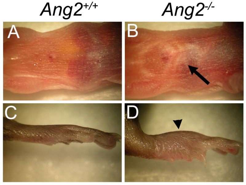
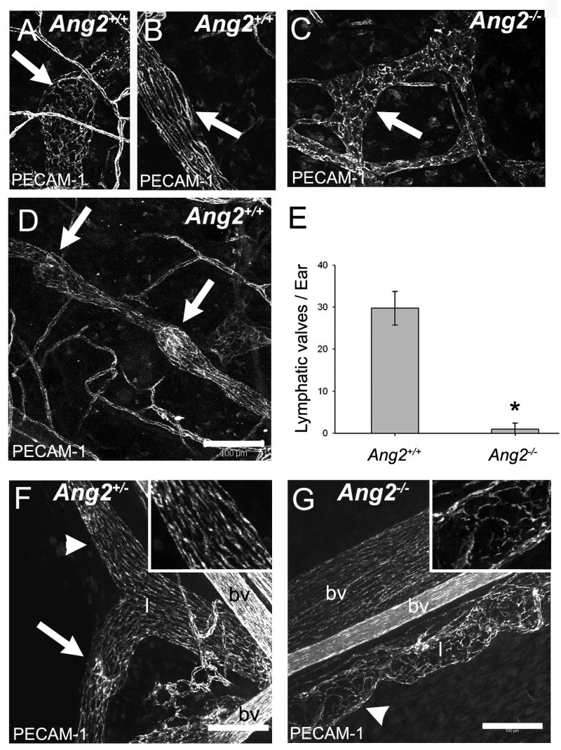
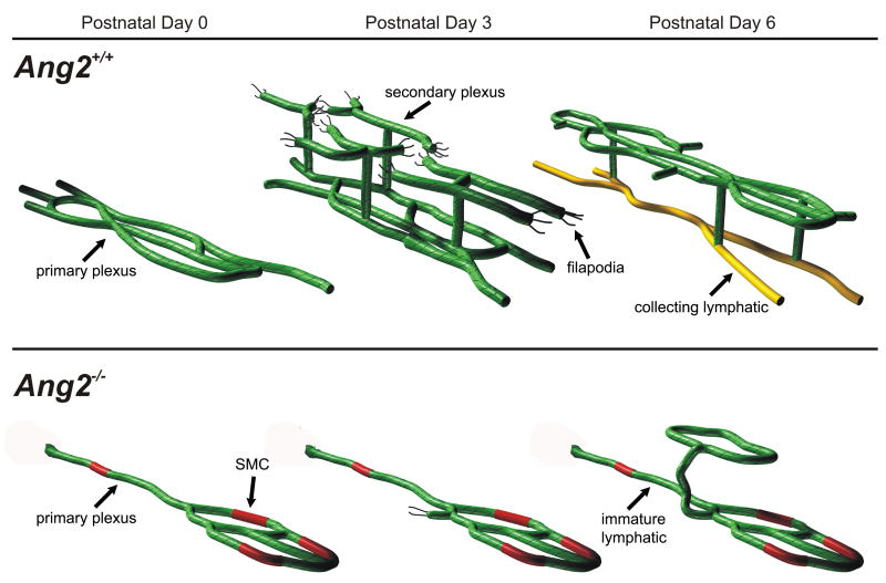
Molecular mechanisms regulating the remodeling of the lymphatic vasculature from an immature plexus of vessels to a hierarchal network of initial and collecting lymphatics are not well understood. One gene thought to be important for this process is Angiopoietin-2 (Ang-2). Ang2(-/-) mice have previously been reported to exhibit an abnormal lymphatic phenotype but the precise nature of the lymphatic defects and the underlying mechanisms have yet to be defined. Here we demonstrate by whole-mount immunofluorescence staining of ear skin and mesentery that lymphatic vessels in Ang2(-/-) mice fail to mature and do not exhibit a collecting vessel phenotype. Furthermore, dermal lymphatic vessels in Ang2(-/-) pups prematurely recruit smooth muscle cells and do not undergo proper postnatal remodeling. In contrast, Ang2 knock-out Ang1 knock-in mice do develop a hierarchal lymphatic vasculature, suggesting that activation of Tie-2 is required for normal lymphatic development. Taken together, this work pinpoints a specific lymphatic defect of Ang2(-/-) mice and further defines the sequential steps in lymphatic vessel remodeling.


Figures

Fig. 1
Fig. 1
Ang2-/- mice on the C57BL/6 genetic background exhibit gross defects of the lymphatic system. (A and B) A newborn Ang2+/+ pup (A) compared to an Ang2-/- littermate (B) displaying chylous ascites (arrow). (C and D) Hind paws of adult Ang2+/+ mice (C) are flat whereas those of adult Ang2-/- mice (D) are edematous (arrowhead).
Fig. 2
Fig. 2
Adult Ang2-/- mice display functional defects of the lymphatic system. (A) Intradermally administered EBD is effectively transported from the injection site to the base of the ear in all Ang2+/+ mice (n = 7). (B) EBD diffuses throughout lymphatic vessels in the ear skin of Ang2-/- mice (n = 5). (asterisks in A and B show EBD injection site)
Fig. 3
Fig. 3
Adult Ang2-/- mice exhibit patterning defects of the dermal lymphatic vasculature and a deficit of LYVE-1 down-regulated collecting lymphatic vessels in the ear. (A) A highly branched hierarchal pattern of lymphatic vessels is observed in ear skin of Ang2+/+ mice after whole-mount immunofluorescence staining for LYVE-1. Initial lymphatic vessels in the periphery of the ear (arrow) express LYVE-1 and transition to LYVE-1 down-regulated regions in the medial portion of the ear (arrowheads). (B-D) LYVE-1 down-regulated (B, arrow) PECAM-1 positive (C, arrow) collecting lymphatic vessels (D, arrow) are present throughout the medial region of ear skin in Ang2+/+ mice. (E) In contrast, all lymphatic vessels in the periphery (arrow) and medial (arrowhead) region of the ear in Ang2-/- mice express the initial lymphatic marker LYVE-1. (F-H) LYVE-1 (F) and PECAM-1 (G) expressing lymphatic vessels (H) are present throughout the immature network of Ang2-/- mice. (A and E used 4X objective; Scale bars D and H = 100 μm)
Fig. 4
Fig. 4
Whole-mount immunofluorescence staining of ear skin and mesentery for PECAM-1 demonstrates that Ang2-/- mice exhibit a deficiency of lymphatic vessels with a collecting vessel phenotype. (A and B) Initial lymphatic vessels comprised of PECAM-1 positive oak leaf-shaped endothelial cells (arrow, A), and collecting lymphatic vessels made up of elongated PECAM-1 positive endothelial cells (arrow, B), are in the ear skin of Ang2+/+ mice. (C) Nearly all lymphatic vessels in Ang2-/- ear skin are comprised of oak leaf-shaped endothelial cells (arrow). (D) PECAM-1 positive collecting lymphatic vessels in the ear skin of Ang2+/+ mice contain intraluminal valves (arrows). (E) Ang2+/+ mice have significantly (asterisk P < 0.001) more lymphatic valves per ear (29.75 ± 2.02, n = 4) than Ang2-/- mice (1.00 ± 0.71, n =4). (F) Mesenteric lymphatic vessels of Ang2+/- mice exhibit a mature collecting vessel phenotype, are comprised of elongated endothelial cells (arrowhead and inset), and contain valves (arrow). (G) In contrast, mesenteric lymphatic vessels of Ang2-/- mice are immature, made up of oak leaf-shaped endothelial cells (arrowhead and inset), and lack valves. (bv = blood vessel, l = lymphatic; Scale bars D,F and G = 100 μm)
Fig. 5
Fig. 5
Smooth Muscle Actin (SMA) positive cells are associated with initial lymphatic vessels in the ear of adult Ang2-/- mice. (A-C) Whole-mount immunofluorescence staining of Ang2+/+ adult ear skin reveals that SMA (arrow, A) positive cells are associated with LYVE-1 (arrow, B) down-regulated collecting lymphatic vessels (arrow, C; n = 3) as well as with blood vessels (arrowhead). (D-F) Conversely, SMA positive cells (arrow, D) are associated with LYVE-1 (arrow, E) positive initial lymphatic vessels (F; n = 5) and blood vessels (arrowheads) in Ang2-/- mice. (Scale bars C and F = 100μm)
Fig. 6
Fig. 6
Jugular lymph sacs develop normally in Ang2-/- embryos. (A) Jugular lymph sacs in E12.5 Ang2+/+ and Ang2-/- embryos express the lymphatic markers LYVE-1, Prox-1, and podoplanin. SMA positive cells surround only arteries and veins in E12.5 Ang2+/+ and Ang2-/- embryos. (B) The area of the right jugular lymph sac is not significantly different between E12.5 Ang2+/+ (74227 μm2 ± 17520.7; n = 3) and Ang2-/- (81492 μm2 ± 38609.9; n = 3) embryos (P = 0.781). (a = artery, jls = jugular lymph sac, v = vein)
Fig. 7
Fig. 7
Ang2-/- pups exhibit defects in postnatal remodeling of the dermal lymphatic vasculature. (A) 90μm confocal z-stack image of LYVE-1 stained ventral skin from a postnatal day (P) 0 Ang2+/+ pup showing a polygonal network of lymphatic vessels. (B) Depth projection of the image from panel A. Lymphatic vessels are in the same plane. (C) Three-dimensional projection (3D) of image from panel A rotated 90 degrees showing a single plexus of lymphatic vessels. (D) Cross section of ventral skin from a P0 Ang2+/+ pup stained for LYVE-1 (green) and counter stained with DAPI (blue). Lymphatic vessels of the primary plexus (arrowheads) are at the border between the dermis and subcutaneous tissue. (E) 90 μm confocal z-stack image of LYVE-1 stained ventral skin from a P3 Ang2+/+ pup. Numerous sprouts and filopodia are present (inset). (F) Depth projection of the image from panel E. Lymphatic vessels are distinct colors and in different planes. (G) Three-dimensional projection (3D) of image from panel E rotated 90 degrees showing two plexuses of lymphatic vessels. (H) Cross section of ventral skin from a P3 Ang2+/+ pup stained for LYVE-1 (green) and counterstained with DAPI (blue). Lymphatic vessels of the primary plexus (arrowhead) are in the subcutaneous tissue whereas vessels of the secondary plexus are in the dermis and at the border between the dermis and subcutaneous tissue. (I) Cross section of ventral skin from a Ang2+/+ P6 pup stained for LYVE-1 (red) and podoplanin (green) showing LYVE-1 positive lymphatic vessels in the dermis and a mature LYVE-1 down-regulated podoplanin positive collecting lymphatic vessel in the subcutaneous tissue (arrowhead). (J) Region arrowhead in panel I is pointing to shown at a higher magnification. (K) Region arrow in panel I is pointing to shown at a higher magnification. (L) 90μm confocal z-stack image of LYVE-1 stained ventral skin from a P0 Ang2-/- pup showing irregular lymphatic vessels. (M) Depth projection of image in panel L demonstrating lymphatic vessels in a single plane. (N) Three-dimensional projection (3D) of image from panel L rotated 90 degrees showing a single plexus of lymphatic vessels. (O) Cross section of ventral skin from a P0 Ang2-/- pup stained for LYVE-1 (green) and counter stained with DAPI (blue). Lymphatic vessels of the primary plexus (arrowheads) are located between the dermis and subcutaneous tissue. (P) 90μm confocal z-stack image of LYVE-1 stained ventral skin from a P3 Ang2-/- pup depicting an irregular network without many filopodia. (Q) Depth projection of image in panel P demonstrating lymphatic vessels in a single plane. (R) Three-dimensional projection (3D) of image from panel P rotated 90 degrees showing a single plexus of lymphatic vessels. (S) Cross section of ventral skin from a P3 Ang2-/- pup stained for LYVE-1 (green) and counterstained with DAPI (blue). Lymphatic vessels of the primary plexus are in the subcutaneous tissue (arrowheads). (T) Cross section of ventral skin from a P6 Ang2-/- pup stained for LYVE-1 (red) and podoplanin (green) showing LYVE-1 positive lymphatic vessels in the subcutaneous tissue (arrowhead). Lymphatic vessels located in the subcutaneous tissue of Ang2-/- mice express LYVE-1 and do not exhibit a collecting vessel phenotype. (U) Region arrowhead in panel T is pointing to shown at a higher magnification.
Fig. 8
Fig. 8
SMCs prematurely associate with lymphatic vessels of postnatal day 0 Ang2-/- pups. Ventral skin tissue sections of postnatal day 0 Ang2+/+ and Ang2-/- pups are stained for LYVE-1 (green) and SMA (red). (A-C) SMCs are not associated (A; arrowheads) with lymphatic vessels (B; arrowheads) in Ang2+/+ pups (C; arrowheads). (D-F) Conversely, SMCs (D; arrow and inset) are prematurely recruited to lymphatic vessels (E; arrow and inset) in the skin of Ang2-/- pups (F; arrow and inset). (bv = blood vessel)
Fig. 9
Fig. 9
Vascular smooth muscle cells and lymphatic vessels in the ventral skin of Ang2-/- pups exhibit β-galactosidase activity during postnatal lymphangiogenic remodeling. (A-C) β-galactosidase positive structures are not present in whole-mount ventral skin preparations from postnatal day 0 (A), 3 (B), or 6 (C) Ang2+/+ pups. (D-F) Only smooth muscle cells surrounding arteries and veins display β-galactosidase activity in the ventral skin of postnatal day 0 (D), 3 (E), and 6 (F) Ang2+/- pups. (G-I) In contrast, smooth muscle cells as well as lymphatic vessels (arrows) in the ventral skin of postnatal day 0 (G), 3 (H), and 6 (I) Ang2-/- pups show β-galactosidase activity.
Fig. 10
Fig. 10
Angiopoietin-1 rescues the lymphatic defects of Ang2-/- mice. (A-C) Representative images of whole-mount ear skin from Ang2+/+ (A), Ang2-/- (B), and Ang2A1/A1 mice (C) stained for LYVE-1. The patterning of lymphatic vessels in Ang2A1/A1 mice is more similar to Ang2+/+ than Ang2-/- mice. (D-I) LYVE-1 down-regulated (D; arrow) PECAM-1 positive (E; arrow) collecting lymphatic vessels (F; arrow) are readily identified in ear skin of Ang2A1/A1 mice and SMA positive cells (G; arrows) associate with LYVE-1 (H; arrows) down regulated collecting lymphatic vessels (I; arrows). (J) The lymphatic vessel density in the ear skin of Ang2A1/A1 mice (0.80 ± 0.037; n = 4) is not as high as Ang2+/+ mice (0.90 ± 0.035; n = 4) but significantly greater than Ang2-/- mice (0.65 ± 0.093; n = 4). (K) Dermal lymphatics in the ear of Ang2A1/A1 mice transport EBD from the injection site (asterisk) without extensive spreading. (*** = P < 0.005; ** = P < 0.01; * = P < 0.05; F and I scale bars 100 μm)
Fig. 11
Fig. 11
Schematic representation of postnatal remodeling of the dermal lymphatic vasculature in normal and Angiopoietin-2 deficient pups. (Top) A primary plexus of LYVE-1 expressing lymphatic vessels is present in the ventral skin at postnatal day 0. By postnatal day 3, sprouts emerge upward from the primary lymphatic plexus and begin to form a secondary plexus exhibiting numerous filopodia (black lines). At postnatal day 6, two plexuses of vessels have appeared in the skin of Ang2+/+ mice. The secondary plexus is comprised of initial lymphatic vessels (green), which transition to LYVE-1 down-regulated collecting lymphatic vessels (yellow) in the primary plexus. (Bottom) In contrast, lymphatic vessels in the skin of Ang2-/- mice fail to mature during lymphangiogenic remodeling. At postnatal day 0, SMCs (red) have been prematurely recruited by the irregular lymphatic network and potentially inhibit lymphatic maturation. At postnatal day 3, only a few sprouts emerge from the lymphatic network, and by postnatal day 6, a hypoplastic secondary plexus has formed. Furthermore, lymphatic vessels in the primary plexus at postnatal day 6 continue to express LYVE-1 and have not acquired a collecting vessel phenotype.

References

    1. Antonelli-Orlidge A, Saunders KB, Smith SR, D'Amore PA. An activated form of transforming growth factor β is produced by cocultures of endothelial cells and pericytes. PNAS. 1989;86:4544–4548. - PMC - PubMed
    1. Baluk P, Fuxe J, Hashizume H, Romano T, Lashnits E, Butz S, Vestweber D, Corada M, Molendini C, Dejana E, McDonald DM. Functionally specialized junctions between endothelial cells of lymphatic vessels. JEM. 2007;204:2349–2362. - PMC - PubMed
    1. Banerji S, Ni J, Wang SX, Clasper S, Su J, Tammi R, Jones M, Jackson DG. LYVE-1, a new homologue of the CD44 glycoprotein, is a lymph-specific receptor for hyaluronan. J Cell Biol. 1999;144:789–801. - PMC - PubMed
    1. Daly C, Pasnikowski E, Burova E, Wong V, Aldrich TH, Griffiths J, Ioffe E, Daly TJ, Fandl JP, Papadopoulos N, McDonald DM, Thurston G, Yancopoulos GD, Rudge JS. Angiopoietin-2 functions as an autocrine protective factor in stressed endothelial cells. PNAS. 2006;103:15491–15496. - PMC - PubMed
    1. Dellinger MT, Hunter RJ, Bernas MJ, Witte MH, Erickson RP. Chy-3 mice are Vegfc haploinsufficient and exhibit defective dermal superficial to deep lymphatic transition and dermal lymphatic hypoplasia. Dev Dyn. 2007;236:2346–2355. - PubMed

Publication types