Review
. 2008 Aug 1;476(1):43-50.
doi: 10.1016/j.abb.2008.05.004. Epub 2008 May 20.

The rotary mechanism of the ATP synthase

Affiliations
Review

The rotary mechanism of the ATP synthase

Robert K Nakamoto et al. Arch Biochem Biophys. .

Abstract

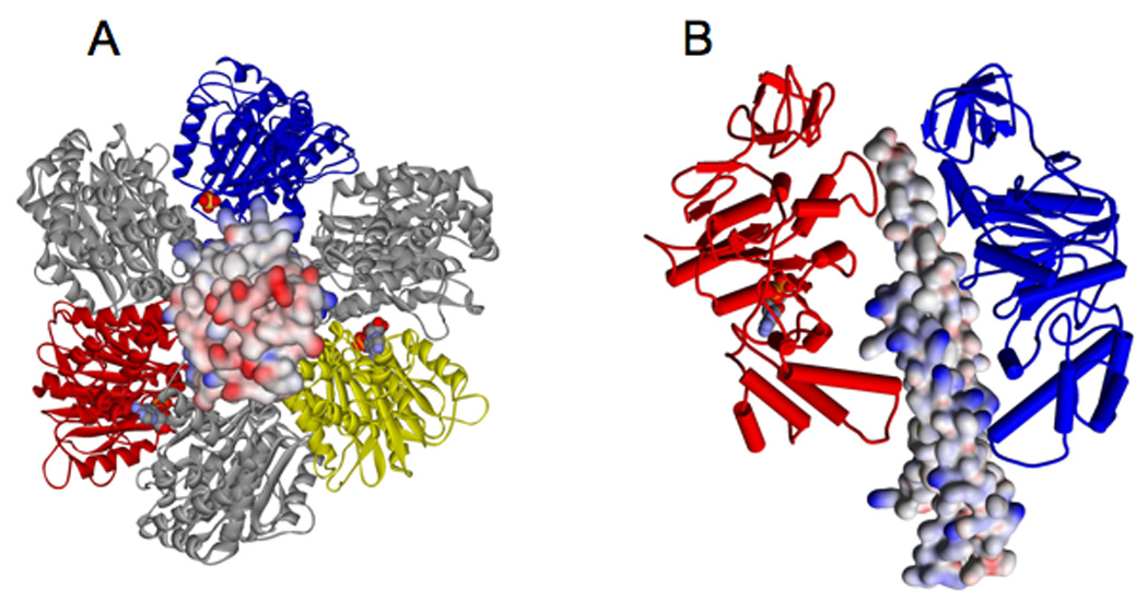
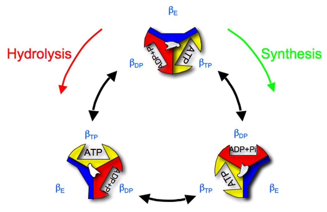
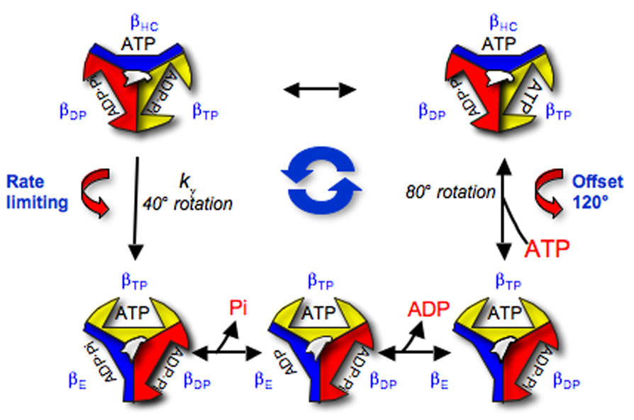
The F0F1 ATP synthase is a large complex of at least 22 subunits, more than half of which are in the membranous F0 sector. This nearly ubiquitous transporter is responsible for the majority of ATP synthesis in oxidative and photo-phosphorylation, and its overall structure and mechanism have remained conserved throughout evolution. Most examples utilize the proton motive force to drive ATP synthesis except for a few bacteria, which use a sodium motive force. A remarkable feature of the complex is the rotary movement of an assembly of subunits that plays essential roles in both transport and catalytic mechanisms. This review addresses the role of rotation in catalysis of ATP synthesis/hydrolysis and the transport of protons or sodium.


Figures

Fig. 1
Fig. 1
Overall subunit organization and structure of the ATP synthase complex (see text for references). The F1 is based on the structures of Stock et al. [74] and Gibbons et al. [27] (α yellow, β red, γ blue, ε brown, and c subunits (black lines) within the membrane (the surfaces of which are indicated by the green rectangles). The subunits whose structures are not completely known are shown graphically. The transport essential carboxylic acid of the c subunits are indicated by the “-“ symbols in red circles. The proposed H+ or Na+ channels are indicated by the yellow (outwards) and green (matrix or cytoplasmic) cylinders, with the conserved and essential a subunit Arg residue indicated by the “+” in blue.
Fig. 2
Fig. 2
Ribbon diagram of the bovine heart mitochondrial F1 based on [2]. A. From the “bottom”, or membrane facing side, of the F1 complex. α subunits are in gray, βTP in yellow, βDP in red, βE in blue, and γ in the surface model showing electrostatic potential (blue is negative, red is positive and white is apolar). B. From the “side” of the complex showing only the γ subunit in relationship to two of the β subunit conformers, βDP (red) with bound ADP (in CPK colors), and βE (blue). Note that the lower portion of the βE subunit is swung outwards which results in an open conformation of the nucleotide binding catalytic site.
Fig. 3
Fig. 3
Key residues contributing to the three catalytic sites. A. Overlay of the residues in βTP (in CPK colors) and βDP sites (in yellow). Bound ATP or ADP and Mg2+ are in black. B. The same amino acids in βE. Note the difference in positions 32 of certain amino acids, in particular αArg376 (E. coli numbering) and αSer347 (red in βTP, green in βDP and βE), which indicate the more open βE conformation [2].
Fig. 4
Fig. 4
The rotation of the β subunit conformers during rotational catalysis. In looking at the F1 from the “bottom” or membrane oriented side of the complex, rotation of the γ subunit is counter-clockwise for ATP hydrolysis, and clockwise in ATP synthesis (see [9] for a review). Note the order of the conformations in hydrolysis is βE→βTP→βDP, and in synthesis is βE→βDP→βTP.
Fig. 5
Fig. 5
Model for partial reaction steps during rotational catalysis based on the kinetic model of Baylis Scanlon et al. [52]. The model explicitly denotes the binding of ATP to the site in the βHC conformer [25], the reversible hydrolysis/synthesis occurring in the βTP site, and the release of product Pi and ADP from βE. Note that the 80° rotation of the γ subunit (eccentric in the middle of the trimer of the β subunit conformers) is associated with ATP binding or release, and the 40° rotation, kγ, is the rate limiting step. The central blue arrows indicate the counter-clockwise rotation in hydrolysis. Notice that the “offset” indicates a 120° rotation in the counter-wise direction in hydrolysis, or 120° rotation in the clockwise direction in synthesis.

References

    1. Nicholls DG, Ferguson SJ. Bioenergetics 3. London, UK: Academic Press; 2002.
    1. Abrahams JP, Leslie AGW, Lutter R, Walker JE. Nature. 1994;370:621–628. - PubMed
    1. Boyer PD. FASEB J. 1989;3:2164–2178. - PubMed
    1. Duncan TM, Bulygin VV, Zhou Y, Hutcheon ML, Cross RL. Proc. Natl. Acad. Sci. U. S. A. 1995;92:10964–10968. - PMC - PubMed
    1. Sabbert D, Engelbrecht S, Junge W. Nature. 1996;381:623–625. - PubMed

LinkOut - more resources