. 2008 Sep 19;283(38):25900-12.
doi: 10.1074/jbc.M707692200. Epub 2008 May 30.

Interleukin (IL) 1beta induction of IL-6 is mediated by a novel phosphatidylinositol 3-kinase-dependent AKT/IkappaB kinase alpha pathway targeting activator protein-1

Affiliations

Interleukin (IL) 1beta induction of IL-6 is mediated by a novel phosphatidylinositol 3-kinase-dependent AKT/IkappaB kinase alpha pathway targeting activator protein-1

Catherine M Cahill et al. J Biol Chem. .

Abstract

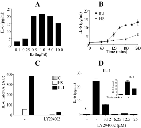
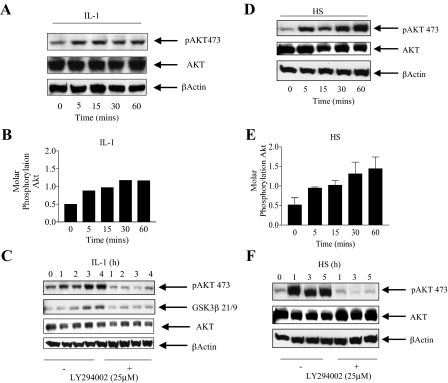
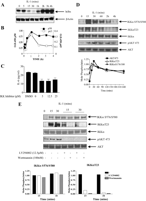
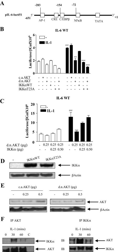
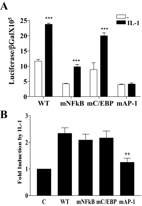
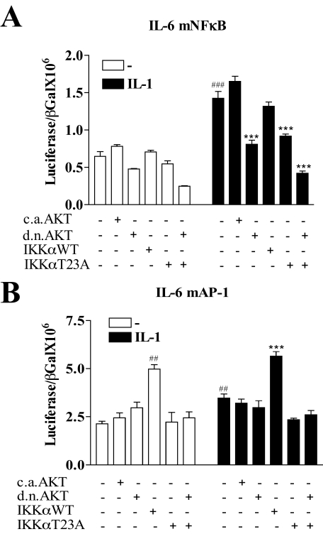
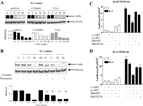
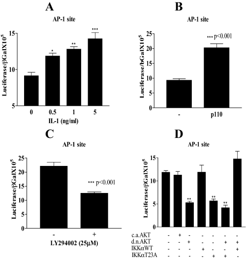
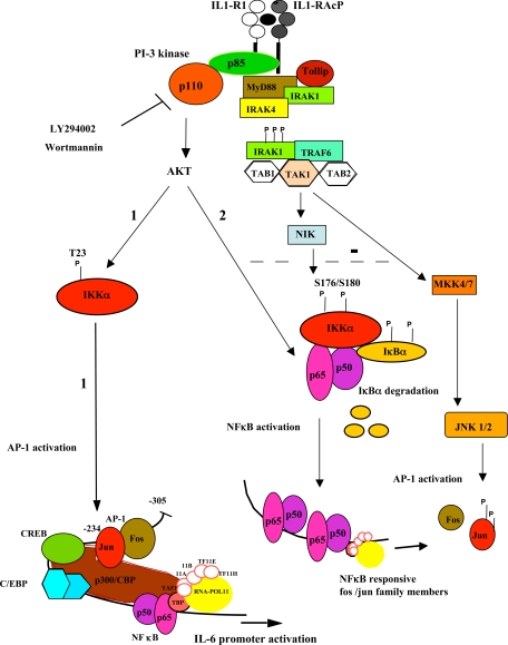
Here we describe a novel role for the phosphatidylinositol 3-kinase/AKT pathway in mediating induction of interleukin-6 (IL-6) in response to IL-1. Pharmacological inhibition of phosphatidylinositol 3-kinase (PI3K) inhibited IL-6 mRNA and protein production. Overexpression of either dominant-negative AKT or IkappaB kinase alpha mutant, IKKalphaT23A, containing a mutation in a functional AKT phosphorylation site, shown previously to be important for NFkappaB activation, completely abrogated IL-6 promoter activation in response to IL-1. However, mutation of the consensus NFkappaB site on the IL-6 promoter did not abrogate promoter activation by IL-1 in contrast to the AP-1 site mutation. IL-1 induces phosphorylation of IKKalpha on the NFkappaB inducing kinase (NIK) phosphorylation sites Ser(176)/Ser(180) and on the Thr(23) site, and although phosphorylation of IKKalphaT23 is inhibited both by LY294002 and wortmannin, phosphorylation of Ser(176)/Ser(180) is not. Neither inhibition of PI 3-kinase/AKT nor IKKalphaT23A overexpression affected IkappaBalpha degradation in response to IL-1. Only partial inhibition by dominant-negative AKT and no inhibitory effect of IKKalphaT23A was observed on an IL-6 promoter-specific NFkappaB site in contrast to significant inhibitory effects on the AP-1 site. Taken together, we have discovered a novel PI 3-kinase/AKT-dependent pathway in response to IL-1, encompassing PI 3-kinase/AKT/IKKalphaT23 upstream of AP-1. This novel pathway is a parallel pathway to the PI 3-kinase/AKT upstream of NFkappaB and both are involved in IL-6 gene transcription in response to IL-1.


Figures

FIGURE 1.
FIGURE 1.
Interleukin 1β induction of IL-6 mRNA and IL-6 release in Caco-2 cells is mediated by a PI 3-kinase-dependent pathway. A, dose response for IL-1 treatment of Caco-2 cells (0–10 ng/ml). Semi-confluent cells were treated with IL-1 and the culture supernatant harvested and assayed by ELISA for IL-6. Data are presented as duplicate experiments measured in triplicate. B, time course of IL-6 produced in response to IL-1 or HS. Cells were treated with 0.5 ng/ml of IL-1 or heat shocked at 43 °C for 1 h and returned to 37 °C. Culture supernatant was harvested at the indicated time points up to 4 h and assayed for IL-6 by ELISA. Mean ± S.E. (n = 3) from three separate experiments. C, cells were treated with IL-1 (0.5 ng/ml) or heat shocked (HS) at 43 °C for 1 h and returned to 37 °C, in the presence or absence of the PI 3-kinase inhibitor LY294002 (25 μm) and harvested at To (C) and at 2 h following initial treatment. Stat-60 was used for mRNA extraction. Quantitative reverse transcriptase-PCR for IL-6 mRNA analysis is presented, mean ± S.E. (n = 2). D, Caco-2 cells were left untreated (C; control), or IL-1 (0.5 ng/ml) alone treated. Treatments were given in the presence or absence of the PI 3-kinase inhibitor LY294002 (3.12 to 25 μm) or wortmannin (100 nm, inset) administered 10 min prior to IL-1 treatment. Tissue culture media were harvested 4 h after IL-1 treatment and assayed for IL-6 by ELISA. Mean ± S.E. (n = 3) from three separate experiments are presented.
FIGURE 2.
FIGURE 2.
Interleukin-1 and heat shock activate AKT in a PI 3-kinase dependent manner. A, Caco-2 cells were treated with IL-1 (0.5 ng/ml) and harvested at the indicated time points. Lysates (70 μg) were run on 10% SDS-PAGE followed by immunoblotting with phospho-specific AKT Ser473. The lysate blots were stripped using 5 ml of Pierce stripping reagent and reprobed using an anti-AKT antibody and an antibody for β-actin. B, the intensity of phosphorylation of AKT over a 1-h time course was estimated using NIH Image software and is expressed as the ratio of phosphorylated protein over total AKT. C, Caco-2 cells were treated with IL-1 (0.5 ng/ml) for up to 4 h in the presence or absence of the PI 3-kinase inhibitor (LY294002, 25 μm) incubated 30 min prior to IL-1 treatment. Cells were harvested and lysed at the indicated hourly time points. AKT was immunoprecipitated and the kinase assay performed using GSK3β peptide. The product of the reaction and the initial lysate were blotted as in A. D, cells were heat shocked (43 °C for 1 h) and harvested at the indicated time points during HS. Cells were lysed, and blotted for AKT Ser473 as in A. E, the intensity of phosphorylation of AKT over the 1-h time course for HS treatment was estimated using NIH Image software as in B. Mean ± S.E. (n = 3) from four separate experiments are presented. F, cells were HS at 43 °C for 1 h in the presence or absence of the PI 3-kinase inhibitor (LY294002, 25 μm) incubated 30 min prior to HS and returned to 37 °C for an additional 4 h. Cells were harvested and lysed at the indicated hourly time points. AKT Ser473 phosphorylation was detected as in A.
FIGURE 3.
FIGURE 3.
Canonical NFκB activation correlates with AKT phosphorylation on phospho-Ser473; requirement for the IKK complex in IL-6 production. A, cells were treated with IL-1 (0.5 ng/ml) and harvested at the indicated time points. Lysates were run on 10% SDS-PAGE and blotted for IκBα. B, nuclear extracts prepared from IL-1-treated cells at the indicated time points were assayed for the binding of p50 and p65 to an NFκB consensus binding site in a Trans-AM ELISA system according to the manufacturer's instructions. Means from duplicate measurements from two experiments are presented. C, cells were treated with IL-1 (0.5 ng/ml) for 4 h in the presence or absence of an IKK complex inhibitor (5–25 μm) (Calbiochem) incubated 30 min prior to IL-1 stimulation. Tissue culture supernatants were harvested and assayed for IL-6 by ELISA. Mean ± S.E. (n = 3) from three separate experiments are presented. D, cells were treated with IL-1 at the indicated time points and total IKKα was immunoprecipitated overnight. Immunoprecipitates were run on a 10% SDS gel and blotted separately for phospho-IKKα Ser176/Ser180, phospho-IKKαT23, and total IKKα. Lysates were run in parallel and blotted for phospho-AKTS473 and total AKT. Molar phosphorylation of IKKα on Ser176/Ser180 and Thr23, together with Akt Ser473 is presented (lower panel). E, effect of the PI 3-kinase inhibitors LY294002 (12.5 μm) and wortmannin (100 nm) on the phosphorylation of IKKα S176/180, IKKαT23, and AKTS473 following IL-1 stimulation at 15 and 30 min. Molar phosphorylation of IKKα on Ser176/Ser180 and Thr23 is presented (lower panel).
FIGURE 4.
FIGURE 4.
Interleukin 1 induction of IL-6 promoter activation is dependent on AKT and on the Thr23 AKT phosphorylation site on IKKα. A, parental IL-6 promoter construct, pIL-6-luc651 containing 651 bases from the transcription start site. Relevant transcription factor binding sites are shown, including NFκB, AP-1, and C/EBPβ. B, cells were transfected with the IL-6 promoter luciferase construct pIL-6-luc651 (0.25 μg) in the presence or absence of a constitutively active AKT (caAKT) (0.125 μg), dominant-negative AKT (dnAKT) (0.125 μg), IKKαWT (0.125 μg) or IKKαT23A (0.125 μg). β-Galactosidase (0.25 μg) was co-transfected for transfection efficiency. Eighteen hours after transfection cells were starved for 3 h and then either untreated (–) or treated with IL-1 (0.5 ng/ml) overnight. Cells were harvested and lysates were assayed for luciferase and β-galactosidase. Results are expressed as the ratio of luciferase over β-galactosidase. Mean ± S.E. (n = 3). ###, p < 0.001 untreated pIL-6-luc651 alone transfected compared with IL-1 treated. Comparison of IL-1-treated transfectants; **, p < 0.01, pIL-6-luc651 alone transfected compared with co-transfection with dnAKT; ***, p < 0.001 pIL-6-luc651 alone transfected compared with co-transfection with IKKαT23A or IKKαT23A + dnAKT. C, cells were transfected with the IL-6 promoter luciferase construct pIL-6-luc651 (0.50 μg) in the presence or absence of dnAKT (0.125 μg) or IKKα WT (0–0.50 μg) together with β-galactosidase (0.25 μg) for transfection efficiency. Results are expressed as the ratio of luciferase over β-galactosidase. Mean ± S.E. (n = 3). ###, p < 0.001 pIL-6-luc651 alone transfected, untreated compared with IL-1 treated. IL-1-treated transfectants, ***, p < 0.001, pIL-6-luc651 alone transfected compared with co-transfection with dnAKT. D, Western blot of lysates from Caco-2 cells transfected with empty vector (–) or plasmids expressing IKKα WT or IKKαT23A (0.25 μg), using polyclonal IKKα antibody and a β-actin antibody for loading control. E, Western blot of lysates from Caco-2 cells transfected with empty vector (–) or plasmids expressing caAKT or dnAKT (0.25 and 0.5 μg) using an antibody against total AKT and β-actin for loading control. F, AKT association with IKKα in response to IL-1. Caco-2 cells were either untreated or treated with IL-1 (0.5 ng/ml) at the indicated time points. Lysates were immunoprecipitated (IP) overnight with anti-IKKα antibody, AKT antibody, or rabbit IgG (C, control). Washed immunoprecipitates were run on 10% SDS-PAGE and the AKT immunoprecipitates (left panel) and IKKα immunoprecipitates (right panel) were blotted for the presence of both IKKα and AKT.
FIGURE 5.
FIGURE 5.
Mutation of the consensus NFκB site affects baseline IL-6 promoter activity, whereas mutation of the AP-1 site significantly impairs its induction by IL-1. Caco-2 cells were transfected with the IL-6 promoter luciferase constructs (0.25 μg), pIL-6-luc651WT (WT), pIL-6-luc651 mutated in the NFκB binding site, pIL-6-luc651 (mNFκB), pIL-6-luc651 mutated in the consensus binding site for C/EBPβ, pIL-6-luc651 (mC/EBPβ), or pIL6–651 mutated in the AP-1 site, pIL-6-luc651 (mAP-1). A β-galactosidase reporter (0.25 μg) was included for transfection efficiency. Eighteen hours after transfection, cells were starved for 3 h and were either untreated or IL-1 treated (0.5 ng/ml) overnight. Lysates were assayed for luciferase and β-galactosidase. A, baseline and IL-1 stimulation of each construct with promoter activation expressed as the ratio of luciferase/β-galactosidase. Mean ± S.E. (n = 3). ***, p < 0.001, pIL-6-luc651WT and mutants, mNFκB, mC/EBP, untreated compared with IL-1 treated. B, -fold induction by IL-1 with untreated, control values set at 1, mean ± S.E. (n = 3). ***, p < 0.001, IL-6-651mAP-1 transfectants compared with pIL-6–651WT transfectants treated with IL-1.
FIGURE 6.
FIGURE 6.
Mutation of the IL-6 promoter NFκB site neither abrogates IL-1 induction nor the inhibitory effect of transfected dominant-negative AKT or IKKαT23A. Caco-2 cells were transfected with the IL-6 promoter luciferase reporter plasmids (0.25 μg) containing either a mutated NFκB site, pIL-6–651mNFκB(A), or a mutated AP-1 site, pIL-6–651mAP-1 (B), in the presence or absence of one of the following expression plasmids (0.125 μg), caAKT, dnAKT, IKKαWT, or IKKαT23A. β-Galactosidase (0.25 μg) was co-transfected for transfection efficiency. Eighteen hours after transfection, cells were starved for 3 h and treated overnight with IL-1β (0.5 ng/ml). Cells were harvested and lysates were assayed for luciferase and β-galactosidase. Promoter activation is expressed as the ratio of luciferase/β-galactosidase. Mean ± S.E. (n = 3). ###, p < 0.001; ##, p < 0.01, pIL-6-luc651 control, untreated transfectants compared with IL-1 treated; ***, p < 0.001, pIL-6-luc651, IL-1 treated compared with co-transfected dnAKT or IKKαT23A (n = 3).
FIGURE 7.
FIGURE 7.
AKT is required for NFκB activation in response to IL-1, independent of IKKαT23 and IκBα degradation. A, cells were transfected with empty vector, pcDNA3, or IKKαT23A. Eighteen hours after transfection cells were serum starved. Cycloheximide (50 μg/ml) was added to prevent protein resynthesis; 1 h thereafter cells were treated with IL-1β. Cells were either untreated or pretreated with the PI 3-kinase inhibitor LY294002 (25 μm) prior to IL-1 (0.5 ng/ml) treatment. Lysates harvested at the indicated time points were blotted for the presence of IκBα. β-Actin is included as a loading control and the ratio of IκBα to β-actin (NIH Image analysis) is plotted below for the three treatment groups. B, cells were treated with IL-1β in the presence or absence of LY294002 (12.5 μm) or wortmannin (100 nm) pretreated 10 min prior to IL-1 treatment. Cell lysates were harvested at the indicated time points and blotted for IκBα. Blots were stripped and reprobed for β-actin as a loading control. The ratio of IκBα to β-actin (NIH Image analysis) is plotted below for the treatment groups. C, Caco-2 cells were transfected with luciferase reporter plasmids containing a 3× NFκB consensus site from the IgκB gene (0.25 μg); or D, the IL-6 promoter NFκB site (0.25 μg), in the presence or absence of one of the following expression plasmids (0.125 μg), caAKT, dnAKT, IKKαWT, or IKKαT23A. β-Galactosidase (0.25 μg) was included for transfection efficiency. Eighteen hours after transfection, cells were starved for 3 h and treated overnight with IL-1β (0.5 ng/ml). Cells were harvested and lysates were assayed for luciferase and β-galactosidase. Reporter activation is expressed as the ratio of luciferase/β-galactosidase. Mean ± S.E. (n = 3). ***, p < 0.001 IL-1-treated NFκB reporter transfectants compared with the co-transfected dnAKT.
FIGURE 8.
FIGURE 8.
AP-1 is the target transcription factor of PI 3-kinase/AKT/IKKα. In A, Caco-2 cells were transfected with the IL-6 promoter-specific AP-1 site luciferase reporter (0.25 μg) together with β-galactosidase (0.25 μg). Eighteen hours after transfection, cells were treated with IL-1 (0, 0.5, 1, and 5 ng/ml) overnight. B, IL-6 promoter-specific AP-1 site reporter (0.25 μg) was transfected alone or co-transfected with p110 subunit of PI 3-kinase (0.125 μg) together with β-galactosidase (0.25 μg) in HT29 cells. C, Caco-2 cells were transfected with the IL-6 promoter-specific AP-1 site reporter (0.25 μg). Eighteen hours after transfection cells were either untreated or treated overnight with the PI 3-kinase inhibitor LY294002 (25 μm). D, Caco-2 cells were transfected with the IL-6 promoter-specific AP-1 reporter (0.25 μg), alone or co-transfected with caAKT, dnAKT, IKKαWT, or IKK T23A (0.125 μg) and combinations of either, dnAKT + IKKαT23A or dnAKT + IKKαWT together with β-galactosidase. Eighteen hours after transfection, cells were starved for 3 h and treated overnight with IL-1 (0.5 ng/ml). Cells were harvested 18 h following treatments and lysates were assayed for luciferase and β-galactosidase. Reporter activation for A–D is expressed as the ratio of luciferase/β-galactosidase. Mean ± S.E., n = 3. **, p < 0.01 AP-1 luciferase alone transfected compared with co-transfection with dnAKT, IKKαT23A, or both.
FIGURE 9.
FIGURE 9.
PI 3-kinase/AKT-dependent pathways involved in IL-1 induction of IL-6. The current model for the binding of IL-1 to its receptor, IL-1R1, and co-receptor, IL-1 receptor accessory protein IL1-RAcP (16). Formation of the signaling module containing MyD88, phosphorylated interleukin-1 receptor-associated kinase (IRAK), and TRAF6 (TNF receptor associated factor), is essential for PI 3-kinase recruitment and AKT activation (21, 73, 74, 81). The kinase TAK1 (TGFβ-activated kinase) activates the NIK-IκB-NFκB pathway and the MAP kinase cascade (MKK4/7 to JNK/AP-1 activation as well as the MKK3/6 to p38 activation (not shown) (, , –84). Possible negative regulation in the Caco-2 cell line between AKT and JNK pathways is shown as a broken line (85, 86). We have identified 2 separate PI-3 kinase-dependent pathways from the IL1-R1 complex to the activation of IL-6 gene transcription. 1, a novel pathway leading to AP-1-dependent induction of IL-6 via IKKαT23. 2, an NFκB-dependent pathway, which indirectly induces IL-6, likely by inducing factors such as AP-1 family members, which are necessary for IL-6 gene transcription in response to IL-1.

References

    1. Hirano, T. (1998) Int. Rev. Immunol. 16 249–284 - PubMed
    1. Nishimoto, N., and Kishimoto, T. (2006) Nat. Clin. Pract. Rheumatol. 2 619–626 - PubMed
    1. Naugler, W. E., Sakurai, T., Kim, S., Maeda, S., Kim, K., Elsharkawy, A. M., and Karin, M. (2007) Science 317 121–124 - PubMed
    1. Rakoff-Nahoum, S., and Medzhitov, R. (2007) Science 317 124–127 - PubMed
    1. Greten, F. R., Eckmann, L., Greten, T. F., Park, J. M., Li, Z. W., Egan, L. J., Kagnoff, M. F., and Karin, M. (2004) Cell 118 285–296 - PubMed

Publication types