. 2008 Sep;95(5):2203-18.
doi: 10.1529/biophysj.108.133199. Epub 2008 May 30.

Dependence of invadopodia function on collagen fiber spacing and cross-linking: computational modeling and experimental evidence

Affiliations

Dependence of invadopodia function on collagen fiber spacing and cross-linking: computational modeling and experimental evidence

Heiko Enderling et al. Biophys J. 2008 Sep.

Abstract

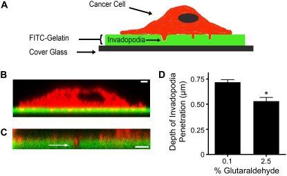
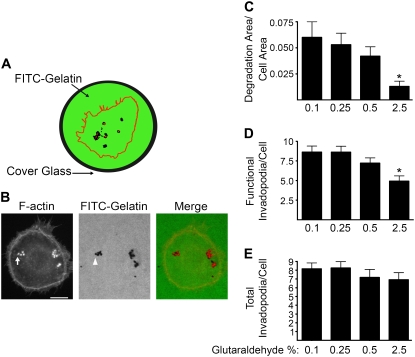
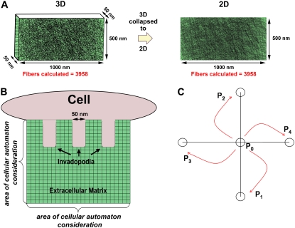
Invadopodia are subcellular organelles thought to be critical for extracellular matrix (ECM) degradation and the movement of cells through tissues. Here we examine invadopodia generation, turnover, and function in relation to two structural aspects of the ECM substrates they degrade: cross-linking and fiber density. We set up a cellular automaton computational model that simulates ECM penetration and degradation by invadopodia. Experiments with denatured collagen (gelatin) were used to calibrate the model and demonstrate the inhibitory effect of ECM cross-linking on invadopodia degradation and penetration. Incorporation of dynamic invadopodia behavior into the model amplified the effect of cross-linking on ECM degradation, and was used to model feedback from the ECM. When the model was parameterized with spatial fibrillar dimensions that closely matched the organization, in real life, of native ECM collagen into triple-helical monomers, microfibrils, and macrofibrils, little or no inhibition of invadopodia penetration was observed in simulations of sparse collagen gels, no matter how high the degree of cross-linking. Experimental validation, using live-cell imaging of invadopodia in cells plated on cross-linked gelatin, was consistent with simulations in which ECM cross-linking led to higher rates of both invadopodia retraction and formation. Analyses of invadopodia function from cells plated on cross-linked gelatin and collagen gels under standard concentrations were consistent with simulation results in which sparse collagen gels provided a weak barrier to invadopodia. These results suggest that the organization of collagen, as it may occur in stroma or in vitro collagen gels, forms gaps large enough so as to have little impact on invadopodia penetration/degradation. By contrast, dense ECM, such as gelatin or possibly basement membranes, is an effective obstacle to invadopodia penetration and degradation, particularly when cross-linked. These results provide a novel framework for further studies on ECM structure and modifications that affect invadopodia and tissue invasion by cells.


Figures

FIGURE 1
FIGURE 1
Electron-micrograph image of cancer-cell invadopodia. Ultrathin vertical-section electron micrograph of an SCC-61 oral squamous carcinoma cell, attached to fibronectin, reveals multiple invadopodia protruding into the underlying matrix. Scale bar = 500 nm.
FIGURE 2
FIGURE 2
The depth of invadopodia penetration is regulated by ECM cross-linking. (A) For the visualization of invadopodia, cancer cells were allowed to adhere to cover glass that was coated with FITC-conjugated gelatin. Cancer cells developed invadopodia that degraded the surrounding ECM, and penetrated into the FITC-gelatin. (B) Confocal Z-section of CA1D breast-cancer cell stained with rhodamine phalloidin, to identify actin filaments (red) attached to FITC-gelatin (green), with multiple invadopodia protruding into the underlying ECM. Scale bar = 2 μm. (C) Representative Z-section, to quantify invadopodia penetration depth, shows a close-up of a CA1D invadopodium (arrow) that degraded and protruded into the FITC-gelatin. Scale bar = 2 μm. (D) Quantification of invadopodia penetration depth of CA1D cells cultured on FITC-gelatin gels that were cross-linked with 0.1% or 2.5% glutaraldehyde buffer. Data are reported as mean ± SE, and are from three independent experiments. *p < 0.05.
FIGURE 3
FIGURE 3
Invadopodia-associated ECM degradation is regulated by cross-linking. (A) Depiction of a typical image from wide-field microscopy of a rhodamine-phalloidin-stained cell (rhodamine-phalloidin stains actin filaments red), cultured for 18 h on FITC-gelatin (green). Functional invadopodia degraded the FITC-gelatin, leaving black areas with no FITC fluorescence. Invadopodia were defined as punctate spots of F-actin staining localized to areas of degradation, and appear as red spots against a black background. (B) Representative wide-field image of a CA1D cell cultured on gelatin that was cross-linked with 0.1% glutaraldehdye buffer. Arrow indicates a cluster of four distinct invadopodia. Arrowhead indicates area of FITC-gelatin degradation from the invadopodia (arrow). This clustering of invadopodia was typical in these cells. Scale bar = 10 μm. (C) Quantification of total area of FITC-gelatin degradation per cell, from experiments using CA1D cells cultured on FITC-gelatin gels that were cross-linked with different percentages of glutaraldehyde buffer. (D) Quantification of number of functional invadopodia (actin puncta associated with FITC-gelatin degradation) per cell. (E) Quantification of total number of invadopodia (all actin puncta, regardless of associated degraded ECM) per cell. Data are reported as mean ± SE, and are from three independent experiments. *p < 0.05, compared with 0.1% glutaraldehyde condition.
FIGURE 4
FIGURE 4
Schematic of cellular automaton model. (A) Calculation of fiber number in the ECM domain. Fiber number was calculated from the number of single-stranded gelatin molecules that were contained in a 50-nm-thick slice of 2.5% gelatin, as might be experienced by an invadopodium in experiments. That fiber number (3958) was then used in 2D simulations. (B) Scale of computational model. We assumed that a cell sat on top of the extracellular matrix, and in simulations we considered only the area of ECM in which the invadopodia protruded beneath the cell. (C) Schematic of the probability distribution of invadopodia moving and protruding into the computational ECM. P1 is the probability of downward movement into the gel. P2 is the probability of upward movement, toward the cell-ECM interface. P3 and P4 are the probabilities of moving left or right, respectively. P0 is the probability of staying stationary.
FIGURE 5
FIGURE 5
Invadopodia penetration in simulated gelatin: effect of a penalty for ECM cross-linking. (A) Enlarged view of a simulated ECM domain that contains 12 fibers with 13 intersections (red circles). Cross-links (blue dots) at intersecting fibers are shown for 50% and 100% cross-link ratios, respectively. (B) An invadopodium (red) invades a matrix with several cross-linked (blue) and unlinked fibers (black). Degraded ECM fibers (white) are also shown. The contents of A and B are not to scale. (C) Sample screen shots from simulations of invadopodia protruding into an ECM domain with 3958 fibers and cross-link ratios cl = 0%, 25%, and 100% at t = 1000 time steps. (D) Penetration depth (black) and ECM degradation (red) for invadopodia protruding into gelatin domains with different cross-linking ratios. Simulations were run for 1000 time steps for each cross-linking variable. Shown is the mean ± SE from 40 invadopodia for each condition. See also Movie S1.
FIGURE 6
FIGURE 6
Inclusion of invadopodia retraction in the model. In these simulations, a retraction rule was implemented in which any invadopodium that does not degrade ECM for ψ >15 time steps becomes retracted. Simulations were initiated with four invadopodia, and were run until all invadopodia were retracted. (A) Sample screen shots at various times from simulations of invadopodia going through ECM domains with 0%, 25%, or 100% cross-linking. Note that retraction (empty stripe where red invadopodium was) occurs before reaching the end of the domain in the 25% and 100% conditions, because of the immobility of invadopodia. (B) Graph of invadopodia lifetime (number of timesteps until retraction). In the 25% cross-linking condition, the average invadopodia lifetime increases because the majority of invadopodia reach the end of the domain without retraction, but with a delay compared to the 0%, 5%, and 10% cross-link conditions. (C) Invadopodia penetration depth (black line) and ECM degradation (red line) at time of retraction, with respect to cross-link ratio. Because simulations were run until no invadopodia were in the domain, penetration and degradation inhibition in cross-linked ECM occurred only when significant retraction occurred (e.g., in domains with cross-link ratios of 50% or 100%). Shown is the mean ± SE from 60 invadopodia for each cross-linking variable.
FIGURE 7
FIGURE 7
Inclusion of new invadopodia formation in the model. Formation of new invadopodia was modeled by implementing a background initiation of new invadopodia with a probability of ζ * 1/1500 (once every 1500 time steps). Under baseline conditions, ζ = 1. Modification of this baseline rate by invadopodia retraction led to either positive (ζ = 1.1) or negative (ζ = 0.75) feedback from ECM conditions. A single invadopodium was initiated at time = 0 in our standard ECM domains, to allow room for additional invadopodia that formed during the simulation. Plot shows the number of newly initiated invadopodia (black) and degraded ECM (red) as a function of ECM cross-linking and ζ, as indicated. These data were taken from simulations that were run for 10,000 time steps. Shown is the mean ± SE from 100 invadopodia for each cross-linking variable. See also Movie S2.
FIGURE 8
FIGURE 8
Invadopodia lifetime is regulated by ECM cross-linking. (A) Images from 2-h movies taken of CA1D cells expressing GFP-ARPC1 cultured on 0.1% cross-linked Texas-red gelatin. Time scale is in minutes. See also Movie S3. (B) Images from 2-h movies taken of CA1D cells cultured on 2.5% cross-linked Texas-red gelatin. Time scale is in minutes. Note the increased number of invadopodia over the course of the movie. See also Movie S4. (C) Quantification of number of newly developed invadopodia that appeared during filming of the movies. (D) Quantification of lifetimes of invadopodia that appeared and disappeared during filming of movies. Data for each condition are reported as mean ± SE, and are taken from six independent experiments with 25 individual cells. *p < 0.05.
FIGURE 9
FIGURE 9
Model implementation with different ECM domains. (A, left to right) images of 25 mg/mL gelatin, 1 mg/mL collagen monomers, 1.67 mg/mL microfibrils, and a macrofibril. (B) Screenshots from simulations of invadopodia migrating into 0% and 100% cross-linked 1 mg/mL collagen monomers. Simulations were run with or without rules incorporating invadopodia dynamics, as indicated, for t = 1000 time steps (without dynamics) or t = 10,000 time steps (with dynamics). The last image from each simulation is shown. Note that with dynamics, the invadopodia are rarely present in the domain, because of a lack of interaction with ECM and rapid retraction. See also Movie S5 and Movie S6. (C) Plots of penetration depth (black) and degradation area (red) for 1 mg/mL collagen monomers (solid line) and 25 mg/mL gelatin (dashed line) without invadopodia dynamics. (D) Plots of penetration depth (black and gray) and degradation area (red and orange) for 1 mg/mL collagen monomers (black and red) and 25 mg/mL gelatin (gray and orange) with invadopodia dynamics. The ζ modifications of initiation probabilities for baseline, negative, and positive feedback are as indicated at right.

References

    1. Linder, S. 2007. The matrix corroded: podosomes and invadopodia in extracellular matrix degradation. Trends Cell Biol. 17:107–117. - PubMed
    1. Weaver, A. M. 2006. Invadopodia: specialized cell structures for cancer invasion. Clin. Exp. Metastasis. 23:97–105. - PubMed
    1. Artym, V. V., Y. Zhang, F. Seillier-Moiseiwitsch, K. M. Yamada, and S. C. Mueller. 2006. Dynamic interactions of cortactin and membrane type 1 matrix metalloproteinase at invadopodia: defining the stages of invadopodia formation and function. Cancer Res. 66:3034–3043. - PubMed
    1. Clark, E. S., A. S. Whigham, W. G. Yarbrough, and A. M. Weaver. 2007. Cortactin is an essential regulator of matrix metalloproteinase secretion and extracellular matrix degradation in invadopodia. Cancer Res. 67:4227–4235. - PubMed
    1. Burgstaller, G., and M. Gimona. 2005. Podosome-mediated matrix resorption and cell motility in vascular smooth muscle cells. Am. J. Physiol. Heart Circ. Physiol. 288:H3001–H3005. - PubMed

Publication types

LinkOut - more resources