. 2008 Sep;95(5):2283-91.
doi: 10.1529/biophysj.107.127423. Epub 2008 May 30.

On the structure of the N-terminal domain of the MscL channel: helical bundle or membrane interface

Affiliations

On the structure of the N-terminal domain of the MscL channel: helical bundle or membrane interface

Irene Iscla et al. Biophys J. 2008 Sep.

Abstract

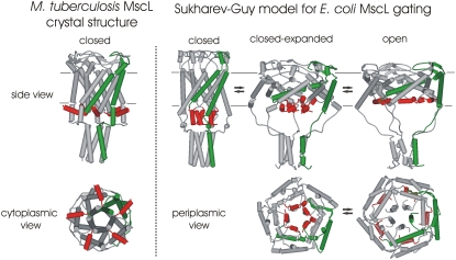
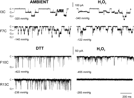
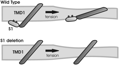
The mechanosensitive channel of large conductance, MscL, serves as a biological emergency release valve protecting bacteria from acute osmotic downshock and is to date the best characterized mechanosensitive channel. A well-recognized and supported model for Escherichia coli MscL gating proposes that the N-terminal 11 amino acids of this protein form a bundle of amphipathic helices in the closed state that functionally serves as a cytoplasmic second gate. However, a recently reexamined crystal structure of a closed state of the Mycobacterium tuberculosis MscL shows these helices running along the cytoplasmic surface of the membrane. Thus, it is unclear if one structural model is correct or if they both reflect valid closed states. Here, we have systematically reevaluated this region utilizing cysteine-scanning, in vivo functional characterization, in vivo SCAM, electrophysiological studies, and disulfide-trapping experiments. The disulfide-trapping pattern and functional studies do not support the helical bundle and second-gate hypothesis but correlate well with the proposed structure for M. tuberculosis MscL. We propose a functional model that is consistent with the collective data.


Figures

FIGURE 1
FIGURE 1
Current models for MscL structure. Schematic representations of M. tuberculosis (left) and E. coli (right) MscL structural models are shown. The most N-terminal region of the channel (amino acids 2 to12) is shown in red, and a single subunit in green for clarity. On the left side of the panel, a model for a closed or nearly closed structure of M. tuberculosis MscL derived from the recently revised crystal structure (15) shows the homopentameric nature of the channel with the N-terminal region positioned at the cytoplasmic interface of the membrane. Side (upper left) and cytoplasmic (lower left) views are shown. The closed, closed-expanded, and open states from the proposed Sukharev-Guy (SG) model for E. coli MscL gating are shown in the three right-most panels (14); side (upper row) and periplasmic (lower row) views are shown. As shown in the middle of these panels (closed-expanded), the model predicts the N-terminal region (called S1) to play the role of a “second gate” that remains closed after most of the expansion of the channel has already occurred. The approximate membrane location is indicated by the parallel gray lines in the side view.
FIGURE 2
FIGURE 2
Deletion of the most N-terminal region of E. coli MscL leads to a protein that is still expressed in the membrane but has a compromised function. (A) The ability of the deletion mutants Δ2–4 and Δ2–12, when expressed in trans, to rescue the MJF455 osmotic downshock-sensitive strain in vivo was compared with that of cells expressing wild-type or empty vector. Deletion mutant Δ2–4 partially rescued the osmotic-lyses phenotype, whereas Δ2–12 proved to be a nonfunctional channel. (B) Protein expression of wild type and both deletion mutants was analyzed by Western blot in whole cell (W), cytosol (C), and membrane fractions (M). Samples were obtained from PB104 cells expressing each of the mutants. Most of the protein was found in membrane fractions.
FIGURE 3
FIGURE 3
In vivo and patch-clamp characterization of S1 cysteine mutants demonstrates that substitutions in the region are well tolerated. (A) Little or no reduction in the stationary phase values was observed for mutants when compared with wild-type MscL (lower dashed line). The dashed line designates the 50% cutoff value used in previous studies to designate a significant functional difference; none of the mutants studied here achieved this value. (B) The ability of the S1 cysteine-substituted mutants to rescue an osmotic-lysis phenotype is shown as a percentage of survival. All of the mutated channels are functional when compared with negative controls (vector only, lower dashed line); however, many of the cysteine substitutions in the S1 region effected only a partial suppression of the osmotic-lysis phenotype when compared with the wild type (upper dashed line). (C) An in vivo SCAM study was performed for the S1 cysteine mutants. The change in their ability to rescue an osmotic-lysis phenotype in the presence of MTSET (black bars) and MTSES (white bars) reagents is shown in the graph as the weighted fold difference ((treated − untreated)/untreated). (D) Single-channel activities of each of the S1 mutants were analyzed by patch clamp in giant spheroplasts. A Δmscl bacterial strain (PB104) with an intact MscS was used. The pressure thresholds for the activation of each of the MscL S1 mutants was compared with that of the internal control MscS and expressed as a ratio.
FIGURE 4
FIGURE 4
In vivo disulfide-trapping experiments show weak intersubunit interactions in S1 cysteine mutants relative to the adjoining S1-TMD1 linker region. (A) Representative Western blots for each of the cysteine-substituted mutants are shown. Samples derived from bacterial cells diluted in a medium of the same osmolarity (m for mock shock) or in water (o for osmotic downshock) were fractionated by SDS-PAGE, and MscL detected by Western blot. (B) The bar graph (left y axis) shows the average and standard error of the percentage total protein that is in the form of dimers for mock shock (dark bars) or osmotically challenged (white bars) samples derived from at least five experiments similar to the examples shown in A. The line graph reflects the inverse distances between α-carbons of neighboring subunits for each residue, as predicted by the model of M. tuberculosis (Tb-MscL, solid triangles) or by the SG model for the closed E. coli MscL (Eco-MscL, open circles). Note that smaller values represent longer predicted distances. Results for the adjacent region (S1-TMD1 linker region, residues R13–D18) derived from similar experiments using an identical protocol (21) are also shown to better determine which model best fits the data. Circled residues are those predicted by the SG model to be in close proximity in the closed and closed-expanded conformations and were shown to yield strong disulfide bridging in membrane preparations (13).
FIGURE 5
FIGURE 5
Single-channel activity of cysteine mutants under different oxidizing conditions. Representative traces of single-channel activity for mutants I3C, F7C, F10C, and R13C shown under different redox conditions. Recordings were made at −20 mV, and by convention openings are shown as downward. No differences were observed in channel activity for I3C and F7C mutants measured before (ambient, left) and after treatment with the oxidant H2O2 (right). Unlike I3C and F7C, F10C and R13C showed variable activity under ambient conditions and were influenced by DTT. Therefore, these membrane patches were treated with DTT for 5 to 35 min (left) before oxidative treatment (right). Note that a decrease in the amplitude of F10C was observed in presence of H2O2, whereas R13C activity was abolished under identical oxidizing conditions. All H2O2-treated traces reflect activities 5 to 15 min after the addition of oxidant.
FIGURE 6
FIGURE 6
Model for the role of the S1 domain in MscL gating. The scheme shows the S1 and TMD1 domains of a single subunit of MscL before (left) and after (right) applying tension to the membrane. The upper graph shows a wild-type MscL where the S1 domain is intact. The two most conserved residues in this region, F7 and F10, are shown as pentagons along this structure, stabilizing the interactions with the membrane. The region serves as an anchor for TMD1 to the cytoplasmic side of the membrane, facilitating and guiding the tilting of TMD1 on membrane-tension-effected bilayer thinning and channel opening. The lower graph shows an S1 deletion mutant of MscL. The lack of this anchor region impairs the proper tilting of the TMD1 domains needed for channel gating, so the channel remains closed. Tilting angles shown for the TMD1 domains in the closed and open states were derived from the crystal structure for Tb-MscL and the open state of E. coli MscL from the SG model, respectively.

References

    1. Hamill, O. P., and B. Martinac. 2001. Molecular basis of mechanotransduction in living cells. Physiol. Rev. 81:685–740. - PubMed
    1. Niu, W., and F. Sachs. 2003. Dynamic properties of strech-activated K+ channels in adult rat atrial myocytes. Prog. Biophys. Mol. Biol. 82:121–135. - PubMed
    1. Sukharev, S., and D. P. Corey. 2004. Mechanosensitive channels: multiplicity of families and gating paradigms. Sci. STKE. 2004:re4. - PubMed
    1. Bass, R. B., P. Strop, M. Barclay, and D. C. Rees. 2002. Crystal structure of Escherichia coli MscS, a voltage-modulated and mechanosensitive channel. Science. 298:1582–1587. - PubMed
    1. Chang, G., R. H. Spencer, A. T. Lee, M. T. Barclay, and D. C. Rees. 1998. Structure of the MscL homolog from Mycobacterium tuberculosis: A gated mechanosensitive ion channel. Science. 282:2220–2226. - PubMed

Publication types

MeSH terms

LinkOut - more resources