. 2008 Jun 10;105(23):8008-13.
doi: 10.1073/pnas.0801758105. Epub 2008 May 30.

Structure and host-cell interaction of SH1, a membrane-containing, halophilic euryarchaeal virus

Affiliations

Structure and host-cell interaction of SH1, a membrane-containing, halophilic euryarchaeal virus

Harri T Jäälinoja et al. Proc Natl Acad Sci U S A. .

Abstract

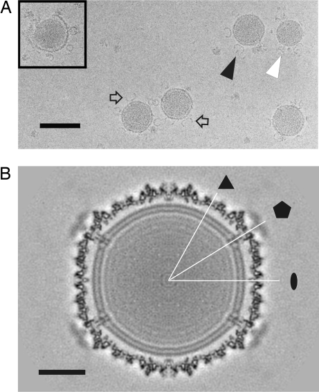
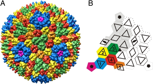
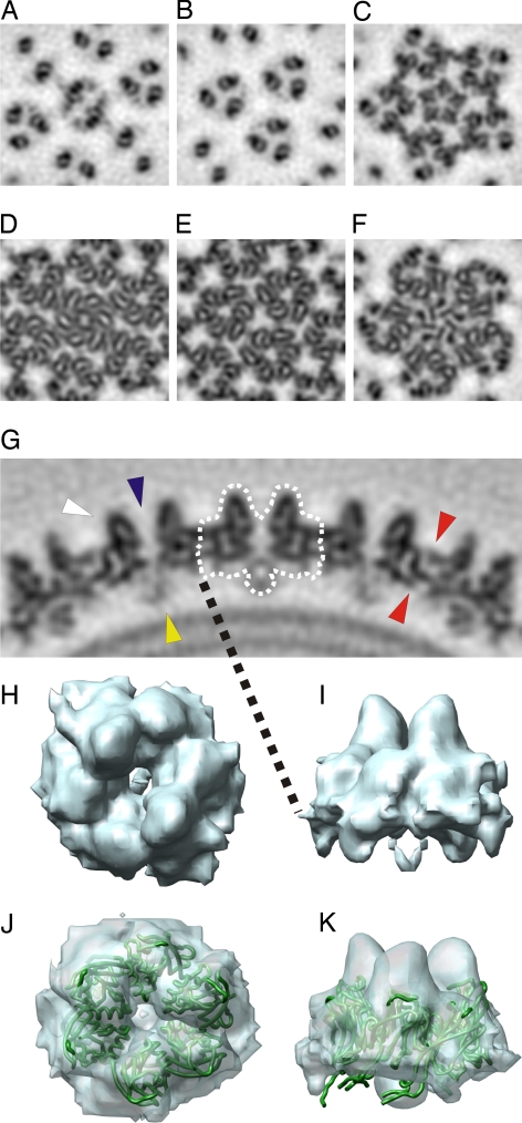
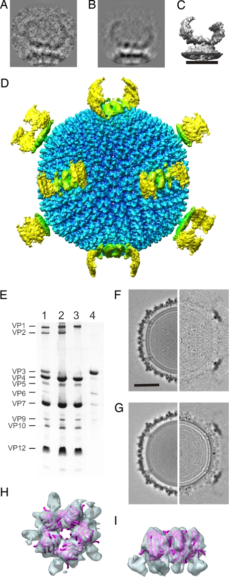
The Archaea, and the viruses that infect them, are the least well understood of all of the three domains of life. They often grow in extreme conditions such as hypersaline lakes and sulfuric hot springs. Only rare glimpses have been gained into the structures of archaeal viruses. Here, we report the subnanometer resolution structure of a recently isolated, hypersalinic, membrane-containing, euryarchaeal virus, SH1, in which different viral proteins can be localized. The results indicate that SH1 has a complex capsid formed from single beta-barrels, an important missing link in hypotheses on viral capsid protein evolution. Unusual, symmetry-mismatched spikes seem to play a role in host adsorption. They are connected to highly organized membrane proteins providing a platform for capsid assembly and potential machinery for host infection.


Conflict of interest statement

The authors declare no conflict of interest.

Figures

Fig. 1.
Fig. 1.
Cryo-EM of SH1. (A) Micrograph at 1.8-μm underfocus showing virions (black arrowhead), bifurcated spikes (arrow), and lipid core particles (white arrowhead). (Inset) Lipid cores are released when the intact virions uncoat. (B) Central section of the icosahedral reconstruction viewed down an icosahedral twofold axis of symmetry. Twofold (ellipse), threefold (triangle), and fivefold (pentagon) symmetry axes are indicated. (Scale bars: A, 100 nm; B, 20 nm.)
Fig. 2.
Fig. 2.
Architecture of the capsid. (A) Isosurface representation of the segmented capsid colored according to icosahedrally independent capsomer type viewed down a threefold symmetry axis. (B) Schematic diagram of a facet of a T = 28 dextro capsid, with one asymmetric unit colored. Transparent triangles and rhombs indicate tower positions in each capsomer. Adjacent facet capsomers are shown with dotted lines. Symmetry axes are indicated with black ellipses (twofold), a triangle (threefold), and pentagons (fivefold).
Fig. 3.
Fig. 3.
Capsomer types. (A–F) Planar cross-sections through the capsomer tips (A–C) and bases (D–F) along twofold (A and D), threefold (B and E), and fivefold (C and F) symmetry axes. (G) A central section along a plane that bisects the type II capsomers. A capsomer is outlined by a dotted white line. A tower barrel (white arrowhead), a channel through the center of the capsomer (blue arrowhead), the globular density connecting the capsomer to the membrane (yellow arrowhead), and some intercapsomer connections (red arrowheads) are indicated. (H and I) Isosurface representation of the averaged type II capsomer from the top (H) and the side (I). (J and K) Isosurface representation of the averaged type III capsomer (blue) fitted with the x-ray structure of the small β-barrel of the PRD1 MCP (PDB ID code 1HX6; green ribbon) viewed from the top (J) and the side (K).
Fig. 4.
Fig. 4.
Analysis of the spike. (A) Example classum used in the vertex reconstruction. (B) Reprojection of the reconstruction in the same view as the example classum in A. (C) Surface rendering of the vertex reconstruction. (Scale bar, 20 nm.) (D) Radially depth-cued isosurface representation of an SH1 montage made from the icosahedral and vertex reconstructions along a twofold axis of symmetry. (E) Coomassie blue-stained Tricine-SDS/PAGE analysis of SH1 (lane 1), SH1-VP36 particles (lane 2), SH1-VP236 particles (lane 3), and the released material from the top of the gradient during production of SH1-VP36 particles (lane 4). (F) Central sections of the SH1-VP36 reconstruction (Left) and the difference map (Right) between SH1 and SH1-VP36. (Scale bar, 25 nm.) (G) Central sections of the SH1-VP236 reconstruction (Left) and the difference map (Right) between SH1 and SH1-VP236. (H and I) Isosurface representation of the pentamer (blue) fitted with the x-ray structure of the β-barrels of the pentameric P31 of PRD1 (PDB ID code 1W8X; pink ribbon) viewed from the top (H) and the side (I).
Fig. 5.
Fig. 5.
Radial cross-sections of the membrane. (A) Peripheral membrane proteins (radius, 33 nm). (B) Transmembrane complex in between the layers (radius, 31 nm). Protein is colored black.

References

    1. Benson SD, Bamford JK, Bamford DH, Burnett RM. Does common architecture reveal a viral lineage spanning all three domains of life? Mol Cell. 2004;16:673–685. - PubMed
    1. Benson SD, Bamford JK, Bamford DH, Burnett RM. Viral evolution revealed by bacteriophage PRD1 and human adenovirus coat protein structures. Cell. 1999;98:825–833. - PubMed
    1. Laurinmäki PA, Huiskonen JT, Bamford DH, Butcher SJ. Membrane proteins modulate the bilayer curvature in the bacterial virus Bam35. Structure (London) 2005;13:1819–1828. - PubMed
    1. Nandhagopal N, et al. The structure and evolution of the major capsid protein of a large, lipid-containing DNA virus. Proc Natl Acad Sci USA. 2002;99:14758–14763. - PMC - PubMed
    1. Rice G, et al. The structure of a thermophilic archaeal virus shows a double-stranded DNA viral capsid type that spans all domains of life. Proc Natl Acad Sci USA. 2004;101:7716–7720. - PMC - PubMed

Publication types

MeSH terms

LinkOut - more resources