. 2008 Jun 3;105(22):7750-5.
doi: 10.1073/pnas.0711344105. Epub 2008 May 30.

A nonneural epithelial domain of embryonic cranial neural folds gives rise to ectomesenchyme

Affiliations

A nonneural epithelial domain of embryonic cranial neural folds gives rise to ectomesenchyme

Marie Anne Breau et al. Proc Natl Acad Sci U S A. .

Abstract

The neural crest is generally believed to be the embryonic source of skeletogenic mesenchyme (ectomesenchyme) in the vertebrate head and other derivatives, including pigment cells and neurons and glia of the peripheral nervous system. Although classical transplantation experiments leading to this conclusion assumed that embryonic neural folds were homogeneous epithelia, we reported that embryonic cranial neural folds contain spatially and phenotypically distinct domains, including a lateral nonneural domain with cells that coexpress E-cadherin and PDGFRalpha and a thickened mediodorsal neuroepithelial domain where these proteins are reduced or absent. We now show that Wnt1-Cre is expressed in the lateral nonneural epithelium of rostral neural folds and that cells coexpressing Cre-recombinase and PDGFRalpha delaminate precociously from some of this nonneural epithelium. We also show that ectomesenchymal cells exhibit beta-galactosidase activity in embryos heterozygous for an Ecad-lacZ reporter knock- in allele. We conclude that a lateral nonneural domain of the neural fold epithelium, which we call "metablast," is a source of ectomesenchyme distinct from the neural crest. We suggest that closer analysis of the origin of ectomesenchyme might help to understand (i) the molecular-genetic regulation of development of both neural crest and ectomesenchyme lineages; (ii) the early developmental origin of skeletogenic and connective tissue mesenchyme in the vertebrate head; and (iii) the presumed origin of head and branchial arch skeletal and connective tissue structures during vertebrate evolution.


Conflict of interest statement

The authors declare no conflict of interest.

Figures

Fig. 1.
Fig. 1.
β-galactosidase+ mesenchymal cells appear precociously in the branchial arches. (A–C) X-Gal staining of E8-E8.5 Ht-PA-Cre/ROSA26 embryos at the 4-somite (A), 8-somite (B), and 10-somite (C) stages. (D) High magnification of outlined region in C, showing β-gal+ epithelial cells lateral to the dorsal ridge of the NF. Labeled cells are present in the BA (white arrowheads), in the frontonasal process (red arrowhead), and around the optic pit (black arrowheads) before any marked cells are present in the NE. The dorsal ridge of NE, comprising the mediodorsal domain of the NF (the NC), is indicated by dashed lines.
Fig. 2.
Fig. 2.
Ht-PA-Cre-recombinase is expressed in the nonneural cranial neural fold epithelium. (A–C and E–G) Double-staining of Cre and E-cad in cranial NF sections of E8 (4–5 som) Ht-PA-Cre transgenic embryos. Red arrowheads indicate cells of the nonneural epithelium that coexpress Cre and E-cad. White arrowheads mark instances of Cre-IR mesenchyme that also express weak cytoplasmic E-cad staining. (C and G) (Inset) Cells of the nonneural epithelium that coexpress Cre and E-cad cells shown at higher magnification. (F and G) Blue arrowheads show cells that appear to be down-regulating cell-surface Ecad during EMT, and intermingling with the neural cells. (D–H) Low-magnification DAPI-stained sections shown for orientation. Planes of section are indicated at right by lines (solid or dashed) on schematic of E8 embryos. Red segments on these lines indicate the location of the double-positive nonneural epithelial cells seen in both left and right NF domains, respectively, lateral to the dorsal ridge.
Fig. 3.
Fig. 3.
Wnt1-Cre-recombinase is expressed in the nonneural cranial neural fold epithelium. (A–C and E–G) Double-staining of Cre and E-cad in cranial NF sections of E8 (4–5 som) Wnt1-Cre transgenic embryos. Red arrowheads indicate instances of the nonneural epithelial cells (C and G) coexpressing Cre and E-cad. White arrowheads indicate Cre-IR mesenchyme with weak cytoplasmic E-cad staining. (C and G) (Inset) Cells of the nonneural epithelium that coexpress Cre and E-cad cells (indicated by asterisks on the arrowheads) shown at higher magnification. (D and H) Red segments indicate the location of the double-positive nonneural epithelial cells in the plane of section.
Fig. 4.
Fig. 4.
Nonneural epithelial cells in the cranial neural fold express PDGFRα. Double-staining of Cre (A, C, E, and G) and PDGFRα (B, C, F, and G) in Ht-PA-Cre (A–D) and Wnt1-Cre (E–H) transgenic embryos. PDGFRα-IR is present as sparse punctate staining at cell surfaces. Red and white arrowheads indicate cells of the nonneural epithelium or underlying mesenchyme, respectively, expressing both Cre and PDGFRα. (C and G) Dashed lines indicate the boundary of E-cad NE. (D and H) Red segments on the lines show the plane of section and the location of the double-positive nonneural epithelial cells.
Fig. 5.
Fig. 5.
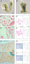
E-cadherin promoter activity is revealed in both nonneural epithelium of the cranial neural folds and the underlying mesenchyme. (A and B) E8 (4–5 som) Ecad-lacZ embryos exhibit β-gal activity in domains of the cranial NFs (white arrowheads) that are distinct from the NE. (C and E) In sections of such embryos, β-gal activity (blue cytoplasm or inclusions) is present in nonneural epithelial cells of the head (red arrowheads) and in underlying mesenchyme (white arrowheads and Insets). (G and I) PDGFRα staining (brown) performed on X-Gal-stained Ecad-lacZ embryos reveals that β-gal+ cells in the nonneural epithelium and underlying mesenchyme coexpress PDGFRα. Instances of such double-positive mesenchymal cells are indicated by white arrowheads. (Insets) Magnified cells in insets denoted by asterisks. (D, F, H, and J) Low magnifications and planes of the sections for C, E, G, and I, respectively. Red segments show the location of β-gal+ or β-gal+/PDGFRα+ cells observed in the head mesenchyme.
Fig. 6.
Fig. 6.
Schematic representation of cranial neural fold complexity during the morphogenetic events leading to neural tube formation and EMT. (A) Based on combined staining of all transgenic embryos, the green shaded area on the diagram suggests the general location of the inferred metablast domain within the NF epithelium. Note that this domain is distinct, at this developmental stage, from the locations of cranial placodes (blue shaded areas) that represent later sources of dispersing cells in the head. n, nasal placode; t, trigeminal placode; o, otic placode; e, epibranchial placodes. (B) Schematic summary of EMT events in metablast and neural crest epithelium. As inferred from immunostaining results, the NF epithelium has distinct domains with sharply defined boundaries: The E-cad+, nonneural cells of NF epithelium are represented by red circles. PDGFRα-IR cells within, and delaminating from, the E-cad+ NF epithelium are represented by green circles. NC cells, which arise in and begin to delaminate from the mediodorsal NE domain, are represented by blue circles. In these diagrams, PDGFRα+ cells in the nonneural epithelium overlying and lateral to the NE (a) delaminate precociously into underlying cell-free interstitial spaces (b). This nascent EM, like somite-derived sclerotomal mesenchyme, then establishes the interstitial environment into which neural crest cells delaminate from the dorsal NE (c). As NFs fuse in the midline to form the NT (d), nonneural (E-Cad+) epithelium (red) separates from the dorsal NE of the nascent NT (blue), EM (green) occupies the interstitial spaces between neural and nonneural epithelia, and NC cells (blue) intermingle with and disperse ventrolaterally among the EM cells, similar to trunk crest cell dispersal among sclerotomal mesenchyme.

References

    1. Thiery JP, Sleeman JP. Complex networks orchestrate epithelial-mesenchymal transitions. Nature Rev Mol Cell Biol. 2006;7:131–142. - PubMed
    1. Le Lievre CS, Le Douarin NM. Mesenchymal derivatives of the neural crest: Analysis of chimaeric quail and chick embryos. J Embryol Exp Morphol. 1975;34:125–154. - PubMed
    1. Hall BK, Hörstadius S. The Neural Crest. London: Oxford Univ Press; 1988.
    1. Le Douarin NM, Kalcheim C. The Neural Crest. 2nd Ed. Cambridge Univ Press; 2000.
    1. Weston JA, et al. Neural Crest and the origin of ectomesenchyme: Neural fold heterogeneity suggests an alternative hypothesis. Devel Dyn. 2004;229:118–130. - PubMed

Publication types