

Evaluation of time domain and spectral domain optical coherence tomography in the measurement of diabetic macular edema
- PMID: 18515567
- PMCID: PMC2574838
- DOI: 10.1167/iovs.08-2113
Evaluation of time domain and spectral domain optical coherence tomography in the measurement of diabetic macular edema
Abstract
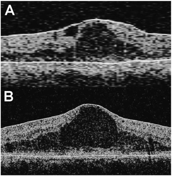
Purpose: To evaluate macular thickness and volume measurements and their intrasession repeatability in two optical coherence tomography (OCT) systems: the Stratus OCT, a time domain system, and the Cirrus HD-OCT, a spectral domain system (both by Carl Zeiss Meditec, Inc., Dublin, CA), in the context of diabetic macular edema (DME).
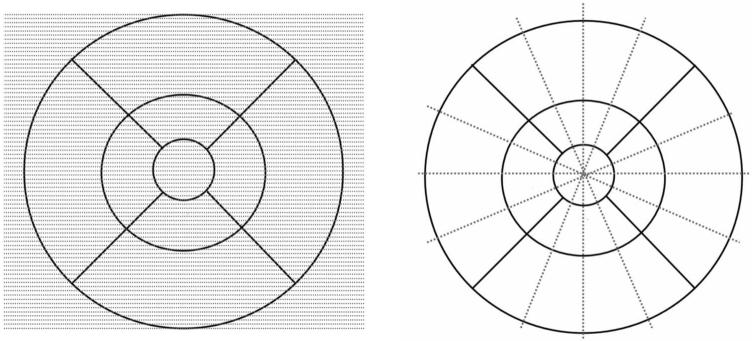
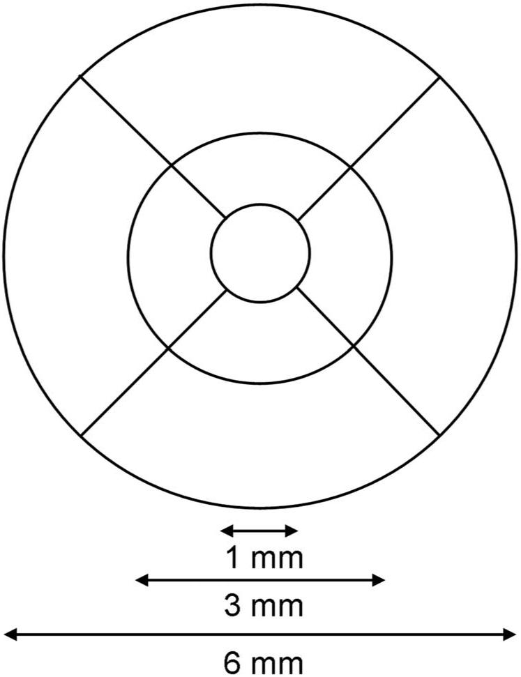
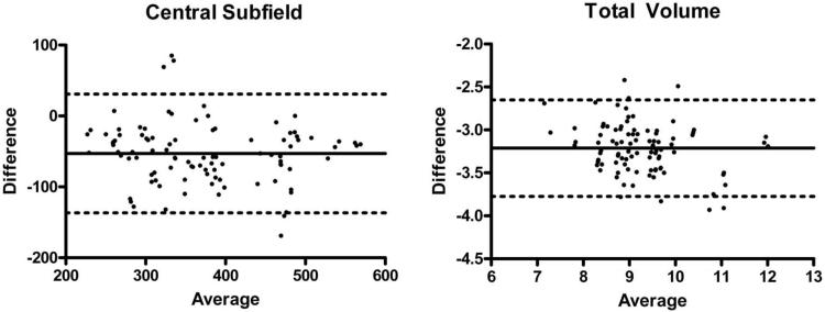
Methods: Thirty-three eyes of 33 diabetic patients with clinically significant macular edema (CSME) were scanned in a single session by a single operator on both OCT systems. Macular thickness measurements of nine standard macular subfields and total macular volume were obtained and analyzed. Bland-Altman plots were constructed to assess agreement in macular measurements. Intraclass correlation coefficients (ICCs), coefficients of repeatability (CR(W)), and coefficients of variation (CV(W)) were used to assess intrasession repeatability.
Results: Macular thickness in nine retinal subfields and macular volume were significantly higher in the Cirrus HD-OCT system compared with the Stratus OCT system. Subfield thickness and total volume measurements, respectively, were 30 to 55 microm and 3.2 mm(3) greater for the Cirrus HD-OCT system compared with the Stratus OCT system. Both Stratus OCT and Cirrus HD-OCT systems demonstrated high intrasession repeatability, with overlapping ranges for CR(W), CV(W), and ICC. Repeatability measures (CR(W) and CV(W)) differed significantly between systems in only one of nine subfields (outer temporal subfield).
Conclusions: Absolute measures of macular thickness and volume in patients with DME differed significantly in magnitude between the Stratus OCT and Cirrus HD-OCT systems. However, both OCT systems demonstrated high intrasessional repeatability. Although the two systems may not be used interchangeably, they appear equally reliable in generating macular measurements for clinical practice and research.
Figures
References
-
- Jaffe GJ, Caprioli J. Optical coherence tomography to detect and manage retinal disease and glaucoma. Am J Ophthalmol. 2004;137:156–169. - PubMed
-
- Otani T, Kishi S, Maruyama Y. Patterns of diabetic macular edema with optical coherence tomography. Am J Ophthalmol. 1999;127:688–693. - PubMed
-
- Haouchine B, Massin P, Tadayoni R, et al. Diagnosis of macular pseudoholes and lamellar macular holes by optical coherence tomography. Am J Ophthalmol. 2004;138:732–739. - PubMed
-
- Sanchez-Tocino H, Alvarez-Vidal A, Maldonado MJ, et al. Retinal thickness study with optical coherence tomography in patients with diabetes. Invest Ophthalmol Vis Sci. 2002;43:1588–1594. - PubMed
-
- Politoa A, Napolitano MC, Bandello F, et al. The role of optical coherence tomography (OCT) in the diagnosis and management of retinal angiomatous proliferation (RAP) in patients with age-related macular degeneration. Ann Acad Med Singapore. 2006;35:420–424. - PubMed
Publication types
MeSH terms
Grants and funding
LinkOut - more resources
Full Text Sources
Other Literature Sources
Medical
