. 2008 Sep;49(9):3821-9.
doi: 10.1167/iovs.07-1470. Epub 2008 May 30.

Accelerated accumulation of lipofuscin pigments in the RPE of a mouse model for ABCA4-mediated retinal dystrophies following Vitamin A supplementation

Affiliations

Accelerated accumulation of lipofuscin pigments in the RPE of a mouse model for ABCA4-mediated retinal dystrophies following Vitamin A supplementation

Roxana A Radu et al. Invest Ophthalmol Vis Sci. 2008 Sep.

Abstract

Purpose: Dietary supplementation with vitamin A is sometimes prescribed as a treatment for retinitis pigmentosa, a group of inherited retinal degenerations that cause progressive blindness. Loss-of-function mutations in the ABCA4 gene are responsible for a subset of recessive retinitis pigmentosa. Other mutant alleles of ABCA4 cause the related diseases, recessive cone-rod dystrophy, and recessive Stargardt macular degeneration. Mice with a knockout mutation in the abca4 gene massively accumulate toxic lipofuscin pigments in the retinal pigment epithelium. Treatment of these mice with fenretinide, an inhibitor of vitamin A delivery to the eye, blocks formation of these toxic pigments. Here the authors tested the hypothesis that dietary supplementation with vitamin A may accelerate lipofuscin pigment formation in abca4(-/-) mice.

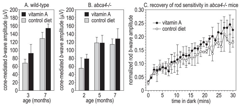
Methods: Wild-type and abca4(-/-) mice were fed normal or vitamin A-supplemented diets. Tissues from these mice were analyzed biochemically for retinoids and lipofuscin pigments. Eyes from these mice were analyzed morphologically for lipofuscin in the retinal pigment epithelium and for degeneration of photoreceptors. Visual function in these mice was analyzed by electroretinography.

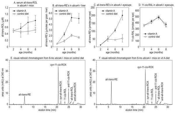
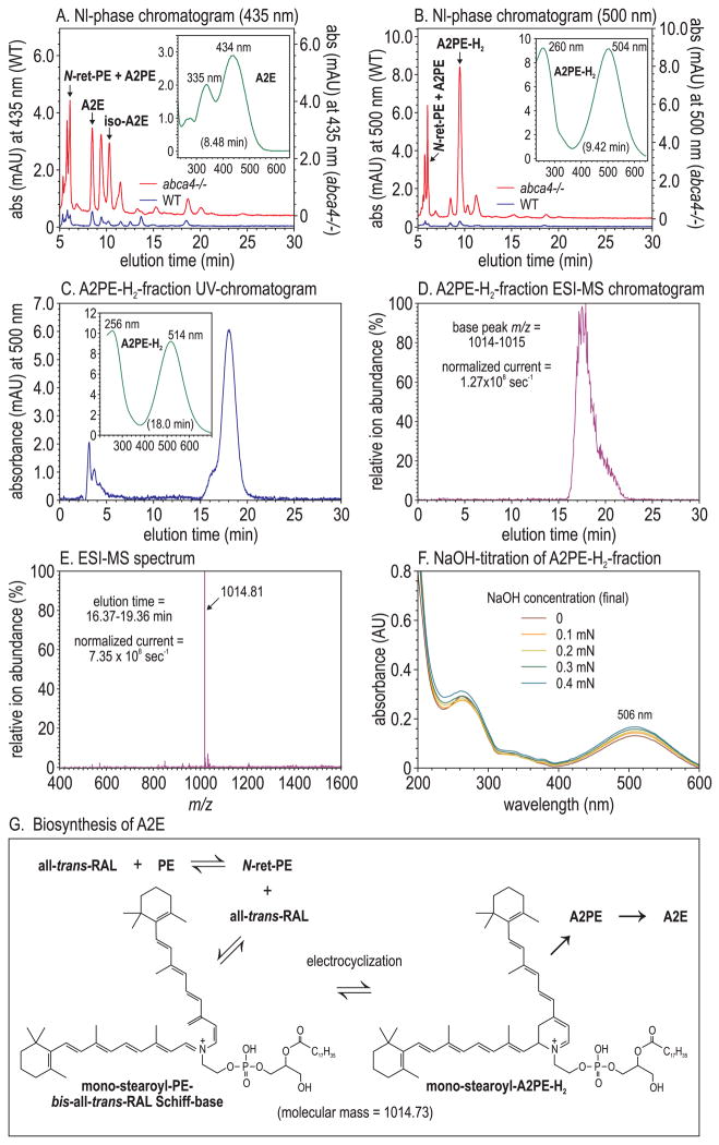
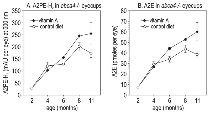
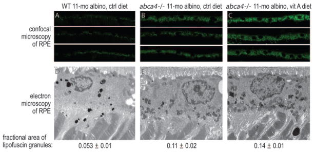
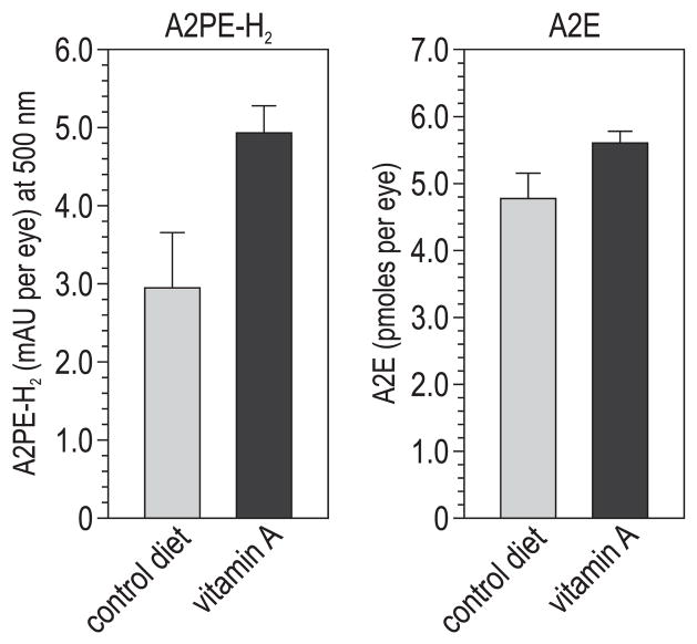
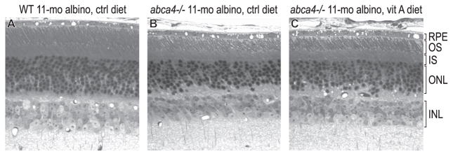
Results: Mice that received vitamin A supplementation had dramatically higher levels of retinyl esters in the liver and retinal pigment epithelium. Lipofuscin pigments were significantly increased by biochemical and morphologic analysis in wild-type and abca4(-/-) mice fed the vitamin A-supplemented diet. Photoreceptor degeneration was observed in 11-month-old albino, but not pigmented, abca4(-/-) mice on both diets.

Conclusions: Vitamin A supplementation should be avoided in patients with ABCA4 mutations or other retinal or macular dystrophies associated with lipofuscin accumulation in the retinal pigment epithelium.


Figures

Figure 1
Figure 1
Retinoids in vitamin A–supplemented and control-fed mouse tissues. (A) Serum all-trans-ROL in abca4−/− mice, expressed in micromoles per liter at the indicated ages. (B) All-trans-RE in abca4−/− livers, expressed in micromoles per gram wet weight of tissue. (C) All-trans-RE in abca4−/− eyecups, expressed in picomoles per eye. (D) 11-cis-RAL in abca4−/− eyecups, expressed in picomoles per eye. All values are shown ± SD (n = 3). (E) Representative normal-phase chromatogram (340 nm) of retinoids from the eye of an 8-month-old abca4−/− mouse on the control diet. Retinaldehydes were stabilized with hydroxylamine to form their cognate oximes (ROX) before chromatography. (F) Representative normal-phase chromatogram of retinoids from the eye of an 8-month-old abca4−/− mouse on the vitamin A–supplemented diet.
Figure 2
Figure 2
Liquid-chromatographic and mass-spectrometric analysis of A2PE-H2. (A) Normal-phase chromatograms at 435 nm eyecup-homogenate extracts from 8-month-old wild-type (blue) and abca4−/− (red) mice. A2E and iso-A2E peaks are labeled. Inset: UV spectrum of the A2E peak. (B) Normal-phase chromatograms at 500 nm of the same samples as in (A). The abca4−/−-dependent 500-nm peak is labeled A2PE-H2. Inset: UV spectrum of the A2PE-H2 peak. (C) Reverse-phase chromatogram at 500 nm of the A2PE-H2 peak-fraction (9.42 minutes) collected during normal-phase chromatography of abca4−/− eyecup homogenates (B). Inset: UV spectrum of the 18-minute (A2PE-H2) peak. (D) ESI-MS chromatogram of the sample in (C) showing the relative abundance of positive ions with 1014 to 1015 m/z. The ion current at 100% is 1.27 × 10⁸/s. (E) MS showing the relative abundance of ions detected during the 16.37- to 19.36-minute elution interval. The ion current at 100% is 7.35 × 10⁸/s. Note the near homogeneity of the 1014.81 ion. (F) Spectra of the A2PE-H2 peak fraction from (B) acquired after the addition of the indicated (final) concentrations of NaOH. (G) Molecular structure of mono-stearoyl-A2PE-H2 in equilibrium with the open-ring isomer (mono-stearyl-phosphatidylethanolamine-bis-all-trans-RAL Schiff base).
Figure 3
Figure 3
Lipofuscin fluorophores in vitamin A–treated and control-fed abca4−/− mouse eyes. (A) A2PE-H2 expressed as milliabsorbance units (mAU) per eye in mice of the indicated ages. (B) A2E expressed as picomoles per eye. All values are shown ±SD (n = 3).
Figure 4
Figure 4
Morphologic analysis of retinas from 11-month-old albino wild-type (WT) mice on the control diet, abca4−/− mice on the control diet, and abca4−/− mice on the vitamin A (vit A)–supplemented diet. (AC) Laser confocal images of RPE from the indicated mice showing lipofuscin autofluorescence. (DF) Electron microscopic images of RPE cells from the indicated mice. Lipofuscin pigment granules are the small, irregularly shaped electron-dense bodies in the RPE cytoplasm. The areas (±SD) of lipofuscin pigment granules divided by total cytoplasmic areas are shown below the electron micrographs. Lipofuscin granules in the wild-type RPE section appear darker because of more intense staining with osmium salts.
Figure 5
Figure 5
Lipofuscin fluorophores in 6-month-old wild-type (129/Sv) mice on the control or vitamin A–supplemented diet. A2PE-H2 levels are shown in milliabsorbance units (mAU) per eye at 500 nm. A2E levels are shown in picomoles per eye. All values are shown ±SD (n = 3).
Figure 6
Figure 6
Light micrographs of retinas from 11-month-old albino mice. (A) Wild-type mouse. (B) abca4−/− mouse fed the control diet. (C) abca4−/− mouse fed the vitamin A–supplemented diet. IS, inner segment; INL, inner nuclear layer; ONL, outer nuclear layer. Note the reduced ONL and OS thicknesses in abca4−/− mice, indicating partial photoreceptor degeneration and OS shortening.
Figure 7
Figure 7
Electroretinographic analysis. (A) Cone-mediated b-wave amplitudes in wild-type (129/Sv) mice of the indicated ages on the control or vitamin A–supplemented diet (n = 3–4). (B) Cone-mediated b-waves in pigmented abca4−/− mice on control or vitamin A–supplemented diets (n = 4–6). (C) Rod-mediated b-wave amplitudes as a fraction of the dark-adapted rod b-wave amplitude at the indicated times in the dark after exposure of 8-month-old abca4−/− mice to a light that bleached approximately 90% of the rhodopsin. All values are shown ±SE (n = 3).

References

    1. Bunker CH, Berson EL, Bromley WC, Hayes RP, Roderick TH. Prevalence of retinitis pigmentosa in Maine. Am J Ophthalmol. 1984;97:357–365. - PubMed
    1. Berson EL, Rosner B, Sandberg MA, et al. A randomized trial of vitamin A and vitamin E supplementation for retinitis pigmentosa. Arch Ophthalmol. 1993;111:761–772. - PubMed
    1. Hartong DT, Berson EL, Dryja TP. Retinitis pigmentosa. Lancet. 2006;368:1795–1809. - PubMed
    1. Klevering BJ, Yzer S, Rohrschneider K, et al. Microarray-based mutation analysis of the ABCA4 (ABCR) gene in autosomal recessive cone-rod dystrophy and retinitis pigmentosa. Eur J Hum Genet. 2004;12:1024–1032. - PubMed
    1. Allikmets R, Singh N, Sun H, et al. A photoreceptor cell-specific ATP-binding transporter gene (ABCR) is mutated in recessive Stargardt macular dystrophy. Nat Genet. 1997;15:236–246. - PubMed

Publication types