. 2008 Dec;49(12):5346-52.
doi: 10.1167/iovs.08-1707. Epub 2008 May 30.

Reduction of the available area for aqueous humor outflow and increase in meshwork herniations into collector channels following acute IOP elevation in bovine eyes

Affiliations

Reduction of the available area for aqueous humor outflow and increase in meshwork herniations into collector channels following acute IOP elevation in bovine eyes

Stephanie A Battista et al. Invest Ophthalmol Vis Sci. 2008 Dec.

Abstract

Purpose: To understand how hydrodynamic and morphologic changes in the aqueous humor outflow pathway contribute to decreased aqueous humor outflow facility after acute elevation of intraocular pressure (IOP) in bovine eyes.

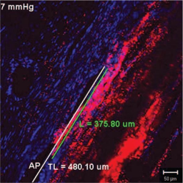
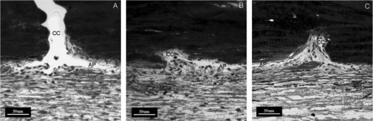
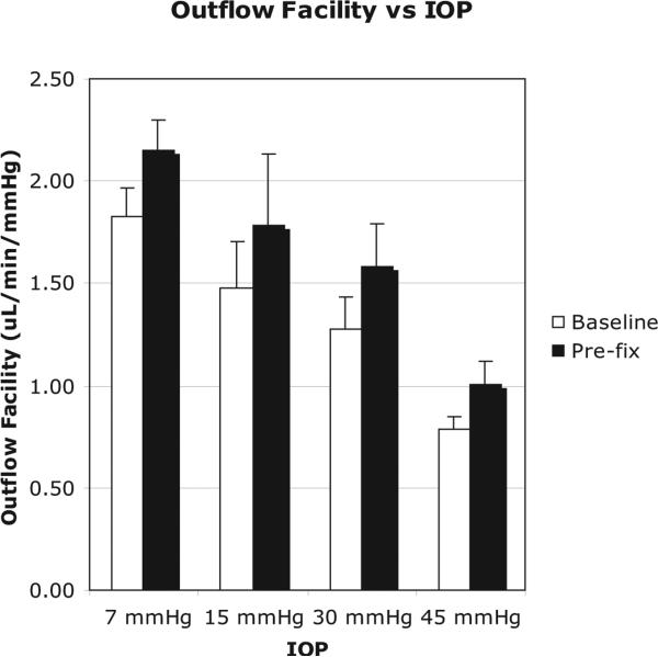
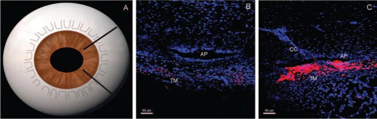
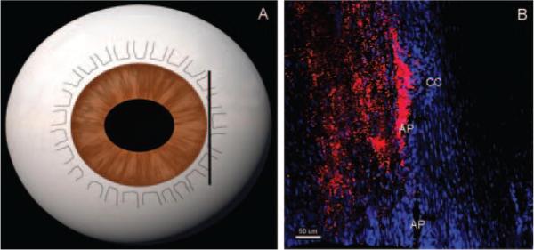
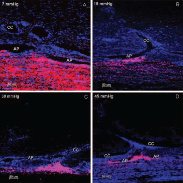
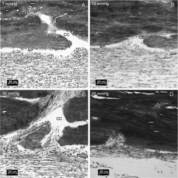
Methods: Enucleated bovine eyes were perfused at 1 of 4 different pressures (7, 15, 30, 45 mm Hg) while outflow facility was continuously recorded. Dulbecco PBS + 5.5 mM glucose containing fluorescent microspheres (0.5 mum, 0.002% vol/vol) was perfused to outline aqueous outflow patterns, followed by perfusion-fixation. Confocal images were taken along the inner wall (IW) of the aqueous plexus (AP) in radial and frontal sections. Percentage effective filtration length (PEFL; IW length exhibiting tracer labeling/total length of IW) was measured. Herniations of IW into collector channel (CC) ostia were examined and graded for each eye by light microscopy.

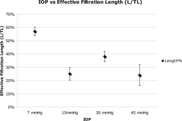
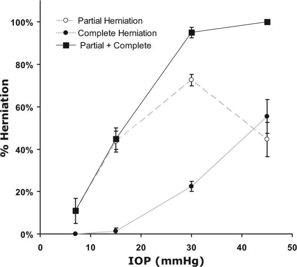
Results: Increasing IOP from 7 to 45 mm Hg coincided with a twofold decrease in outflow facility (P < 0.0001), a 33% to 57% decrease in PEFL with tracer confined more to the vicinity of CC ostia, progressive collapse of the AP, and increasing percentage of CC ostia exhibiting herniations (from 15.6% +/- 6.5% at 7 mm Hg to 95% +/- 2.3% at 30 mm Hg [P < 10(-4)], reaching 100% at 45 mm Hg).

Conclusions: Decreasing outflow facility during acute IOP elevation coincides with a reduction in available area for aqueous humor outflow and the confinement of outflow to the vicinity of CC ostia. These hydrodynamic changes are likely driven by morphologic changes associated with AP collapse and herniation of IW of AP into CC ostia.


Figures

Figure 1
Figure 1
Measurement of effective filtration length. Effective filtration length was calculated as L/TL, the ratio of the length of the tracer labeling (L, green line) to the total length of the IW (TL, white line) of AP.
Figure 2
Figure 2
Grading system for the CC ostia obstructed by herniated tissue. Light microscopic images with AP and CC ostia were graded as open (A), partially obstructed (B), or completely obstructed (C) by herniated tissue.
Figure 3
Figure 3
Relationship between outflow facility (μL/min/mm Hg) and IOP (mm Hg). As IOP increased from 7 mm Hg to 45 mm Hg, the outflow facility decreased significantly (P = 0.0024).
Figure 4
Figure 4
Tracer distribution in radial sections at 15 mm Hg. In radial sections (A), tracer distribution was dependent on proximity to the collector CC. Radial sections that do not contain a CC ostium (B) have less tracer or no tracer labeling compared with radial sections containing a CC ostium, which have more tracer labeling (red, C) along the AP. TM, trabecular meshwork.
Figure 5
Figure 5
Tracer distribution in frontal sections at 15 mm Hg. In frontal sections, tangential to the corneoscleral limbus and perpendicular to the ocular surface (A), segmental tracer distribution concentrated near the CC ostium was observed (B). Less tracer labeling was seen along the AP away from CC ostium. Scale bar, 50 μm.
Figure 6
Figure 6
Distribution of fluorescent microspheres near CC ostia at four IOPs. At 7 mm Hg (A), less segmental distribution of microspheres (red) was found along the inner wall of the AP. In contrast, at pressures of 15 mm Hg (B), 30 mm Hg (C), and 45 mm Hg (D), there was an increasing segmental pattern of microsphere distribution with a greater concentration of microspheres in the trabecular meshwork near the CC ostia. Triangular distribution of microspheres was seen when more trabecular meshwork tissue was herniated into CC ostia (C) compared with a more diffuse pattern when less trabecular mesh-work tissue (B, D) herniated into CC ostia. Collapse of AP was seen with increasing IOP. Scale bar, 50 μm.
Figure 7
Figure 7
Relationship between effective filtration length and IOP. Effective filtration length (%) measured as a function of IOP. As IOP increased, the percentage of effective filtration length decreased.
Figure 8
Figure 8
Light microscopy of the aqueous plexus and CCs at four different pressures. At 7 mm Hg (A), the aqueous plexus (AP) was open. At 15 mm Hg (B), the AP was open and the IW tissue and JCT tissue were partially herniated into the CC ostia. At 30 mm Hg (C) and 45 mm Hg (D), the AP was collapsed adjacent to the CC ostia. There was a more dramatic herniation of the IW and JCT into the CC ostia. Scale bar, 50 μm.
Figure 9
Figure 9
Percentage ostia obstructed by herniated tissue as a function of IOP. As IOP increased, the number and degree of CC ostia obstructed by herniated IW and JCT increased.

References

    1. Quigley HA. Number of people with glaucoma worldwide. Br J Ophthalmol. 1996;80:389–393. - PMC - PubMed
    1. Grant WM. Clinical tonography. Trans Am Acad Ophthalmol Otolaryngol. 1951:774–781. - PubMed
    1. Grant WM. Further studies on facility of flow through the trabecular meshwork. AMA Arch Ophthalmol. 1958;60:523–533. - PubMed
    1. AGIS Investigators The Advanced Glaucoma Intervention Study (AGIS), 7: the relationship between control of intraocular pressure and visual field deterioration. Am J Ophthalmol. 2000;130:429–440. - PubMed
    1. Grant WM. Experimental aqueous perfusion in enucleated human eyes. Arch Ophthalmol. 1963;69:783–801. - PubMed

Publication types

Substances