

An autonomous CDR3delta is sufficient for recognition of the nonclassical MHC class I molecules T10 and T22 by gammadelta T cells
- PMID: 18516039
- PMCID: PMC2768525
- DOI: 10.1038/ni.1620
An autonomous CDR3delta is sufficient for recognition of the nonclassical MHC class I molecules T10 and T22 by gammadelta T cells
Abstract
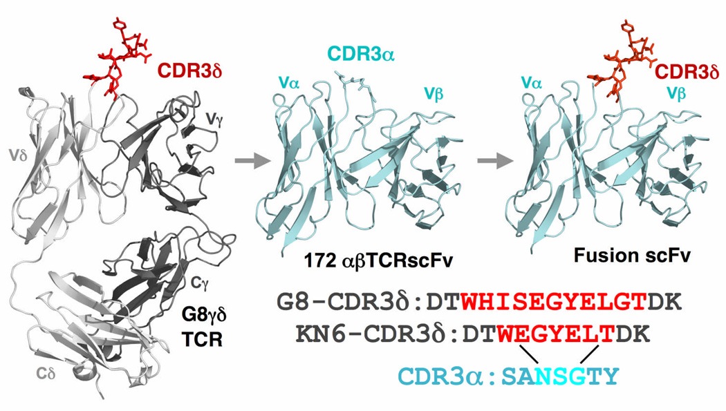
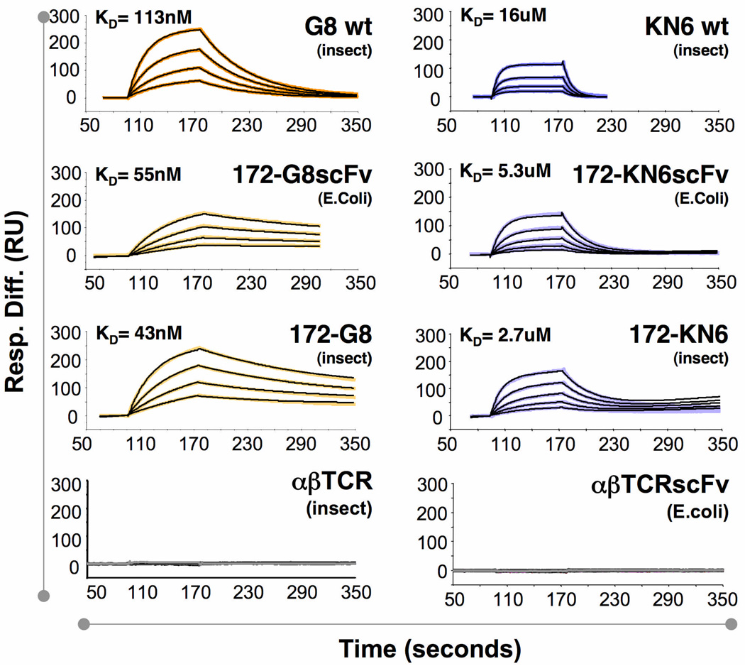
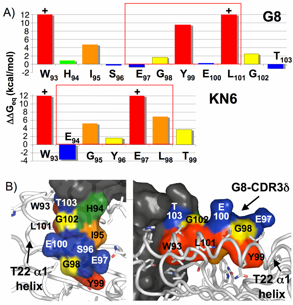
It remains unclear whether gammadelta T cell antigen receptors (TCRs) detect antigens in a way similar to antibodies or alphabeta TCRs. Here we show that reactivity between the G8 and KN6 gammadelta TCRs and the major histocompatibility complex class Ib molecule T22 could be recapitulated, with retention of wild-type ligand affinity, in an alphabeta TCR after grafting of a G8 or KN6 complementarity-determining region 3-delta (CDR3delta) loop in place of the CDR3alpha loop of an alphabeta TCR. We also found that a shared sequence motif in CDR3delta loops of all T22-reactive gammadelta TCRs bound T22 in energetically distinct ways, and that T10(d), which bound G8 with weak affinity, was converted into a high-affinity ligand by a single point mutation. Our results demonstrate unprecedented autonomy of a single CDR3 loop in antigen recognition.
Figures
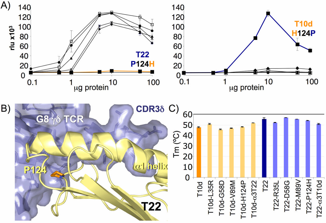
 =α3, ●=T22 and
  =negative control, m157) and T10d (right; ◆=L35R, □=G58D, ▲=V89M, ■=H124P,
  =α3, ●=T10d and
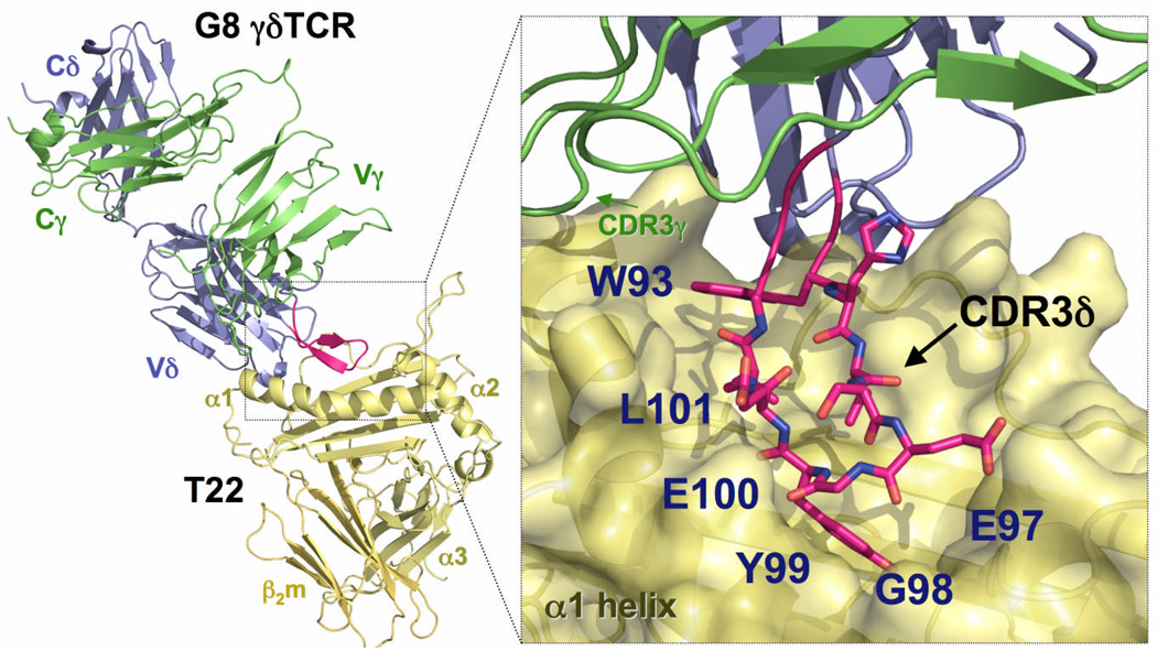
  =negative control, m157). Graphs depicting P124H and H124P mutants are drawn in orange and blue, respectively. All stimulation assays were performed in triplicate. b) Location of position 124 in the interface between the G8 γδ TCR and T22. The proline is colored in orange, and is located on the edge of the platform domain of T22, in direct contact with the G8 γδ TCR. c) Melting temperatures (Tm) of wild-type and mutant T22 and T10d. At least two independent measurements were made for each sample; error bars represent the standard error of these measurements.
References
-
- Adams EJ, Chien YH, Garcia KC. Structure of a gammadelta T cell receptor in complex with the nonclassical MHC T22. Science. 2005;308:227–231. - PubMed
-
- Allison TJ, Winter CC, Fournie JJ, Bonneville M, Garboczi DN. Structure of a human gammadelta T-cell antigen receptor. Nature. 2001;411:820–824. - PubMed
-
- Battistini L, et al. Phenotypic and cytokine analysis of human peripheral blood gamma delta T cells expressing NK cell receptors. J Immunol. 1997;159:3723–3730. - PubMed
-
- Ferrick DA, et al. Differential production of interferon-gamma and interleukin-4 in response to Th1- and Th2-stimulating pathogens by gamma delta T cells in vivo. Nature. 1995;373:255–257. - PubMed
Publication types
MeSH terms
Substances
Grants and funding
LinkOut - more resources
Full Text Sources
Other Literature Sources
Research Materials
