Review
. 2008 May 30;4(5):e1000018.
doi: 10.1371/journal.ppat.1000018.

Viral control of mitochondrial apoptosis

Affiliations
Review

Viral control of mitochondrial apoptosis

Lorenzo Galluzzi et al. PLoS Pathog. .

Abstract

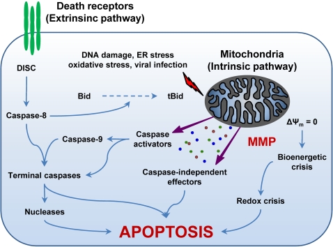
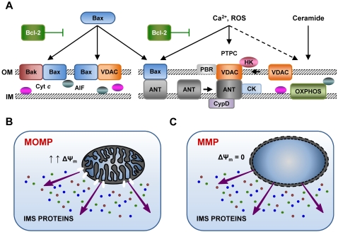
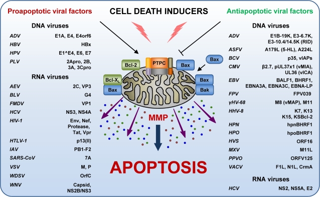
Throughout the process of pathogen-host co-evolution, viruses have developed a battery of distinct strategies to overcome biochemical and immunological defenses of the host. Thus, viruses have acquired the capacity to subvert host cell apoptosis, control inflammatory responses, and evade immune reactions. Since the elimination of infected cells via programmed cell death is one of the most ancestral defense mechanisms against infection, disabling host cell apoptosis might represent an almost obligate step in the viral life cycle. Conversely, viruses may take advantage of stimulating apoptosis, either to kill uninfected cells from the immune system, or to induce the breakdown of infected cells, thereby favoring viral dissemination. Several viral polypeptides are homologs of host-derived apoptosis-regulatory proteins, such as members of the Bcl-2 family. Moreover, viral factors with no homology to host proteins specifically target key components of the apoptotic machinery. Here, we summarize the current knowledge on the viral modulation of mitochondrial apoptosis, by focusing in particular on the mechanisms by which viral proteins control the host cell death apparatus.


Conflict of interest statement

The authors have declared that no competing interests exist.

Figures

Figure 1
Figure 1. The Extrinsic and the Intrinsic (Mitochondrial) Pathways of Apoptosis.
The extrinsic apoptotic pathway involves the activation of death receptors at the cell surface, followed by a caspase cascade that eventually leads to the execution of cell death. In contrast, different proapoptotic stimuli initiate the intrinsic pathway by triggering mitochondrial membrane permeabilization (MMP). Following MMP, intermembrane space proteins are released into the cytosol, the mitochondrial transmembrane potential (Δψm) is dissipated, and the bioenergetic and redox-detoxifying functions of mitochondria are compromised. The resulting bioenergetic and redox crises, associated with the activation of both caspase-dependent and -independent executioner mechanisms, commit the cell to death. The two pathways are interconnected by the BH3-only protein Bid, whose truncated form (tBid) is generated by caspase-8 and can target mitochondria to trigger MMP. For a more detailed description of the intrinsic and extrinsic pathways of apoptosis please refer to the Introduction and to . DISC, death-inducing signaling complex; ER, endoplasmic reticulum.
Figure 2
Figure 2. Different Models of Mitochondrial Membrane Permeabilization (MMP).
Large pores formed by the oligomerization of proapoptotic Bcl-2 proteins (e.g., Bax, Bak) and/or the voltage-dependent anion channel (VDAC) may promote selectively mitochondrial outer membrane permeabilization (MOMP). In this case, specific intermembrane space (IMS) proteins are liberated in the cytosol, but the mitochondrial transmembrane potential (Δψm) is (at least initially) retained (A,B). On the contrary, some proapoptotic stimuli, such as calcium (Ca2+) overload, reactive oxygen species (ROS), and the lipid second messenger ceramide, favor MMP by inducing the permeabilization of the inner mitochondrial membrane (IM) via the activation of the permeability transition pore complex (PTPC). When the PTPC opens, Δψm is immediately lost and an unregulated entry of solutes and water into the mitochondrial matrix occurs. This results in the osmotic swelling of mitochondria, followed by rupture of both mitochondrial membranes and the unspecific release into the cytosol of IMS proteins (A,C) (please refer to the sections “MMP Regulation by Bcl-2 Family Proteins” and “MMP Regulation by the PTPC” for additional details). Notably, antiapoptotic proteins from the Bcl-2 family play a role in both models. AIF, apoptosis-inducing factor; ANT, adenine nucleotide translocase; CK, creatine kinase; CypD, cyclophilin D; Cyt c, cytochrome c; HK, hexokinase; OXPHOS, oxidative phosphorylation complexes; PBR, peripheral-type benzodiazepine receptor.
Figure 3
Figure 3. Control of Mitochondrial Membrane Permeabilization (MMP) by Viral Proteins.
A number of viral polypeptides modulate apoptosis, either by favoring or inhibiting MMP. This control can be exerted directly at mitochondria, or on upstream/downstream steps of the apoptotic cascade (please refer to the section “Viral Modulation of Mitochondrial Apoptosis” for further details). ADV, adenovirus; AEV, avian encephalomyelitis virus; ASFV, African swine fever virus; BLV, bovine leukemia virus; BCV, baculovirus; CMV, cytomegalovirus; EBNA, Epstein–Barr nuclear antigen; EBV, Epstein–Barr virus; Env, envelope glycoprotein complex; FMDV, foot-and-mouth disease virus; FPV, fowlpox virus; γHV-68, γ-herpesvirus 68; HBV, hepatitis B virus; HBx, HBV X protein; HCV, hepatitis C virus; HHV-8, human herpesvirus 8; HIV-1, human immunodeficiency virus 1; HPN, herpesvirus pan; HPO, herpesvirus papio; HPV, human papillomavirus; HTLV-1, human T lymphotropic virus 1; HVS, herpesvirus saimiri; IAV, influenza A virus; KSBcl-2, Kaposi sarcoma Bcl-2; M, matrix protein; MXV, myxoma virus; NS, non structural protein; ORF, open reading frame; P, phosphoprotein P; PPVO, parapoxvirus ORF virus; PTPC, permeability transition pore complex; PLV, poliovirus; RID, receptor internalization and degradation complex; SARS-CoV, severe acute respiratory syndrome coronavirus; VACV, vaccinia virus; vIAPs, viral inhibitor of apoptosis proteins; vICA, viral inhibitor of caspase-8 activation; vMAP, viral mitochondrial antiapoptotic protein; Vpr, viral protein R; VSV, vesicular stomatitis virus; WDSV, Walleye dermal sarcoma virus; WNV, West Nile virus.

References

    1. Kroemer G, Galluzzi L, Brenner C. Mitochondrial membrane permeabilization in cell death. Physiol Rev. 2007;87:99–163. - PubMed
    1. Chipuk JE, Kuwana T, Bouchier-Hayes L, Droin NM, Newmeyer DD, et al. Direct activation of Bax by p53 mediates mitochondrial membrane permeabilization and apoptosis. Science. 2004;303:1010–1014. - PubMed
    1. Wang HG, Pathan N, Ethell IM, Krajewski S, Yamaguchi Y, et al. Ca2+-induced apoptosis through calcineurin dephosphorylation of BAD. Science. 1999;284:339–343. - PubMed
    1. De Maria R, Lenti L, Malisan F, d'Agostino F, Tomassini B, et al. Requirement for GD3 ganglioside in CD95- and ceramide-induced apoptosis. Science. 1997;277:1652–1655. - PubMed
    1. Ricci JE, Munoz-Pinedo C, Fitzgerald P, Bailly-Maitre B, Perkins GA, et al. Disruption of mitochondrial function during apoptosis is mediated by caspase cleavage of the p75 subunit of complex I of the electron transport chain. Cell. 2004;117:773–786. - PubMed

Publication types

Substances