. 2008 May 30;4(5):e1000085.
doi: 10.1371/journal.pgen.1000085.

Systematic identification of genes that regulate neuronal wiring in the Drosophila visual system

Affiliations

Systematic identification of genes that regulate neuronal wiring in the Drosophila visual system

Jürg Berger et al. PLoS Genet. .

Abstract

Forward genetic screens in model organisms are an attractive means to identify those genes involved in any complex biological process, including neural circuit assembly. Although mutagenesis screens are readily performed to saturation, gene identification rarely is, being limited by the considerable effort generally required for positional cloning. Here, we apply a systematic positional cloning strategy to identify many of the genes required for neuronal wiring in the Drosophila visual system. From a large-scale forward genetic screen selecting for visual system wiring defects with a normal retinal pattern, we recovered 122 mutations in 42 genetic loci. For 6 of these loci, the underlying genetic lesions were previously identified using traditional methods. Using SNP-based mapping approaches, we have now identified 30 additional genes. Neuronal phenotypes have not previously been reported for 20 of these genes, and no mutant phenotype has been previously described for 5 genes. The genes encode a variety of proteins implicated in cellular processes such as gene regulation, cytoskeletal dynamics, axonal transport, and cell signalling. We conducted a comprehensive phenotypic analysis of 35 genes, scoring wiring defects according to 33 criteria. This work demonstrates the feasibility of combining large-scale gene identification with large-scale mutagenesis in Drosophila, and provides a comprehensive overview of the molecular mechanisms that regulate visual system wiring.


Conflict of interest statement

The authors have declared that no competing interests exist.

Figures
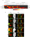
Figure 1
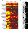
Figure 1. Classification of visual system wiring mutants.
(A) Diagnostic phenotypic defects for the four major mutant classes, scored on a scale from 0 (no defect, black) to 4 (most severe defect, yellow). “R8 defects” is an average of all R8 phenotypes (Table S1). (B) Wild-type visual system anatomy and examples of mutants in each class. From left to right, images show: DV axons, whole-mount larval eye-brain complexes stained with mAb24B10 to visualize all R-axons (red) and anti-β-galactosidase to visualize dorsal and ventral axons expressing an omb-τlacZ reporter (green); R1–R6 axons, adult brain sections stained with anti-β-galactosidase to visualize R1–R6 axons expressing an Rh1-τlacZ reporter; R8-axons, confocal sections of adult brains stained with mAb24B10 (red) and anti-GFP to visualize R8-axons expressing an Rh6-GFP reporter (green); R7-axons, confocal sections of adult brains stained with mAb24B10 (red) and anti-GFP to visualize R7 axons expressing an Rh4-GFP reporter (green). tai and enok illustrate stalling and ventral mistargeting of dorsal omb-τlacZ axons, respectively (arrowheads). In cdk8 clones, some R1–R6 axons project through the lamina and across the optic chiasm into the medulla (arrowhead). R8-and R7-axons are disorganized in gogo clones, and some R8-axons extend to the R7 target layer (arrowheads). For the larval eye-brain complexes, dorsal is up and anterior left; for adult brain sections, anterior is up and lateral left. Scale bars, 50 µm.
Figure 2
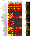
Figure 2. Axon growth genes.
(A) Full phenotypic analysis of mutants in the axon growth class, scored for all defect criteria as in Figure 1A. (B) Whole-mount larval eye-brain complexes stained with mAb24B10 to visualize all R-axons (red) and anti-β-galactosidase to visualize dorsal and ventral axons expressing an omb-τlacZ reporter (green). Arrowheads indicate delayed or stalled axons; arrow indicates polar axons misrouted to the equatorial regions of the optic lobe. Scale bar, 50 µm. (C) Quantification of stalling defects, scored by counting the percentage of larval eye-brain complexes in which at least some omb-τlacZ axons failed to extend fully within the optic lobe, as visualized by X-gal stainings, (n).
Figure 3
Figure 3. Topographic mapping genes.
(A) Phenotypic analysis of enok and Br140 mutations, scored for all defect criteria as in Figure 1A. (B) Whole-mount larval eye-brain complex of wild-type and eyFLP clones of enok and Br140. (i and i′) Staining of the optic lobe with mAb24B10 to visualize all R-axons (red) and anti-β-galactosidase to visualize dorsal and ventral axons expressing an omb-τlacZ reporter (green). Left panels (i) show both channels; right panels (i′) show the green channel only. Arrowheads indicate ventral misrouting of dorsal omb-τlacZ axons in the enok and Br140 mutants, which occurs at the surface of the optic lobe. (ii) Staining of the eye disc with the mitotic marker anti-phospho H3 (green). Arrowheads indicate the position of the morphogenetic furrow. In both wild-type and mutant discs, mitotic cells are observed in a dispersed pattern ahead (left) of the furrow and in a narrow zone just behind it. (iii) Staining of the eye disc with anti-elav to visual R-cell nuclei (red) and anti-β-galactosidase to visualize dorsal cells expressing an mrr-lacZ reporter (green). (iv) Staining of the eye disc with anti-elav to visual R-cell nuclei (red) and anti-β-galactosidase to visualize ventral cells expressing an fng-lacZ reporter (green). Expression of the fng-lacZ reporter is greatly reduced in the enok and Br140 eye discs (arrowheads), but remains in the antennal disc (asterisks). (C) Quantification of dorsal-to-ventral mistargeting, scored by counting the percentage of larval eye-brain complexes in which at least some (“partial”) or all (“complete”) dorsal omb-τlacZ axons projected ventrally within the optic lobe, as visualized by X-gal stainings, (n).
Figure 4
Figure 4. Lamina targeting genes.
(A) Phenotypic analysis of mutants in the lamina targeting class, scored for all defect criteria as in Figure 1A. (B) Horizontal sections through the optic lobes of adult heads, stained with anti-β-galactosidase to visualize R1–R6 axons expressing an Rh1-τlacZ reporter. Arrowheads indicate R1–R6 axons extending through the lamina into the medulla in whole-eye eyFLP clones of selected mutants.
Figure 5
Figure 5. Medulla targeting genes.
(A) Phenotypic analysis of mutants in the medulla targeting class, scored for all defect criteria as in Figure 1A. (B) Horizontal confocal sections of adult optic lobes, stained with anti-GFP to visualize R8 axons expressing an Rh6-mCD8-GFP reporter (green) and mAb24B10 to visualize all R axons (red). Animals carried whole-eye eyFLP clones of the indicated mutants. Arrowheads indicate R8-axons that overshoot their correct target layer and extend to or beyond the R7 target layer.

References

    1. Clandinin TR, Zipursky SL. Making connections in the fly visual system. Neuron. 2002;35:827–841. - PubMed
    1. Hiesinger PR, Zhai RG, Zhou Y, Koh TW, Mehta SQ, et al. Activity-independent prespecification of synaptic partners in the visual map of Drosophila. Curr Biol. 2006;16:1835–1843. - PMC - PubMed
    1. Clandinin TR, Lee CH, Herman T, Lee RC, Yang AY, et al. Drosophila LAR regulates R1-R6 and R7 target specificity in the visual system. Neuron. 2001;32:237–248. - PubMed
    1. Newsome TP, Asling B, Dickson BJ. Analysis of Drosophila photoreceptor axon guidance in eye-specific mosaics. Development. 2000;127:851–860. - PubMed
    1. Martin KA, Poeck B, Roth H, Ebens AJ, Ballard LC, et al. Mutations disrupting neuronal connectivity in the Drosophila visual system. Neuron. 1995;14:229–240. - PubMed

Publication types

Substances

LinkOut - more resources